Congenital Scoliosis Flashcards
(13 cards)
1
Q
Congenital Scoliosis
A
Congenital scoliosis is the failure of normal vertebral development during 4th to 6th week of gestation
- caused by developmental defect in the formation of the mesenchymal anlage
2
Q
Epidemiology
A
prevalence in general population estimated at 1% to 4%
3
Q
Causes
A
- most cases occur spontaneously
- maternal exposures
- diabetes
- alcohol
- valproic acid
- hyperthermia
- genetic
- uncertain
4
Q
Associated conditions
A
- may occur in isolation or with associated conditions
- with associated systemic anomalies, up to 61%
- cardiac defects - 10%
- genitourinary defects - 25%
- spinal cord malformations
- with underlying syndrome or chromosomal abnormality
- VACTERL syndrome
- in 38% to 55%
- characterized by vertebral malformations, anal atresia, cardiac malformations, tracheo-esophageal fistula, renal, and radial anomalies, and limb defects
- Klippel-Feil syndrome
- VACTERL syndrome
short neck, low posterior hairline, and fusion of cervical vertebrae
5
Q
Prognosis
A
- most rapid in the first 3 years of life
- determined by the morphology of vertebrae. Rate of progression from greatest to least is:
- unilateral unsegmented bar with contralateral hemivertebra >
- greatest potential for rapid progression (5 to10 degrees/year)
- unilateral unsegmented bar >
- fully segmented hemivertebra >
- unincarcerated hemivertebra >
- incarcerated hemivertebra >
- unsegmented hemivertebra >
- block vertebrae
- little chance for progression (<2 degrees/year)
- unilateral unsegmented bar with contralateral hemivertebra >
- presence of fused ribs increases risk of progression
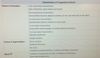
6
Q
Classification of Congenital Scoliosis
A
7
Q
Radiographs
A
- Radiographs
- recommended views
- AP and lateral plain films usually sufficient to confirm diagnosis
- recommended views
- CT
- indications
- judicious use recommended due to radiation exposure
- 3D CT useful to better delineate posterior bony anatomy and define type for surgical planning
- indications
8
Q
MRI Indications
A
indications
- all patients with congenital scoliosis prior to surgery to evaluate for neural axis abnormality (found in 20-40%) including
- Chiari malformation
- tethered cord
- syringomyelia
- diastematomyelia
- intradural lipoma
- technique
sedation required in infants so may be delayed if no surgery is planned and no neuro deficits
9
Q
Additional medical studies
10
Q
Nonoperative
A
observation and bracing
-
indications for observation
- absence of documented progression, ie:
- incarcerated hemivertebrae
- nonsegmental hemivertebrae
- some partially segmented hemivertebrae
- absence of documented progression, ie:
-
bracing
- not indicated in primary treatment of congenital scoliosis (no effectiveness shown)
- may be used to control supple compensatory curves, but effectiveness is unproven
11
Q
Operative
A
- in situ arthrodesis, anterior/posterior or posterior alone
- hemiepiphysiodesis
- osteotomy
- hemivertebrectomy
- spinal column shortening resection
12
Q
operative treatment measures
A
-
in situ arthrodesis, anterior/posterior or posterior alone
-
indications
- unilateral unsegmented bars with minimal deformity
-
indications
-
hemiepiphysiodesis
-
indications
- intact growth plates on the concave side of the deformity
- patients less than 5 yrs. with < 40-50 degree curve
- mixed results
-
indications
-
osteotomy
- osteotomy of bar
-
hemivertebrectomy
- hemivertebrae with progressive curve causing truncal imbalance and oblique takeoff
- often caused by a lumbosacral hemivertebrae
- patients < 6 yrs. and flexible curve < 40 degrees best candidates
- hemivertebrae with progressive curve causing truncal imbalance and oblique takeoff
-
spinal column shortening resection
-
indications
- deformities that present late and have severe decompensation
- rigid, severe deformities
- pelvic obliquity, fixed
-
indications
13
Q
Complications
A
- Crankshaft phenomenon
- a deformity caused by performing posterior fusion alone
- Short stature
- growth of spinal column is affected by fusion
- younger patients affected more
- growth of spinal column is affected by fusion
- Neurologic injury
- surgical risk factors include
- overdistraction or shortening
- overcorrection
- harvesting of segmental vessels
- somatosensory and motor evoked potentials important
- surgical risk factors include
- Soft-tissue compromise
- nutritional aspects of care essential to ensure adequate soft tissue healing
