CPA #2 Flashcards

(118 cards)

1
Q

Shoulder Anatomy

A
2
Q

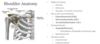
How to screen for TART: Structural of the shoulder?

A

Shoulder height

Spine of scapula

Angle of scapula

Shoulder position in sagittal plane

3
Q

What should you palpate for shoulder TART?

A

Joints: Glenohumeral, SC, AC

Myofascial: upper trapezius, levator scapulae, supraspinatus, deltoids, pectoralis, rhomboids

4
Q

ROM Flexion and Abduction of Shoulder

A

180

5
Q

ROM extension - shoulder

A

60

6
Q

ROM horizontal adduction shoulder

A

130-140

7
Q

ROM horizontal abduction shoulder

A

40-55

8
Q

ROM internal and external rotation shoulder

A

90

9
Q

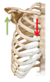
Downward rotation of scapular motion testing

A

Turning on an anterior/posterior axis so that the scapula rotates in the frontal pane to tilt the glenoid fossa downward

10
Q

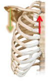
Upward rotation of scapular motion testing

A

Turning on an anterior/posterior axis so that the scapula rotates in the frontal plane to tilt the glenoid fossa upward

11
Q

Elevation of scapular motion testing

A

Superior/cephalad glide in a vertical direction along the coronal plane

Upper trapezius and levator scapulae

12
Q

Depression of scapular motion testing

A

Inferior/caudal glide in a vertical direction along the coronal plane

Lower trapezius, lower rhomboids

13
Q

Abduction (protraction) of scapular motion testing

A

Away from the spine, combined with a lateral tilt around the thorax

Serratus anterior

14
Q

Adduction (retraction) of scapular motion testing

A

Moving closer to the spine

Rhmboids and middle trapezius

15
Q

Backward tilt of scapular motion testing

A

Turning on a horizontal axis so that the posterior surface faces downward and the inferior angle is anterior

16
Q

Forward tilt of scapular motion testing?

A

Turning on a horizontal axis so that the posterior surface faces upward and the inferior angle protrudes

17
Q

Muscle Energy Steps Review

A
18
Q

MET vs ART review

A
19
Q

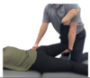
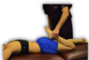
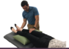
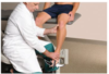
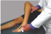
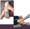
Hamstring Restriction Muscle Energy Practice

A

Diagnosis: left hip extension SD, left hamstring tenderpoint, left hamstring restriction

Physician: Standing, same side

Patient: Supine

Tx:

  1. Flex pt’s leg with knee extended. Support pt’s leg with arms or shoulder.
  2. Have patient puch heel towards table a/g physician counterforce for 3-5 seconds.
  3. Stop counterforce when pt relaxes
  4. Wait 1-2 seconds till tissues relax, take leg to next restrictive barrier.
  5. Repeat 3-5 times, ending with final push toward restrictive barrier.
  6. Reposition patient to neutral and reassess.
20
Q

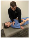
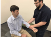
GH Joint Flexion/Extension SD MET

A
  1. Stabilize shoulder girdle with one hand, contact elbow with the other.
  2. Engage RB in flexion/extension based on diagnosis.
  3. Apply principles and steps of MET to the motions of the GH joint.
  4. Reassess
21
Q

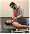
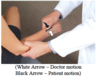
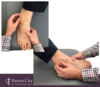
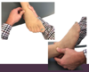
GH Joint IR/ER SD MET

A
  1. Stabilize shoulder girdle with one hand, contact wrist with the other.
  2. Engage RB in IR/ER based on diagnosis.
  3. Apply MET
  4. Reassess
22
Q

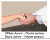
GH Joint AB/ADduction SD MET

A
  1. Stabilize shoulder girdle with one hand, contact elbow with the other
  2. Engage RB in AB/ADduction based on diagnosis.
  3. Apply MET
  4. Reassess
23
Q

GH Articulatory Tx: Spencer’s Technique

A

Patient: Lateral recument with shoulder to be treated up

Physician: Standing at side of table facing pt

Tx:

  1. Extension (every)
  2. Flexion (fine)
  3. Compression circumduction (cat)
  4. Traction circumduction (takes)
    5a. Adduction and ER (an)
    5b. Abduction (an)
  5. IR (indoor)
  6. [Pump] traction with inferior glide (pee)
24
Q

What does scapular elevation lead to in SC joint?

A

Inferior movement

Termed: SC ABduction

25
What does scapular depression lead to in SC joint?
Superior motion Termed: SC ADduction
26
In 90 degrees shoulder flexion, what does protraction of the scapular lead to at the SC joint?
**Posterior** glide of the clavicle at the SC joint Termed: SC Flexion
27
What does retraction of the scapula lead to at the SC joint?
**Anterior** glide of the clavicle at the SC joint Termed: SC extension
28
SC Joint Abduction/Adduction Dx
1. Pt is supine, examiner places **index finger on clavicle head** next to the sternum 2. The pt then **shrugs shoulder upward (abduction)** and an **inferior/caudal** movement should be palpated with normal motion at SC joint 3. Pt then lower shoulders **downward (Adduction)** and a **superior/caphalad** movement should be palpated
29
SC Joint Flexion/Extension Dx
1. Pt is supine, examiner places index finger on the clavicular head next to the sternum; pt **flexes shoulder to 90 degrees and reaches for ceiling forcefully (flexion)** 2. A **posterior** movement of the clavicle should be palpated with normal motion at SC joint 3. Patient then lowers arm back toward the table **(extension)** 4. An **anterior** movement of the clavicle should be palpated
30
SC Elevated/Adduction SD Articulatory Tx
1. Pt lying supine with neck fully flexed by physician 2. Physician places thumb over sternal end of clavicle exerting an inferior/caudal pressure 3. Pt instructed to inhale and exhale fully. During exhalation, the physician springs the clavicle inferiorly to release restriction
31
SC Joint - Articulary Technique Dx: Clavicle anterior and superior glide
1. Pt supine, examiner opposite side of SD 2. Pt helps to gap SC joint by ADducting the arm of the same side as the SD (using opposite hand to aid in the motion). Physician's opposite hand may be placed on the table under pt's axilla to create a fulcrum forthe pt to adduct a/g 3. Articulatory springing is applied **laterally, posteriorly, and inferiorly** over medial end of clavicle uding hypothenar eminence 4. Reassess
32
SC Elevated/ADducted SD MET
1. Pt supine, examiner on same side as affected shoulder 2. Dr. places one hand on proximal clavicular head. With the other, grasp pt's wrist and hold arm extended and internally rotated 3. Pt is instructed to raise arm a/g Dr.'s hand toward the ceiling (flexion at the shoulder) for 3-5 seconds then relax. Joint then brought to new barrier. Repeat until no new barriers or full ROM restored.
33
SC Horizontal Extension SD MET
1. Pt supine, examiner on side of affected shoulder 2. One hand on restricted clavicle, other hand placed behind axilla to cover axilla. Pt holds physician's shoulder with hand of affected shoulder 3. Flex the calvicle toward the manubrium until movement is palpated in the SC joint by pulling scapula anteriorly 4. Posterior force simultaneously applied to proximal calvicle from anterior to posterior to engage RB 5. Apply MET by having pt pull their shoulder down toward the table
34
AC Joint Dx Reference point: Distal end of clavicle
**Inspect for asymmetry:** superior/inferior; step-off **Palpate** **Motion screen:** cross arm adduction test; +test: pain **Assess glide** springing inferiorly on distal clavicle →Resistant to springing inferiorly: clavicle superior →Presence of sprining inferiorly: clavicle inferior
35
AC - Superior Clavicle ART
1. Pt supine with Dr. on same side 2. Dr.'s fingers monitoring AC joint and other fingers on superior aspect of clavicle; the other hand graps the pt's forearm proximal to the wrist 3. Apply a traction force in a caudad direction to gap AC joint (use enough force to register a change with the monitoring hand) 4. While maintaining the traction fforce, maximally flex the arm 5. Reassess
36
AC Joint - Direct - seated ART Dx: Right clavicle superior glide
1. Grasp elbow or forearm of dysfunctional side 2. Grasp dysfunction clavicle between thumb and fingers of free hand (**thumb on posterior/superior surface of distal clavicle - NOT on scapula)** 3. Apply anterior/inferior pressure with thumb on lateral (or posterior) aspect of clavcile while flexing pt's elbow, extending and adducting the humerous (to gap AC j) 4. Dr. holds clavcile antero-inferior (with thumb). Shoulder is extended into a circulatory sweep, posterior, superior, then anteromedial while maintaining adduction and capsular tension 5. Recheck
37
External and internal rotation ofthe humerous leads to what rotation of the clavicle at the AC joint?
External and internal rotation (respectively)
38
AC Joint IR/ER Evaluation
1. Pt seated, physician stands behind pt 2. One hand contacts and stabilizes the calvicular side of the joint with index finger over the AC joint, noting if patient has tenderness 3. Note asymmetry of joint gap compared to opposite side 4. Flex, abduct (approx 45 degrees) to maximally engage the AC component of GH rotation 5. IR and ER to assess for 90 degrees 6. Note restriction of motion and ease of motion 7. Name dysfunction based on the direction of ease of motion (IR or ER)
39
AC Internal Rotation SD MET
1. Pt seated, physician stands behind pt 2. Dr. places hand on clavicle just medial to AC joint while grasping wrist with other hand 3. Add compressive force to stabilize clavicle/AC joint while flexing, abducting and **ER** to RB 4. Apply the principles of MET by having the pt **IR** a/g Dr.'s resistance for 3-5 seconds 5. Repeat 3-5 times or until motion is fully restored 6. Reassess
40
AC External Rotation SD MET
1. Pt seated, physician stands behind pt 2. Dr. places hand on clavicle just medial to AC joint while grasping wrist with other hand 3. Add compressive force to stabilize clavicle/AC joint while flexing, abducting and **IR** to RB 4. Apply the principles of MET by having the pt **ER** a/g Dr.'s resistance for 3-5 seconds 5. Repeat 3-5 times or until motion is fully restored 6. Reassess
41
Principles governing the elbow joint
1. SD is found in the minor gliding motions of the joint, not the major motions 2. SD of the ulnohumeral joint is usually primary and SD of the radioulnar joints is usually secondary 3. Impaired function of any joint of the arm produces compensatory changes in nearby/distal joints (ie can cause secondary SD) Note\*\*: There is reciprocal or couple motion between the elbow and forearm/wrist. Reference point for naming dysfunction is distal ulna.
42
What is anterior radial head elbow SD?
East of motion with anterior glide and supination
43
What is posterior radial head elbow SD?
Ease of motion with posterior glide and pronation
44
Elbow Extension SD
Patient seated. Physician seated or standing. 1. Place elbow into flexion barrier 2. Pt gently attempts to extend eblow for 3-5 seconds while physician applies isometric counterforce 3. Pt relaxes 4. Repeat 5. Reassess
45
Elbow Flexion SD
Patient seated, standing, or supine; shoulder flexed to 90; elbow extended. Physician: seated or standing 1. Elbow into extension barrier 2. Pt flexes a/g Dr. counterforce 3. Pt relaxes 4. Repeat until SD is alleviated 5. Reassess
46
Elbow ADduction SD
Pt: seated, supine or standing; elbow flexed to 30 Physician: seated or standing 1. Place elbow in abduction barrier 2. Pt attempts to adduct elbow for 3-5 seconds with physician giving counterforce 3. Pt relaxes 4. Repeat 5. Reassess
47
Elbow ABduction SD
Pt: seated, standing, or supine; elbow flexed to 30 Dr: seated or standing 1. Place elbow into adduction barrier 2. Pt abducts a/g Dr. counterforce 3. Pt relaxes 4. Repeat 5. Reassess
48
Anterior Radial Head SD
Patient is seated and physician stands facing pt 1. Physician graps the pt's hand on the side of dysfunction, contacting the dorsal aspect of the distal radius with the thumb 2. Other hand is palm up with the thumb resting a/g the anterior and medial aspect of radial head 3. Physician pronates pt's forearm to edge of RB. Pt attempts to supination while physician applies counterforce 4. Isometric contraction helf for 3-5 seconds then pt relaxes 5. Physician then pronates pt's forearm to edge of new RB while exaggerating the posterior rotation of the radial head with the left hand 6. Repeat 7. Reassess
49
Posterior Radial Head SD
Pt is seated, physician stands in front of and to the side of the pt's dysfunctional arm 1. Physician "handshakes" the pt's dysfunctional arm, contacting the palmar aspect of the distal radius with the index finger 2. Other hand is palm up with the thumb resting a/g the posterolateral aspect of the radial head 3. Physician supinates forearm, until edge of RB 4. Pt attempts pronation w/ physician counterforce 5. Held for 3-5 seconds, pt then relaxes 6. Physician then supinates pt's forearm to new RB while exaggerating the anterior rotation of the radial head with the other hand 7. Repeat 8. Reassess
50
Radiocarpal Flexion SD
Patient seated w/ physician standing facing pt 1. Extend wrist to edge of RB 2. Pt flexes wrist a/g counterforce 3. Relax 4. New extension RB 5. Repeat 6. Reassess
51
Radiocarpal Extension SD
Patient seated w/ physician facing pt 1. Pt wrist to edge of flexion RB 2. Pt extends wrist a/g counterforce 3. Pt relaxes 4. Repeat 5. Reassess
52
Radiocarpal Adduction SD
Pt seated w/ physician standing and facing pt 1. Wrist is abducted (radial deviation) to edge of RB 2. Pt adducts a/g counterforce 3. Pt relaxes after 3-5 seconds 4. Physician takes pt to new abduction RB 5. Repeat 6. Reassess
53
Radiocarpal Abduction SD
Pt seated w/ physician standing and facing pt 1. Wrist is adducted (ulnar deviation) to edge of RB 2. Pt abducts a/g counterforce 3. Pt relaxes after 3-5 seconds 4. Physician takes pt to new adduction RB 5. Repeat 6. Reassess
54
Flexor Retinacula MFR
Pt sits on table with physician standing facing pt 1. Dr. interlaces fingers of both hands, applying a thenar eminence contact across distal radius and ulna on teh dorsal side and the wrist retinaculum on the volar side 2. Dr. maintains anteroposterior compression over the wrist while the pt actively flexes and extends the fingers 3. The pt repeats flexion and extension effors several times, mobilizing flexor tendons under the flexor retinaculum while the Dr.'s hand maintain compression resulting in distraction 4. Reassess
55
Wrist Isotonic MET
Pt seated, standing or supine Physician seated or standing 1. Physician crosses thumbs and contacts the tissue over the pt's pisiform and trapezium 2. While pt tries to flex the wrist, the Dr. applies pressure with both thumbs in a lateral direction 3. Physician lightens force slowly to allow pt to overcome the physician's force 4. Repeat 5. Reassess
56
Figure 8 Wrist Articulation
Patient seated. Physician seated or standing 1. Place the pt's wrist between the wrists of operator (perpendicularly) 2. Move the wrist in a figure 8 motion repetitively until SD is alleviated
57
Metacarpophalangeal Joint SD ART
Pt seated, standing or supine Physician seated or standing 1. Physician evaluates the motion at the metacarpophalangeal joint in flexion, extension, abduction, adduction, clockwise and counterclockwise circumduction 2. When a restriction is felt, gentle repetitive motion is made through the RB toward the anatomic barrier 3. Continue articulation until SD is alleviated
58
Proximal and Distal Interphalangeal Joint SD ART
Pt seated, standing or supine Physician seated or standing 1. Physician evaluates the motion at the DIPs and PIPs joints in flexion, extension, abduction, adduction, clockwise and counterclockwise circumduction 2. When a restriction is felt, gentle repetitive motion is made through the RB toward the anatomic barrier 3. Continue articulation until SD is alleviated
59
Hip Extension SD Muscle Energy: Hamstring Hypertonicity
Patient supine 1. Physician stabilizes contralateral ASIS w/ one hand; other hand grasps pt's leg above ankle and flexes at hip until barrier is reached 2. Pt pushes leg downward toward floor a/g physician's counterforce for 3-5 seconds 3. Pt relaxes; new RB 4. Repeat 5. Reassess for TART
60
Hip Extension SD Muscle Energy: Gluteus Hypertonicity
Pt lateral recumbent with affected side up 1. Physician stabilizes same side PSIS with one hand; other hand grasps pt's leg above ankle and flexes at hip until RB is reached; foot placed on physician's thigh 2. Pt pushes foot downward a/g physician's thigh/counterforce for 3-5 seconds 3. Pt relaxes; pushed to new RB 4. Repeat 5. Reassess for TART
61
Hip Flexion SD Muscle Energy
Pt lateral recumbent with affected side up 1. Physician faces the pt and places cephalad hand on inferior lateral angle to stabilize and monitor 2. Caudad hand proximal to the pt's knee cap adn extends the pt's leg at the hip until the RB is reached 3. Pt pushes leg anteriorly a/g counterforce 4. Pt relaxes; pushed to new RB 5. Repeat 6. Reassess for TART
62
Hip External Rotation SD Muscle Energy
Patient supine or ptone with hip and knee flexed to 90 1. Physician interally rotates pt's hip to RB 2. Pt externally rotates hip a/g counterforce 3. Pt relaxes; pushed to new RB 4. Repeat 5. Reassess for TART
63
Hip Internal Rotation SD Muscle Energy
Patient supine or ptone with hip and knee flexed to 90 1. Physician externally rotates pt's hip to RB 2. Pt internally rotates hip a/g counterforce 3. Pt relaxes; pushed to new RB 4. Repeat 5. Reassess for TART
64
Hip Abduction SD/IT Band Restriction Muscle Energy
Pt supine 1. Physician stabilizes contralateral LE just above the ankle with one hand; other hand adducts other LE to test for hypertonic abductor 2. Approx RB 3. Pt pushes LE laterally a/g counterforce for 3-5 seconds 4. Pt relaxes; pushed to new RB 5. Repeat at least 3-5 times or until no new barriers are attained 6. Reassess for TART
65
Hip Adduction SD Muscle Energy: Hypertonic Long Adductor of Lower Extremity
Pt supine with both legs straight at hip and knee Assessment: Physician uses one hand to abduct the leg that is being tested and stablizies joint proximal to pt's knee using own hip; places other hand on opposite knee to stabilize leg 1. Physican abducts pt's leg until RB met 2. Pt gently pushes the knee of affected leg into physician's hip 3. Pt relaxes; pushed to new RB 4. Repeat 5. Reassess for TART
66
Hip Adduction SD Muscle Energy: Hypertonic Short Adductor of Lower Extremity
Pt supine with non-tested leg straight at knee and hip Assessment: tested leg is ER and flexed at the knee with the foot resting a/g the thigh; for dx: physician places cephalad hand on pt's opposite hip and caudad hand medial to the pt's knee of the tested leg 1. Physician abduts pt's leg until RB met 2. Pt gently pushes knee of affected leg into physician's hand 3. Pt relaxes; pushed to new RB 4. Repeat 5. Reassess for TART
67
Hip Abduction SD/IT Band Restriction Soft Tissue/Prone
Patient: Prone Physician stands opposite of affected IT Band. 1. Physician uses caudad hand to grab foot or ankle and flexes knee to 90⁰. Cephalad hand will contact pt’s lateral thigh. 2. Physician pushes pt’s foot and lower leg out laterally while simultaneously engaging the IT Band by compressing cephalad hand into pt’s IT Band and pulling posteromedially.
68
What are the antomical landmarks when inspecting the knee?
Patella Tibial plateau Femoral condyle Collateral ligaments ACL/PCL Tibial tuberosity Fibular head \*\*ALWAYS INSPECT BILATERALLY\*\*
69
Observation and Palpation for TART - Knee
70
ROM - Flexion - Knee
145-150 (Tibiofemoral Joint) Major motion
71
ROM - Extension - Knee
0 Tibiofemoral Joint Major motion
72
Minor motions of the Tibiofemoral Joint
Internal rotation External rotation Anterior/posterior glide Abduction/Adduction
73
Define posterior glide of knee
As knee **flexes**, tibia **glides posteriorly** on femur
74
Define anterior glide of knee
As knee **extends**, tibia **glides anterioly** on femur
75
Patellar reflex
L4
76
Achilles reflex
S1
77
Review MET vs ART
78
Indications and Contraindications MET and ART
79
Post-Isometric Relaxation vs Reciprocal Inhibition (word form)
80
Post-Isometric Relaxation vs Reciprocal Inhibition (picture form)
81
Extended Tibiofemoral SD MET/ART
Patient prone Physician standing on dysfunctional side 1. **Flex** knee to RB 2. Place hand proximal to ankle of same side 3. Pt extends knee a/g counterforce 4. Pt relaxes; pushed to new RB 5. Repeat 6. Reassess
82
Flexed Tibiofemoral SD MET/ART
Patient supine with pillow under lower leg Physician standing on dysfunctional side 1. **Extend** knee to RB 2. Place caudad hand under pt's calcaneus and cephalad hand over patella 3. Pt **flexes** knee a/g counterforce (isometric contraction) 4. Pt relaxes; pushed to RB 5. Repeat 6. Reassess
83
Extended and Flexed Tibiofemoral SD Reciprocal Inhibition
1. Dx SD using TART 2. Place pt at RB 3. Have pt exert a force toward the barrier (away from name) - 3-5 seconds; 3-5 times 4. Return pt to neutral position 5. Reassess TART
84
Dx ABduction/ADduction Tibiofemoral SD
Patient supine; flex knee to 30 with hip flexion of 90 Physician standing at the side of table on dysfunctional side; facing the head of the table 1. ABduction SD: prefers knee moving outward; ankle moving inward 2. ADduction SD: prefers knee moving inward; ankle moving outward \*Variation: grasp the tibial plateu in both hands and hold distal LE between forearm and rib cage (valgus force at knee=abduction; varus stress=adduction)
85
MET/ART ABduction and ADduction Tibiofemoral SD
Patient supine, flex hip/knee to 90 Physician standing at end of table on dysfunctional side, facing towards head of table 1. Grasp tibial plateau in both hands and hold distal LE b/w forearm and rib cage (or place leg on thigh) 2. Engage RB; have pt apply equal counterforce a/g physician 3. Pt relaxes; pushed to new RB 4. Repeat/Reassess
86
ROM External Rotation Tibiofemoral Joint
10 Motion leads to anteromedial glide of tibia on femur
87
Primary restraints of ER of Tibiofemoral Joint
MCL/LCL will be taut (tight) ACL/PCL will be lax
88
ROM - Internal Rotation of Tibiofemoral Joint
10 Motion leads to posteriolateral glide of tibia on femur
89
Primary restraints of IR of Tibiofemoral Joint
ACL/PCL will be taut MCL/LCL will be lax
90
Chart w/ knee SD and ER/IR with glides
91
Dx IR/ER Tibiofemoral SD
Patient supine; hip flexed to 45; knees flexed to 90 Physician: seated, facing dysfunctional leg 1. Place hands around tibial plateau 2. ER tibia to assess for restriction of motion 3. IR tibia to assess for restriction of motion Variation: pt supine; flex hip and knee to 90; grasp tibial plateau in both hands and hold distal LE between forearm and rib cage; induce ER towards RB by physician sidebending away from midline; induce IR towards RB by physician sidebending toward midline
92
MET/ART for External Rotation Tibiofemoral SD
Patient seated with legs off table Physician seated, facing dysfunctional leg 1. Grasp **lateral** aspect of pt's foot and ankle with one hand; other hand contacts the **medial** tibial plateau to monitor motion 2. **Dorsiflex and IR** the distal tibia to RB 3. Pt **ER** a/g resistance 4. Repeat/reassess
93
MET/ART for Internal Rotation Tibiofemoral SD
Patient seated with legs off table Physician seated, facing dysfunctional leg 1. Grasp **medial** aspect of pt's foot and ankle with one hand; other hand contacts the **medial** tibial plateau to monitor motion 2. **Dorsiflex and ER** the distal tibia to RB 3. Pt **IR** a/g resistance 4. Repeat/reassess
94
Dx Proximal Fibular Head SD
Patient: seated or supine with hip flexed to 45 and knee flexed to 90 (modification: pt seated, legs hanging off table) Physician: seated at end of table on side of dysfunction, facing head of table 1. Grasp proximal fibular head b/w index finger and thumb 2. Translate anterior/posterioly noting any ease and restriction of motion
95
What glide does the Fibular Head do during foot pronation?
Anterior glide Pronation: dosriflexion, eversion, abduction
96
What glide does the Fibular Head do during foot supination?
Posterior glide Supination: plantarflexion, inversion, adduction
97
MET/ART for Posterior Fibular Head SD
Patient supine or seated, hip and knee flexed to 90 Physician: standing on dysfunctional side, facing pt 1. Cephalad hand holds fibular head b/w thumb and index finger or with the 1st MCP joint 2. Caudad hand grasp foot and engage RB by moving the foot in **pronation (dorsiflexion+eversion+abduction)** 4. Have pt **move foot medially** a/g resistance 5. Repeat/reassess
98
MET/ART for Anterior Fibular Head SD
Patient supine or seated, hip and knee straight or flexed to 90 Physician: standing on dysfunctional side, facing pt 1. Cephalad hand holds fibular head b/w thumb and index finger or place the thenar eminence on the anterior aspect of the fibular head 2. Caudad hand grasp foot and engage RB by moving the foot into **supination** (**plantarflexion, inversion, adduction**) 4. Have pt **move foot laterally** a/g resistance 5. Repeat/reassess
99
Anatomical landmarks of the foot/ankle
Medial and lateral malleolus Tenonds/ligaments Tarsal bones Metatarsal bones MTP, PIP, DIP
100
ROM - Dorsiflexion
15-20
101
ROM Plantarflexion
55-65
102
ROM - Inversion ankle
20
103
ROM Eversion ankle
10-20
104
Arches of foot - picture
105
Dx Distal Tibiofibular Joint
1. Patient supine 2. Flex knee to allow pt to plant heel on table surface (stabilize forefoot with medial hand) 3. Using a pincer grasp to the lateral malleolus: →Anterior glide: apply an anterior motion →Posterior glide: apply a posterior motion Dx: \*Anterior lateral malleolus favors anterior motion \*Posterior lateral malleolus favors posterior motion
106
Distal Fibula Anterior ART
Patient supine Physician stands at the foot of table 1. Stabilize pt's foot, wrapping fingers around calcaneus, and engage **dorsiflexion** RB 2. Thumb of lateral hand contacts the anterior aspect of the distal fibula with other thumb on top 3. Egage in RB and use articulatory technique until motion improves 4. Reassess TART
107
Distal Fibular Posterior ART
Patient prone Physician standing at the foot of the table 1. Stabilize pt's foot, wrapping fingers around calcaneus and engage **plantarflexion** RB 2. Thumb on lateral hand contacts the posterior aspect of the distal fibula with other thumb on top 3. Engage RB and use articulatory technique until motion improves 4. Reassess TART
108
Dorosiflexed Talus MET
1. Stabilize pt's ankle with one hand at the level of the malleoli 2. Place the other hand over the dorsum on the pt's foot 3. Bring the pt's foot into the plantarflexion RB 4. Pt brings foot into dorsiflexion a/g resistance 5. Pt relaxes; pushed to new RB 6. Repeat/reassess
109
Plantarflexed Talus MET
1. Stabilize pt's ankle with one hand at the level of the malleoli 2. Engage RB by contacting the sole of the pt's forefoot and **dorsiflexing** the foot 3. Pt pushes through their forefoot a/g resistance 4. Pt relaxes; pushed to new RB 5. Repeat/reassess
110
ROM inversion/eversion of Talocalcaneal (subtalar) Joint
Eversion/anteromedial glide: 5 Inversion/posterolateral glide: 5
111
Articulatory with Traction Dx: Right talus eversion with anteriomedial glide Dx: Right talus inversion with posterolateral glide
1. Grasp pt's hell with one hand and grasp the talus and dorsum of foot with the other 2. Maintain traction on calcaneus and articular inversion and eversion with a "figure 8" maneuver until no new RBs are met or quality of ROM normalizes 3. Reassess TART
112
Most likely preferences of cuboid, navicular, and cuneiforms
Cuboid = eversion glide with plantar glide Navicular = inversion glide with plantar glide Cuneiforms = plantar glide only
113
Dx Plantar Glide SD
TART changes and resistance to pressure applied to the plantar source of these bones is diagnostic
114
Plantar Guide SD MET | (Cuboid, navicular, cuneiform)
1. Grasp pt's foot with both hands on dorsum of oot, lifting the foot off the table, and dorsiflex to engage PB 2. Adjust inversion and eversion, dorsiflexion and plantarflexion to engage RB 3. Cross thumbs on plantar surface with one pad on cuboid and other on navicular with a separating force 4. Pt pushes foot into thumb; relaxes; pushed to new RB 5. Repeat/reassess
115
Cuneiform Dx
Inspection/palpation bilaterally Joint motion: dorsal/plantar glide Moving from medial to lateral dx and compare anterior and posterior glide in the 1st, 2nd, 3rd cuneifrom. Commonly, the SD present favors plantar glide; dorsal glide SD is often associated with hypertonic plantar fascia.
116
MTPs, PIPs, and DIPs ranges of motion
Flexion/extension Adduction/abduction IR/ER
117
Dx MTP and IP joints
Inspection/palpation bilaterally _3 axes of motion_ Plantar and dorsal glide Medial and lateral glide IR and ER (rotatory glide)
118
MTP/IP Joint SD ART
Patient seated or supine Physician seated or standing 1. Physician evaluates motion at the MTP joint in flexion, extension, adduction, abduction, clockwise and counterclockwise circumduction 2. When a restriction is felt, gentle repetitive motion is made through the RB toward the anatomic barrier 3. Continue articulation until SD is alleviated 4. Reassess TART