Cryptosporidia, Toxoplasma, Besnoidea, Sarcosystis Flashcards
(67 cards)

Cryptosporidium
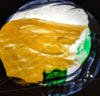
Tracheal surface
The cryptosporidia are on the surface of the epithelium on the top of the image

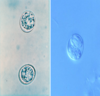
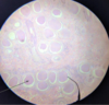
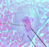
Cryptosporidium in jejunum of calf
upper arrow: normal gut part
lower arrow: Destroyed by cryptosporidium
MIDTERM SOS

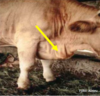
Cryptosporidia
Artificial infection
Trachea tissue

Cryptosporidium
Destroyed gut part

Cryptosporidium
Kinyoun and Ziehl Neelsen
The oocysts are purple red colour or whiteish
Can have thick or thin wall

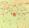
Cryptosporidium Parvum (oocyst)
Ziegl Nelsen staining

Cryptosporidium Parvum

Cryptosporidium
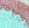
Gut
The first top arrow shows the destroyed part of the epithelium that’s why is thinner. The villi of the gut are destroyed

Toxoplasma - Tachyzoites
Stained with immunofluorescent method
What is the difference of a Bradyzoite and a Tachyzoite in toxoplasma?
Bradyzoite : In cyst and slower
Bradyzoite can affect any cell but NO RBC
Tachyzoite: Thinner, longer and faster


Toxoplasma
Tachyzoites

Toxoplasma Gondii
Tachyzoites
Giemsa staining

Toxoplasma Gondii
Bradyzoite in cells

Toxoplasma Gondii
Bradyzoite in cells

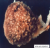
Neospora caninum tissue cyst in brain
Difference with Toxoplasma : No white wall around
What is the difference of Toxoplasma and Sarcocyst?
Sarcocyst is bigger than Toxoplasma

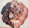
Toxoplasma cyst in brain

Toxoplasma cyst
Cross section, Vessels are seen below!

Toxoplasma cyst
Brain cells around

Toxoplasma cyst
Hematoxilin Eosin staining

- Brain cell of mouse because it is cross section of it
- Vessel
- Toxoplasma cyst

Brain cell

Toxoplasma - Bradyzoites
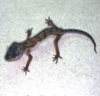
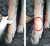
Reptilian Cryptosporidiosis
C.saurophilum
Wasting syndrome