CVS and Respiratory Anatomy Flashcards

(68 cards)

1
Q

What is the lymphatic drainage of the breast?

A
  1. breast medial to the nipple drains through intercostal spaces to lymph nodes within the thorax (internal mammary lymph nodes)
  2. lateral breast drains to lymph nodes in the axilla
2
Q

What forms the anterior axillary fold?

A

The pectoralis major

3
Q

what nervs innervate the pectoral muscles

A

Pectoralis Major:

  • Lateral and medial pectoral nerve

Pectoralis Minor

  • Medial pectoral nerve
4
Q

What innervates serratus anterior and why is this clinically relevalnt

A
  • Long thoracic nerve
  • Part of the function of SA is to hold the scapula against the ribcage
  • if this muscle is paralysed you get ‘wing’
5
Q

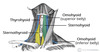
where is the cephalic vein and why is it clinically relevant

A

it’s where pacemakers are inserted through a catheter that is threaded through to the tip of the right atrium and SA node

6
Q

What are the articulations of the clavicle

A

Medially with the menubrium of the sternum: sternoclavicular joint

Laterally with the acromion of the scapula: acromioclavicular joint

7
Q

which ribs attach to serratus anterior

A

upper 8

8
Q

what innervates all 3 layers of the intercostal muscles

A

intercostal nerves (T1-T11)

9
Q

External intercostals - which direction do they run, and what is their action

A
  • 11 pairs
  • run inferiorly and mediall y
  • they elevate the ribs increasing thoracic volume
10
Q

Internal and Innermost intercostals

A
  • run inferiorly and laterally
  • they reduce thoracic volume by depressing the ribcage
11
Q

what is the main role of the intercostal muscles

and what is the clinical relevance

A

contrct just enought so that they don’t get sucked in/blown out with negative and positive pressures of breathing

in some patients, the pressure needed to breathe overcome the intercostal muscles and you can obserce ‘intercostal recession’ which is an important sign of advanced resp distress

12
Q

what can the LIMA sometimes be used for

A

coronary bypass because it runs so close to the LAD

13
Q

Label structures in this Left Lung hilum

A
14
Q

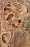
Label this Right Lung hilum

A
15
Q

Label this lovely bit of heart

A
16
Q

Label THIS lovely bit of heart

A
17
Q

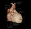
Label this third lovely bit of heart

A
18
Q

LTLBOH

A
19
Q

Does the phrenic nerve run anterior or posterior to the hilum of the lung?

A

Anterior

20
Q

where does the left vagus branch to form the left recurrant laryngeal?

A

Under the arch of the aorta. The left recurrant laryngeal passes under the ligamentum arteriosum

21
Q

What is the clinical significance of the nerve roots of the phrenic nerve?

A

painful stimulation of the diaphragm will be felt in the dermatomes supplied by the phrenic nerve nerve roots (C3, 4, and 5) this is typically in the side of neck and shoulder tip

22
Q

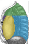
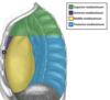
Surfaces of the heart:

A
  • Diaphragmatic: inferior
  • Sterno-costal: anterior
  • Base: posterior
23
Q

What does the AVN recieve its blood supply from?

A

The posterior interventricular artery

24
Q

What percentage of people have their posterior interventricular artery supplied by the left coronary, right coronary and how many by both?

A
  • 90% supplied by the right coronary
  • 30% supplied by the circumflex of the left coronary
  • 20% supplied by arteries from each

i.e. 70% of people are right dominant, 10% of people are left dominant, 20% of people are codominant

25
What is the crista terminalis
* This is a ridge of modified muscle that seperates the trabeculatedaurocle from the smooth walled atirum * The SA node is found in the upper half of the crista terminalis
26
What is the most posterior part of the heart
The left atrium
27
In what percentage of people is there a patency in the fossa ovalis
23% of adults
28
How might a heart valve fail?
* During systole the pressure in the heart is higher inside than outside * any reduction in blood flow will affect the inside more than the outside * the papillary muscles may die as a result and a dead one will rupture * this will lead to sudden failure of the valve as it is not held in position during systole and there is regurgitation
29
How many leaflets does the aortic valve have?
3
30
Which is the only valve in the heart that doesn't have 3 cusps? How many does it have
The mitral - it has 2
31
how many leaflets does the pulmonary valve have
3
32
What artery supplies the SA node?
The sinoatrial nodular artery * in 60% of hearts this is a branch of the RCA * in ~40% of hearts it is a branch of the left circumflex which artery supplies the SA node is not related to dominance
33
stellate ganglion
A fusion of the first thoracic and inferior cervical ganglia It lies anterior to the neck of the first rib and the C7 transverse process It lies posterior to the common carotid, internal jugular and phrenic nerve
34
Label this heckin diagram
35
to what do the different colours correspond
The border between the superior and inferior mediastinum is an imaginary line backwards from the sternal angle. NB the inferior mediastinum is subdivided into anterior, middle and posterior
36
at the level of which vertebral body does the descending thoracic aorta start
lower edge of T4
37
Azygous system of veins
* drains blood from the body walls and mediastinal viscera * empties into SVC * Formed from: * Azygous vein * Hemiazygous vein * Accessory hemiazygous vein
38
Where do the right and left vagus nerve pass through the diaphragm relative to the oesophagus
Pass through with the oesophagus at the level of T10 Left in front of the oesophagus and the right behind
39
briefly describe the journey of the thoracic duct:
from cystern of chyli it goes up on the posterior right side of the oesophagus it drains all the lymph from the lower half of the body into the blood stream at the confluence of the left subclavian vein and the internal jugular at the left side of the neck so it crosses the body - right to left
40
Where does the greater splanchnic nerve arise from and what does it supply
Ganglia on the sympathetic chain from T5-9 it supplies the foregut
41
where does the lesser splanchnic nerve arise from and what does it supply?
Ganglia on the sympathetic chain from T10-11 and it supplies the midgut
42
Where does the least splanchnic nerve arise from and what does it supply?
Ganglia on the sympathetic chain from T12 and it supplies the hindgut
43
where does the sympathetic nerve upply to the head and neck come from? Why is this clinically relevant?
through the T1 ganglion of the sympathetic chain (the stellate ganglion) Damage to this ganglion will cause loss of sympathetic innervation to the face and eye No facial sweating, drooping eye lid, constricted pupil and eye slightly draw in. HORNERS
44
which nerve enters the deep surface of the sternocleidomastoid
CN XI Spinal accessory nerve
45
What are the infrahyoid muscles?
Group of 4 muscles located inferior to the hyoid and between the two SCMs Attachements are obvious from the names * Superficial * Sternohyoid * Omohyoid: arises from scapula (omo) * Deep * sternothyroid * thyrohyoid
46
Label this bad boy
47
arterial supply to the thyroid gland
two sets of paired arteries * Superior thyroid artery * first branch to come off the external carotid * supplies superior and anterior portions * Inferior thyroid artery * arises from thryocervical trunk of subclavian * supplies posterior inferior aspects
48
venous drainage of the thyroid gland
* Superior, middle veins drain into the internal jugular * The inferior drains into the brachiocephalic
49
what does the carotid sheath contain?
carotid artery jugular vein vagus nerve
50
whats going on here?
These are scans of the thyroid the left one is normal and the right one has a thyroxine producing tumour due to negative feedback, overproduced thyroxine leads to very low TRH and TSH and therefore the healthy throid is not stimulated and doesn't produce much thyroxine
51
how can you tell the difference between a lump in the thyroid and a lump in a lymph node?
thyroid one will elevate with swallowing
52
How many ganglia does the cervical portion of sympathetic chain have
3 Superior, middle and inferior in some cases the inferior is fused with the first thoracic and forms the stellate ganglion
53
at the level of which vertebral body does the common carotid bifurcate?
At around the level of the 4th cervical vertebra
54
55
what does the glossopharyngeal nerve do?
* general sensation anf taste to the posterior 1/3 of the tongue * general sensation to the oropharynx * tympanic branch supplies middle ear and the eustachian tube * also supplies motor to stylopharyngeus muscle
56
stylopharyngeus
* supplied by the glossopharyngeal nerve * takes origin from the styloid process of the skull
57
through which nerve does the carotid sinus signal to the brain?
the glossopharyngeal
58
what does the superior laryngeal nerve innervate?
* Internal branch: * sensation to the larynx above the vocal cords * is the afferent nerve of the cough reflex * External branch: * motor to cricothyroid muscle * this is the only muscle of the larynx not innervated by the RLN
59
what does the recurrant laryngeal nerve innervate
* Motor to all muscles except the cricothyroid * Sensation to the larynx below the glottis
60
what is this nerve, what are its branches called, what do they innervate and what structure is this all happening in?
Facial nerve - Two Zulus Buggered My Cat the facial nerve enters the deep posterior aspect of the parotid gland before it splits - cancer of the parotid can cause facial paralysis
61
where does the duct from the parotid gland drain into the mouth?
a papilla adjascent to the second molar
62
why might patients who've had a brain ste, stroke have trouble swallowing?
* Glossopharyngeal provides sensation to back of pharynx * Vagus provides motor and sensory innervation to the larynx * Damage to either of these may result in loss of control of swallowing * The larynx may remain open during swallowing * Fluid can pass into the lungs
63
where is the SA node located
on the medial side of the junction between the SVC and the right atrium
64
describe the histological structure of the heart vlaves
* thick collagen fibres with occasional strands of elastic tissue * both surfaces are covered by endothelial cells
65
describe the divisions of cellular component of blood
* cells = 45% of blood * 44% is erythrocytes * White cells and platelets constitute 1% * Of that 40-75% are neutrophils * Lymphocytes 20-50% * Eosinophils ~5% * Monocytes 1-5% * Basophils ~1%
66
what can an eosionphil also be called?
an acidophilic lymphocyte NB in histology they can be identified by their dark pink granules
67
what can a basophil also be called
a basophilik leukocyte
68
from which cells are platelets derived?
megakaryocytes