Derm Flashcards

(103 cards)

1
Q

Papules and paulopustules. OPen and closed comedones.

A

Acne vulgaris

2
Q

Acne pathogenesis

A

Microcomedone

Comedone

Inflammatory papule/pustule

Nodule

3
Q

What must be present to Dx acne?

A

Comedones.

4
Q

Facial flushing, telangiectases, central face edema, burning, stinging; spares periorbital areas

A

Erythematotelangiectatic rosacea (ETR)

5
Q

Central face erythema with papules/pustules; edema of skin; less often burning, stinging; flushing less severe; spares periorbital areas

A

Papulopustular rosacea (PPR)

6
Q

Patulous follicular orifices, thickened skin, nodularities; and in men, phyma - rubbery thickening of skin nose, chin, forehead, eyelids, or ears

A

Phymatous rosacea

7
Q

Initial presentation in ~20%; more often occur after an above type; most often blepharitis; also conjunctivitis, iritis, scleritis, hypopyon, keratitis

A

Ocular rosacea

8
Q

Possible rosacea triggers

A

Hot or cold temperature, exercise, cosmetics

sunlight, spicy food, topical irritants

wind, alcohol, menopausal flushing

hot drinks, emotions, medications

9
Q

Rosacea stage 1

A

Persistent erythema with telangiectases

10
Q

Rosacea stage 2

A

Persistent erythema, telangiectases, papules, tiny pustules

11
Q

Rosacea stage 3

A

Persistent deep erythema, dense telangiectases, papules, pustules, nodules, rarely persistent “solid” edema of central face

12
Q

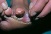
Chronic diseas of unknonw etiology. Very tender, red, inflamed nodules/abscesses. May contain double comedones.

Distributed to axilla, breast, groin.

A

Hidradenitis Suppurativa

13
Q

May occur minutes afte exposure. lesions range from erythema to vesiculation to necrosis.

A

Irritant contact dermatitis

14
Q

Delayed, cell-mediated hypersensitivity rxn

A

Allergic contact dermatitis

15
Q

Poorly defined erythematous patches with or without scales. Found on flexor surfaces. Usually begins in infancy.

A

Atopic dermatitis

16
Q

Solid plaque of lichenification, arising from a confluence of small papules. Excoriations are often present.

A

Lichen simplex chronicus

17
Q

Sudden onset of many deep-seeded, pruritic, clear, tapioca-like vesicles

A

Dyshidrosis

18
Q

Management for dyshidrosis

A

Burow wet dressings

19
Q

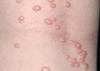
Chronic, pruritic, inflammatory dematitis in the form of coin-shpaed plaques composed of grouped small papules and vesicles on an erythematous base

A

Nummular eczema

20
Q

Common, chronic dermatitis characterized by redness and scaling and occuring where sebaceous glands are active

A

Seborrheic dermatitis

21
Q

T-cell mediated papulosquamous disease

A

Psoriasis

22
Q

Common areas for psoriasis breakouts

A

Extensor surfaces

sacrogluteal region

scalp

palms/soles

*Often bilateral and symmetric

23
Q

Three most common psoriasis subtypes

A

Chronic stable plaque psoriasis

Guttate psoriasis

Palmoplantar pustular psoriasis

24
Q

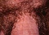
What do you call psoriasis that presents on the groin or genitals?

A

Inverse psoriasis

25
What test should you perform if you suspect guttate psoriasis?
Throat culture (looking for strep)
26
Raindrop psoriasis
Guttate
27
Middle to older aged patient with history of mild chronic stable plaque psoriasis who gets a nasty URI and has sudden guttate flare. Prognosis?
Good prognosis for return to baseline after treatment with antibiotics
28
Young adult without prior history of psoriasis gets nasty URI and has sudden guttate flare Prognosis?
Guttate lesions often slow to improve with slow shift into chronic plaque psoriasis.
29
Velvity thickening anf hyperpigmentation related to heredity, obesity, endocrine disorders, drug administration, and maligancy
Acanthos Nigricans
30
Bullous automimmune disease usually seen in the elderly.
Bullous pemphigoid
31
Often the first location for bullous pemphigoid to appear
Lower legs
32
Oval, slightly raised plaques. Salmon-colored and often begins with a Heald patch
Pityriasis Rosea
33
This lesions is a: purple polygonal pruritic planar papule
Lichen planus
34
Comon rxn pattern of blood vessels in the dermis with secondary epidermal changes. Most cases related to HSV infection.
Erythema multiforme
35
Necrotic epidermis and sloughing covering \<10% BSA
Stevens-Johnson Syndrome
36
Epidermal necrosis and sloughing \>30% BSA
Toxic epidermal Necrolysis
37
Located in derma-epidermal junction Macules or may be slightly raised
Junctional nevus
38
Located in papillary dermis Raised lesions
Compound nevus
39
Epithelial lining of a hair follicle Assymptomatic May grow to max 3 cm Central pore Usually face, neck, upper trunk
Epidermoid cyst
40
Benign subcutaneous tumor- between skin and underlying muscle layer
Lipoma
41
Most common benign epithelial tumors. Warty plaque with a "stuck on" appearance
Seborrheic keratosis
42
What do you call the light brown lesion that typically surrounds a seborrheic keratosis?
Solar lentigo
43
Single/multiple dry rough areas on sun exposed parts of body May develop into Squamous Cell CA
Actinic Keratosis
44
Which of the following is precancerous? Solar lentigo Actinic keratosis
Actinic keratosis
45
sharply demarkated, scaling, or hyperkeratotic, macule or papule, or plaque. Pink or red. May have small erosions. Capable of metastasis.
Squamous cell carcinoma
46
Pearly nodule with telangiectasis. Caused by sun damage. This is the most common CA in the humans.
Basal cell carcinoma
47
Sometime present as Red firm nodules Sometimes scaly with a crust Sometimes they bleed Often appear like a sore that does not heal What is it?
Squamous cell carcinoma
48
Slightly raised pink patch, scaling Develops from keratinocytes of the skin and mucosal membranes Slow growing, develops into squamous cell carcinoma
Bowen's disease
49
This variant of SCC is a rapidly growing nodule with a central crater
Keratocanthoma
50
Name the four types of melanoma
Superficial spreading - Most common type Nodular Lentigo maligna Acral Lentiginous Melanoma
51
Precursors to melanoma
Dysplastic nevi Congenitalnevomelanocytic nevus
52
A-asymmetric B-border (irregular) C-color (viariegated) D-Diameter (\>6mm) E-evolution (this grows fast)
Superficical spreading melanoma
53
Uniformly flat macule with geographic shape
Lentigo maligna melanoma
54
Acral lentiginous melanoma
55
Totally white macules that enlarge and can affect all of the skin. Associated with autoimmune and/or endocrine disorders
Vitiligo
56
Macular hyperpigmentation, usually of the face. Often associated with pregnancy.
Melasma
57
Sharply defined, edematous wheals. Can be erytheamtous or blanched with an erythematous border. Can be localized or general.
Uritcaria
58
Butterfly rash Diffuse or patchy alopecia ulcers or necrotic lesions on oral mucosa
Systemic lupus erythematosus
59
Raynaud phenomenon Non-pitting edema of hands and feet Sclerodactyly Periorbital edema followed by fibrosis and loss of normal face lines Cutaneous calcification
Scleroderma
60
Subcutaneous inflammatory, bright red to bluish nodules that follow a course of involved arteries. Become confluent to create painful SubQ plaques.
Polyarteritis nodosa
61
Develop over bone prominences as a result of external compression, shearing, and friction.
Pressure ulcers
62
Nonblanching erythema of intact skin
Stage 1 pressure ulcer
63
Necrosis, superficial or partial thickness.
Stage 2 pressure ulcer
64
Deep necrosis, crateriform ulceration with full thickness skin loss.
Stage three pressure ulcer
65
Full thickness necrosis with involvement of of supporting structures like muscle or bone.
Stage four pressure ulcer
66
Multifocal systemic tumor or endothelial cell origin. Ecchymotic-like macules. Can evolve into pathces/papule/plaques/nodules/tumors that are violaceous, red, pink ,or tan.
Kaposi sarcoma
67
This can mimic virtually all the morphologic expressions in dermatology.
Drug eruptions
68
Cutaneous eruption that mimics a measle-like viral exanthem
Exanthematous drug rxn
69
Dermatophytic infection of the feet
Tinea pedis
70
Tinea manuum
71
Large, scaling, well demarcated, dull red/tan/brown plaques. Associated with tinea pedis, and tinea unguium.
Tinea cruris
72
Small to large scaling, sharply marginated plaques with or without pustules or vesicles, usually at the margins. Annular configuration.
Tinea corporis
73
Most common agent of tinea corporis
T. rubrum
74
Large, round, hyperkeratotic plaque of alopecia. Can last for weeks to months.
Tinea capitis
75
Confluent, beefy red psutules on erythematous base. Satelite lesions
Cutaneous candidiasis
76
Three types of cutaneous candidiasis
Intertrigio Ocluded skin Diaper dermatitis
77
White-to-creamy plaques on oral mucosa. Caused by C. albicans
Oral candidiasis
78
Intertrigio at the angles of the lips Erythema, slight erosion Sometimes white colonies
Angular cheilitis
79
Sharply demarcated macules. Light brown lesions on light-skin, hypopigmented on dark skin. Caused by Malassezia furfur
Pityriasis Versicolor
80
Pearly white or skin-colored papules or nodules. Round or oval and umbillicated. Occur in children and sexually active adults
Molluscum contagiosum
81
Course and prognosis of molluscum contagiosum
In healthy people, MC resolves spontaneously w/o scarring but may take up to 2 years. In immunocompromised patients MC can progress desite aggressive treatment. Can become disfiguring.
82
Firm, hyperkeratotic, vegitative papules. Caused by HPV.
Verruca valgaris
83
Flat wart
Verruca plana
84
Plantar wart
Verruca plantaris
85
Generalized cutaneous eruption associated with a primary systemic infection
Infectious exanthems
86
Course, prognosis, and management of infectious exanthems
Usually self-resolves in ~10 days. Tx is supportive
87
This infectious agent typically presents with grouped vesicles arising on an erythematous base on keratinized skin or mucous membranes
Herpes simplex virus
88
Wear gloves so you don't get a...
Herpetic whitlow
89
Vesicular lesions evident in successive crops. Papule or wheals quickly evolve into dewdrop vesicles. Vesicles quickly become umbilicated, evolve into papules and crust.
Varicella
90
Okay, you had chicken pox. Mom sent you to school so your friends would get chicken pox too. Now you have a painful herpetiform lesion following a dermatomal pattern and your cursing the bed sheet that hurts to drape over yourself. You have...
Herpes zoster.
91
Get the permethrin 1%.
Pediculosis capitis
92
Check the seams of your clothes. I think you have...
Pediculosis corporis
93
I don't want to be the one to have to tell you this, but you've got...
Pediculosis pubis.
94
Gray or skin-colored ridges, linear or wavy. Common in between your fingers. All because you shared a sleeping bag on that camping trip.
Scabies! You've got scabies!
95
Okay, so you've got scabies. No one's here to judge you. Let me give you an Rx for...
Permetherin 5%
96
This common STD is most commonly associated with HPV 6 and 11
Condyloma acuminatum
97
No apparent inflammation to the skin Non-scarring, hair follicle intact
Alopecia areata
98
Chronic dermatitis of proximal nail fold and and matrix. Loss of cuticle. Separation of nail plate from proximal nail fold.
Paronychia
99
Onychomycosis aka Tinea unguium
100
Small vesicles or erosions with honey-colored crusts. S. aureas.
Impetigo
101
Bacterial skin infection involving the upper dermis that characteristically extends into the superficial cutaneous lymphatics. It is a tender, intensely erythematous, indurated plaque with a sharply demarcated border. It has a well-defined margin.
Erysipelas
102
Progressive local necrosis Associated w/ fever, headache, malaise, arthralgia & N/V Initial bite may be painless but pain typically ↑ over the next 2-8 hrs and may become severe
Brown recluse
103
Generalized muscular pain/spasms & rigidity Target lesion +/- diaphoresis at affected site (20% of patients)
Black widow bite