derm Flashcards
(421 cards)
Name the primary lesion.

Macula
This is a small spot that is not palpable & that is < 1 cm.
What is a macula?
What is a large spot that is not palpable & that is > 1 cm
A patch
Name the primary lesion.

Patch
What is a small superficial bump that is elevated & that is < 1 cm?
Papule
Name the primary lesion.

Papule
Name the primary lesion.

Plaque
What is a large superficial bump that is elevated & > 1 cm
Plaque
Name the primary lesion.

Nodule
What is a small bump with a significant deep component & is < 1 cm
Nodule
Name the primary lesion.

Tumour
What is a large bump with a significant deep component & is > 1 cm
Tumour
Name the primary lesion.

Vesicle
What is a small fluid-filled bubble that is usually superficial & that is < 0.5 cm
Vescile
Name the primary lesion.

Bulla(e)
What is a large fluid-filled bubble that is superficial or deep & that is > 0.5 cm
Bulla(e)
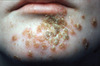
Name the primary lesion.

Pustule
What is pus containing bubble often categorized according to whether or not they are related to hair follicles
Pustule
Follicular pustule rash that is superficial, and generally multiple follicles
Folliculitis
Follicular pustule rash that is a deeper form of folliculitis.
Furuncle
A deeper follicular pustular rash that involves multiple follicles coalescing
Carbuncle
What may a nonfollicular pustule rash indicate, as opposed to a follicular rash.
Indicates systemic infection as opposed to local.
Name the primary lesion.

Cyst
What is a primary lesion?
Lesions that appear as a direct result of the pathologic process.