Dermatopathology Flashcards

1
Q

What are the functions of skin?

A
  • Enveloping barrier
  • Protection from environment
  • Motion and form
  • Appendages
  • Regulation of temperature
  • Storage unit
  • Indicator
  • Immune regulation
  • Pigmentation
  • Defense against microorganisms
  • Sensory function
  • Secretion and excretion
  • Metabolism (Vit. D3 synthesis)
  • Blood pressure
2
Q

What are the layers of the epidermis?

A
  1. Stratum corneum- keratinized cell layer
  2. Stratum lucidum- translucent cell layer
  3. Stratum granulosum- granular cell layer
  4. Stratum spinosum- prickle cell layer
  5. Stratum basale- basal cell layer
3
Q

What are the cell types of the epidermis?

A
  • Keratinocytes
  • Melanocytes
  • Langerhans cells
  • Merkel cells
  • Mast cells
4
Q

What are intercellular adhesions between keratinocytes?

A

Desmosomes (“adherens junctions”)

5
Q

What are adhesions from keratinocytes to the basement membrane?

A
  • Hemidesmosomes
  • Focal adhesions
6
Q

What comprises the dermo-epidermal junction?

A
  • Lamina lucida (bullous pemphigoid antigen, laminin, proteoglycan)
  • Lamina densa (type IV collagen)
  • Sub-lamina densa zone (type I, III, VII collagen)
7
Q

What makes up the dermis?

A
  • Collagen fibers
  • Fibroblasts
  • Dermal dendrocytes
  • Melanocytes
  • Mast cells
8
Q

What makes up the subcutis/hypodermis/panniculus?

A
  • Adipocytes
  • Skeletal muscle cells
  • Endotheliocytes
  • Smooth muscle cells
  • Nerves/ganglia
9
Q

What makes up the adnexa of skin?

A
  • Hair follicles
  • Arrector pili muscles
  • Sebaceous glands
  • Apocrine sweat glands
  • Eccrine sweat glands
10
Q

What are the phases of the hair cycle?

A
  1. Telogen
  2. Eary anagen
  3. Anagen
  4. Anagen
  5. Early catagen
  6. Catagen
11
Q

What are the blood and lymph plexuses of the skin?

A
  • Superficial (subpapillary)
  • Middle (mid-dermal)
  • Deep (subcutaneous)
12
Q

What are some challenges associated with skin diseases?

A
  • Diverse problems with similar clinical presentation
  • Conditions producing multiple patterns
  • Stereotypical response of chronic skin conditions
  • Pruritic conditions with secondary (self-inflicted) lesions
  • Misunderstanding (representing) control for cure
  • Frustrated owners
  • Need for a bigger team (owner, practitioner, pathologist, microbiologist)
13
Q

Describe this lesion.

A

Fluid-filled vesicle/cyst over the level of the surface of the skin

14
Q

Describe this lesion.

A

Pustules, raised over the surface of the skin

15
Q

Describe this lesion.

A

Solid, raised growth; papule (notice the hairs)

16
Q

Describe this lesion.

A

Nodule; histiocytoma

17
Q

Describe this lesion.

A

Hyperpigmentation within the level of the skin

18
Q

Describe this lesion from a horse.

A

Indentation; ulcer

19
Q

Describe this lesion.

A

Crust

20
Q

Describe this lesion.

A

Dry; fissures

21
Q

Describe this lesion.

A

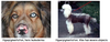
Loss of hair, hyperpigmentation

22
Q

Describe this lesion.

A

Sharp demarcation, hemorrhage, scab, ulcer

23
Q

What is the difference between a primary lesion and a secondary lesion?

A
  • Primary lesion- direct result of injury
  • Secondary lesion- develop from primary lesions over time due to healing, traumatization, secondary infection, treatment…
24
Q

This primary lesions is flat, has (dis) coloration, and is

A

Macule (color change within skin)

25
Q

This primary skin lesion is like a macule, but > 1 cm.

A

Patch

26
Q

This primary skin lesion is a solid elevation < 1 cm

A

Papule (epidermal, dermal)

27
Q

This primary skin lesion is a round-topped solid elevation > 1 cm.

A

Nodule (superficial, deep)

28
Q

This primary skin lesion is a flat-topped solid elevation > 1 cm.

A

Plaque (superficial, deep)

29
Q

This primary skin lesion is an edematous elevation in the dermis.

A

Wheal

30
Q

This primary skin lesions is a fluid-filled elevation

A

Vesicle

31
Q

This primary skin lesion is a fluid-filled elevation > 1 cm.

A

Bulla

32
Q

This primary skin lesion is a cell-filled elevation (cavity), usually with granulocytes.

A

Pustule

33
Q

This is a multi-chambered pustule.

A

Pock

34
Q

These are tightly clustered pustules.

A

Impetigo

35
Q

This primary/secondary skin lesion is defined as hair loss.

A

Alopecia

  • Thin hair coat
  • Over 30% hair loss before clinical
  • Primary: ex. follicle dysplasia like color dilution
  • Secondary: ex. trauma
36
Q

This primary/secondary skin lesion is defined as flakes of keratinized layer.

A

Scale (flake, squame)

  • Primary: hyperkeratosis (seborrhea)
37
Q

This primary/secondary skin lesion is defined as keratin cylinders extending from follicles.

A

Follicular cast

38
Q

This primary/secondary skin lesion is defined as the plugging of hair follicles by keratin and sebum.

A

Comedone

  • “Black heads”
  • Ex. follicular hyperkeratosis (also epidermal hyperplasia)
39
Q

This primary/secondary skin lesion is defined as too much or too little pigment.

A

Pigmentary disturbances (hypermelanosis, [hypo]leukoderma, [hypo]leukotrichia)

40
Q

This secondary lesion is a ring of scales that progresses outward.

A

Epidermal collarette (“collar of crusts”)

41
Q

This secondary lesion are dried consolidations/exudate on the skin (serum, cells…).

A

Crust

42
Q

This secondary lesion is a loss of epidermis (basement membrane intact).

A

Erosion

43
Q

This secondary lesion is a loss of epidermis and basement membrane zone.

A

Ulcer

44
Q

This secondary lesion is a linear loss of epidermis; traumatic abrasion.

A

Excoriation

45
Q

This secondary lesion is a crack.

A

Fissure

46
Q

This secondary lesion is thickened, hardened, rough skin.

A

Lichenification

47
Q

This secondary lesion is fibrous tissue replacing dermis/subcutis.

A

Scar (thin or thick)

48
Q

This secondary lesion is a draining tract.

A

Fistula

49
Q

What is the epidermal reaction to injury where keratin builds up on top of the skin surface?

A

Hyperkeratosis

50
Q

Which type of hyperkeratosis is characterized by anuclear keratinocytes and hypergranulosis?

  1. Orthokeratotic
  2. Parakeratotic
A
  1. Orthokeratotic (keratin loses nuclei by apoptosis and piles up on the skin surface; more common)
51
Q

Which type of hyperkeratosis is characterized by keratinocytes with nuclei and hypogranulosis?

  1. Orthokeratotic
  2. Parakeratotic
A
  1. Parakeratotic (keratin retains nuclei, blue dots in picture)
52
Q

What is the epidermal reaction to injury where the epidermis builds up with keratin and also cell layers of the skin?

A

Epidermal hyperplasia (if mostly stratum spinosum = acanthosis)

53
Q

What is the epidermal reaction to injury where there is abnormal development?

A

Dysplasia

  • Can be due to repeated irritation (mechanical or chemical)
54
Q

What is the epidermal reaction to injury where there is premature keratinization?

A

Dyskeratosis (feature of dysplasia)

  • Can be due to repeated irritation (mechanical or chemical)
55
Q

What is the difference between epidermal hyperplasia and dysplasia?

A
  • Epidermal hyperplasia
    • Often secondary = indication of chronicity
    • Sometimes primary problem e.g. seborrhea or infections with papillomavirus
  • Dysplasia + dyskeratosis
    • Abnormal differentiation = severe chronic irritation
    • (Pre) neoplastic
56
Q

What is the epidermal reaction to injury where there is programmed/contained cell death?

A

Apoptosis (can be completely physiological)

57
Q

What is the epidermal reaction to injury where there is cell death > release of cellular components > inflammation?

A

Necrosis

58
Q

What is the epidermal reaction to injury resulting in spongiosis (space between cells is expanded)?

A

Intercellular edema (vesicle formation in epidermis)

59
Q

What is the epidermal reaction to injury resulting in ballooning and vacuolar degeneration?

A

Intracellular fluid accumulation = degeneration

  • Basal = vacuolar (autoimmune diseases)
  • Superficial = ballooning (viral infections, metabolic)
60
Q

What is the epidermal reaction to injury where melanocytes, melanosomes, melanin transfer?

A

Hyperpigmentation

  • Congenital (ex. mole)
  • Acquired = chronicity
    • Skin disease (e.g. sunlight)
    • Part of systemic disease (e.g. endocrinopathies)
61
Q

What is the epidermal reaction to injury such as leukodermia and/or leukotrichia?

A

Hypopigmentation

  • Genetic or acquired
  • Congenital (albino)
62
Q

What is the epidermal reaction to injury where melanophages are in the superficial dermis due to damaged stratum basale and basement membrane zone (chronic irritation and/or inflammation)?

A

Pigmentary incontinence (will look paler when the pigment gets lost into the dermis)

63
Q

What are dermal reactions to injury of collagen?

A
  • Hyalinization
  • Lysis
  • Atrophy (thinner skin)
    • Catabolic conditions
  • Dysplasia (skin folds)
    • Genetic defect
64
Q

What are dermal reactions to injury where the end stage is pachyderma/ elephantiasis (thick and hard dermis)?

A

Fibroplasia > fibrosis > sclerosis (too much fibroblasts > too much collagen)

65
Q
A
66
Q

What are dermal reactions to injury of elastin?

A
  • Solar elastosis (UVB radiation)
  • Atrophy
67
Q

What abnormal deposits can be formed due to dermal reactions to injury?

A
  • Mucin
    • Genetic: Mucinosis
    • Acquired: Myxedema
  • Calcium (ex. Cushings)
  • Amyloid (tumorous presentation)
  • Lipids
68
Q

What inflammatory dermal reaction to injury is characterized by more widely spaced collagen fibers, dilated lymphatics, and serum lakes?

A

Edema (perivascular or interstitial)

  • Ex. wheal = localized edema
69
Q

What inflammatory dermal reaction to injury can be acute vs. chronic and superficial vs. mid-dermal vs. deep?

A

Cellular infiltrate (perivascular, interstitial or lichenoid)

70
Q

Depending on the type(s) of infiltrating cells, how would you describe inflammation (type/nature/quality)?

A
  • Suppurative
  • Eosinophilic (+/- mastocytic)
  • Lymphoplasmacytic
  • Histiocytic/granulomatous
  • Necrotizing
  • Fibrosing
71
Q

What are some subcutaneous reactions to injury?

A
  • Atrophy
  • Sclerosis
  • Necrosis
  • Panniculitis (steatitis) [img]
  • Subcutis = panniculus = hypodermis
72
Q

What are follicular reactions to injury?

A
  • Hyperkeratosis
  • Atrophy
  • Arrest in phase of hair cycle
  • Dysplasia
    • Incomplete/abnormal hair development
    • Can be associated with color dilution
  • Folliculitis- cellular infiltrates
  • Furunculosis- follicular destruction leads to massive inflammation due to released keratin
73
Q

This type of follicular response to injury is characterized by cellular infiltrates

A

Folliculitis

74
Q
A
75
Q

What is the clinical presentation of follicular disease?

A
  • Thin hair coat- alopecia/hypotrichia
    • Over 30% hair loss before clinically noticeable
  • Color dilution of hair
76
Q

What are some glandular reactions to injury?

A
  • Sebaceous glands
    • Atrophy
    • Dilation- cystic
    • Hyperplasia
    • Sebaceous adenitis
  • Apocrine/epitrichial glands
    • Dilation
    • Hidradenitis
77
Q

What are some vascular reactions to injury?

A
  • Endothelial swelling
  • Fibrinoid degeneration, vasculitis
  • Thrombosis/embolism
  • Extravasation/diapedesis of erythrocytes (hemorrhage)
78
Q

What is the rational to a problem based approach with dermatopathology?

A
  • Dissimilar etiologies have confusingly similar clinical presentation
  • Many common diseases can present with widely dissimlar appearances
  • Pruritic disease can be confusing due to self-inflicted lesions and secondary infections
79
Q

What are the steps to a problem based approach in dermatopathology?

A
  1. Gathering objective and subjective data
  2. Create a problem list including abnormalities that require further investigation or discussion with owner
  3. Working differential diagnosis list
  4. Diagnostic plan to rule in or rule out most likely differentials
  5. Redefine problem based on further testing
  6. Therapeutic plan and client education
80
Q

True or False. Skin is the only organ that is visible in its entirety and readily accessible for diagnostics and treatment.

A

True

81
Q

As a clinician, how would you go about a dermatopathology case in a clinical patient?

A
  1. Detailed history
  2. General physical exam (may be a systemic problem)
  3. Dermatologic exam
    • Systematic approach to look at the entire skin including mucocutaneous junctions and mucous membranes
    • Identify primary lesions (use for biopsy)
    • Recognize secondary lesions with diagnostic significance
    • Evaluate inducible pruritus
    • Otoscopic examination of external ear canal
82
Q

How would you describe the morphology of a skin lesion?

A
  • Consistency
  • Location and distribution
  • Color
  • Size
  • Configuration
  • Demarcation
  • Nature/quality
  • Extent
83
Q

How would you describe the location of a skin lesion?

A

Body area (topography)

  • Ventral
  • Dorsal
  • Pinna
  • Plantar
84
Q

How would you describe the distribution of a skin lesion?

A
  • Generalized vs. local
  • Bilateral symmetric vs. asymmetric (symmetric most likely an endocrine problem)
  • Regular vs. irregular
  • Focal vs. multifocal vs. disseminated (sky full of sheep clouds)
  • Multifocal confluent vs. diffuse (everywhere)
85
Q

How would you describe the color of a skin lesion?

A

Red, pink, yellow, green, white, brown, grey, black, white

86
Q

How would you describe the size & extent of a skin lesion?

A
  • Measure (ideally metric)
  • “Miliary” (millet) vs. “dime-sized”
  • Relative involvement of % body surface
  • Superficial vs. deep
87
Q

How would you describe the demarcation of a skin lesion?

A
  • Discrete/well vs. poorly demarcated
  • Confluent
  • Expansile/infiltrative
88
Q

How would you describe the form/shape of a skin lesion?

A
  • Raised vs. flat vs. depressed
  • Round vs. oval vs. rhomboid vs. dicoid vs. irregular
89
Q

How would you describe the configuration of a skin lesion?

A
  • Annular (eg. dermatomycoses)
  • Solitary (eg. lick dermatitis, tumors)
  • Confluent (eg. demodicosis)
  • Clustered/grouped (eg. folliculitis)
  • Tortuous, garlands (eg. scabies)
  • Arciform/curved (eg. seborrhea)
  • Linear (eg. lightning bolt)
  • Concentric (eg. dermatomycoses)
90
Q

How would you describe the quality of a skin lesion?

A
  • Dry vs. wet vs. greasy vs. tacky
  • Oozing vs. bleeding vs. purulent
  • Odor?
91
Q

How would you describe the consistency of a skin lesion?

A
  • Osseous, hard
  • Indurated vs. tough vs. leathery vs. lichenification
  • Soft
  • Doughy (edema feel)
  • Fluctuating
  • Puffy, crepitating
92
Q

What is the diagnostic assay/procedure in which the epidermis and crust is taken off to observe mites such as Sarcoptes and Cheyletiella?

A

Superficial scraping

93
Q

What is the diagnostic assay/procedure in which the hair follicle is taken off to observe mites such as Demodex?

A

Deep scraping (we want hyperemia and slight hemorrhage)

94
Q

What is the diagnostic assay/procedure in which a slide is gently dabbed on the skin?

A

Impression smear

95
Q

What is the diagnostic assay/procedure in which hair is pulled out and observed under the microscope?

A

Trichogram

96
Q

What is the diagnostic assay/procedure in which the skin is observed by fluorescence for dermatophytoses?

A

Wood’s lamp

97
Q

What is the diagnostic assay/procedure used to identify a fungal or bacterial infection by observing its growth?

A

Culture

98
Q

Cytology is a diagnostic assay/procedure in which samples by the collection method of ______________ or _____________ are observed under the microscope.

A

Impression smear or fine needle aspirate

99
Q

What is the diagnostic assay/procedure in which a slide is used to press the skin to see if reddening will disappear?

A

Diascopy (hemorrhage > red will not go away)

100
Q

What is the diagnostic assay/procedure used to observe fleas and mites?

A

KOH/flea comb (KOH to look for mites)

101
Q

True or false. Biospy is the “last resort” diagnostic assay/procedure in a dermatopathology case.

A

False (chronic lesions look the same- not just clinically)

HISTORY IS VERY IMPORTANT FOR BIOPSIES!

[submission sheet with signalment, patient history, results from PE, description of lesions and distribution, other diagnostic tess (results/pending), ID of biopsy site(s) and technique, treatment (what, how long, currently, results)]

102
Q

How would you take samples for biopsy of the following:

A
  • Excisional: complete, with margins
  • Incisional: transition (normal and abnormal)
  • Punch biopsy: entire punch filled with lesion, multiple, >/= 6 mm dense, early and advanced lesions
103
Q

How would pathologists process/trim samples from biopsies?

A
  • Excisional: cross section and lateral margins based on 3 micrometer thin sections
  • Incisional: transverse section?
  • Punch biopsy: in the direction of hair growth
104
Q

True or false. You must surgically prep the site before taking a biopsy to remove scale (flake, squame) and crust.

A

False!

105
Q

Put in order of clinical importance in small animal practice. Dermatitis due to:

  1. Parasitic
  2. Fungal
  3. Viral infections
  4. Bacterial
A
  1. Bacterial, 2. Fungal, 1. Parasitic, 3. Viral infections
106
Q

What are 3 portals of entry for infectious diseases of the skin?

A
  1. Pores
  2. Hematogenous
  3. Direct (penetration)
107
Q

Name 5 bacterial skin diseases.

A
  1. Superficial pyoderma: pyogenic infection
  2. Deep pyoderma: pyogenic infection
  3. Bacterial granuloma: implanation of saprophytes
  4. Toxin producing bacteria (systemic or localized): vascular damage; necrosis
  5. Pododermatitis