Dermatopathology Flashcards
(114 cards)
Oculocutaneous albinism, retained visual acuity, immunodeficiency, early dementia
Chediak-Higashi syndrome
- Presence of giant lysosome-related organelles (melanosomes, platelet-dense granules, neutrophil granules)
Disorders with complete absence of melanocytes (3)
- Vitiligo
- Piebaldism
- Waardenburg syndrome
Decreased visual acuity
Lung and GI problems
Absence of dense bodies within platelets
AlbinismHermansky Pudlak syndrome
Hermansky Pudlak syndrome
Absence of stage III/IV melanosomes
Photophobia WITH decreased visual acuity
NO dementia
Albinism
Oculocutaneous albinism
Melanocytes with an INCREASED number of normal-sized melanosomes
Silvery-grey hair
Type 1 has neurologic impairment
Type 2 requires HSCT
Griscelli syndrome
Newborn with clustered vesicles in an arcuate and polycyclic array
What type of EB is this?
EB simplex, Dowling-Meara (EBS-DM) subtype
- Split seen within basal keratinocytes
- Clumping of tonofilaments within keratinocytes’ cytoplasm
What type of EB shows absence of anchoring fibrils due to collagen VII defect?
Dystrophic epidermolysis bullosa (DEB)
What type of EB shows perinuclear stellate inclusions?
Autosomal dominant EB
Reduced number of hemidesmosoms is seen in what type of EB?
Junctional EB
Ziehl-Neelson stain is used to identify what type of bacteria?
Acid-fast bacteria (i.e., mycobacteria)
PCT may have identical histopathologic features to what EB? What type of collagen is targeted in that form of EB?
What findings on DIF can help differentiate the two conditions?
What are the various porphyrin abnormalities in PCT? (3)
(Non-inflammatory) EB acquisita (autoantibodies against type VII collagen)
- DIF for PCT shows perivascular IgG > IgM and complement in the upper dermis
- DIF for EBA shows linear BMZ deposition
- In PCT, there is elevated urine, stool, and serum uroporphyrin.

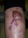
What type of EB is this?
On electron microscopy, what cytological structures would be absent in the specimen?
What gene mutation is present?
Recessive dystrophic EB (RDEB)
Absence of anchoring fibrils
- Compound heterozygous mutation within the COL7A1 gene, leading to a truncated collagen VII protein that in most cases is nonfunctional

Absence of anchoring filaments, hemidesmosomes, and sub-basal dense plates is seen in what type of EB?
Herlitz subtype of junctional epidermolysis bullosa (JEB)
- Anchoring fibrils are still present in JEB probably accounting for less severe scarring compared with RDEB
Multiple sclerotic fibromas (storiform arrangement of thickened collagen bundles with interspersed spindled cells) are associated with what disease?
What cancers are associated? (2)

Cowden disease
- Autosomal dominant mutation of PTEN gene
- Oral papillomas, trichilemmomas, acral keratosis, sclerotic fibromas
- Benign and malignant breast and thyroid tumors
- GI polyps, skeletal abnormalities
What syndrome is associated with acrochordons, multiple fibrofolliculomas, and trichodiscomas?
What is the genetic mutation?
What cancer is associated? What lung finding may occur?
Birt-Hogg-Dube (BHD) syndrome
Mutation in BHD gene
Associated with renal cell carcinoma and pneumothorax
Gardner syndrome is caused by what genetic mutation?
What are the three skin findings of this syndrome?
APC gene
Epidermoid cysts, osteomas, desmoid tumors
Associated with colorectal adenocarcinoma
What CD is “langerin”?
What is this a marker of?
CD207
Marker of Birbeck granules
What is the best tissue sample for transmission electron microscopy when evaluating for an inherited blistering disease?
A clinically induced blister at the time of biopsy.
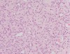
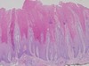
Pretibial myxedema histology
Increased mucin within the upper dermis that separates collagen bundles.
The overlying epidermis may demonstrate acanthosis, papillomatosis, and hyperkeratosis.

Muir-Torre syndrome is associated with mutations in which mismatch repair genes? (4)
25% of patients will have what type of skin cancer?
>60% of patients will have what internal malignancy?
- Caused by mutations in mismatch repair genes including MLH1, MSH2, MSH6, and PMS2
- Twenty-five percent of patients with MTS also have keratoacanthomas
- More than 60% of patients will have associated colorectal adenocarcinoma
KRT1 and KRT10 mutations cause what skin disease?
Epidermolytic ichthyosis, in which the pathology is not characterized by acantholysis but rather epidermolytic hyperkeratosis
What gene mutation and organelle is involved in Darier disease (keratosis follicularis)?
What gene mutation and organelle is involved in Hailey-Hailey disease (familial benign chronic pemphigus)?
- Darier disease (keratosis follicularis) → ATP2A2 mutation, encoding the endoplasmic reticulum Ca2+ ATPase
- Hailey-Hailey disease (familial benign chronic pemphigus) → ATP2C1 gene, which encodes the Golgi-associated Ca2+ ATPase
Think of the disease names, mutations, and organelles involved in ALPHABETICAL order. And the organelles involved are just one letter away from the first letter of the name!!
You are called by the neurology team to perform a skin biopsy for the workup of cerebral autosomal dominant arteriopathy with subcortical infarcts and leukoencephalopathy. You perform a 4-mm punch biopsy of the arm.
In what medium do you place the specimen?
Cerebral autosomal dominant arteriopathy with subcortical infarcts and leukoencephalopathy (CADASIL) is a neurologic disorder caused by mutations in NOTCH3. Patients with CADASIL have migraines, recurrent strokes, and progressive dementia. Transmission electron microscopy (TEM) of a skin biopsy is the gold standard to detect pathologic findings.
Placing the skin specimen in the correct medium, glutaraldehyde for TEM, is critical for accurate diagnosis.
What media should be used for preservation of gout crystals or for fat immunostaining?
Alcohol