Doggo Flashcards
(16 cards)
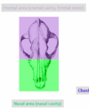
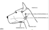
Label the green and purple area

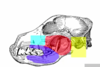
Label the coloured part of this photo
List the 3 compartments of the frontal sinus
rostal
medial
Lateral extend to the front process
What is present in the nasal cavity
-there are 3 conchae
ventral has lots of fold and is very develped
dorsal is very reduced
middle is extended
-3 meatus

Explain the maxillary sinus
Maxillary sinus also known at the infraorbital area
its over the last 3 superoir molars
isn’t spaceious
Label the salivary glands on this dog


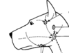
Discuss the 2 main nerves of the face and explain their branches and if they are motor or sensory
Nerves
- facial nerve; mtor nerve of the face
- Trigmenial nerve consists of;
opthalmic nerve (sensory nerve of the orbit) and becomes the frontal, lacrymla and palpebronasal nerve
maxiallry nerve; sensory for the superoir lip, teeth and upper jaw
mandibular nerve; motor for masticators and sensory for tongue, teeth and lower jaw
Label the following lymph nodes
Discuss the structures present in the neck
Neck
no jugular groove
jugular vein= site of vein puncture
common cartoid and internla jugular vein
vago-sympathetic vein
sternocephalicus and cutaneous muscle
trachea and oesphagus (located dorsally to the trachea and then on the left)
Explain what 3 organs are present in the thoraxic cavity and state their individual locations
Thorax
- diaphragm; inserts on the upper 1/2 of rib 13 and lower 1/4 of rib 12 and extends to rib 7 on the left and rib 6 on the right
- heart; located inbw the 3rd-7th rib (middle mediastinum), lying in the sternum
- lung; ;left is smaller because of the heart both sides have 3 lobes but right has an accessory lobe, cadual edge of thr lungs are upper end of 12th rib to rib cartilage of 7th
Explain what is present in the abdomen
Abdomen;
cutaneous muscle
tuncia (membranes/ layers)
external and internal oblique muscles
rectus and transversus abdominis muscle
transersalis fascia
peritoneum
GIT
Label the following coloured msucles
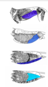
dark dark blue- extenrla oblique muscle
pretty light blue- internal oblique muscle
gross middle blue colour- rectuc abdminis muscle

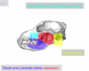
Label this photo


Explain the following
greater omentum
greater omentum; covers the intestines when you first open a body up and is attached to the greater curvature of the stomach and joins the pancreas
Explain the location of the following
liver
stomach
spleen
coelic artery
duodenum
jenunum
cranial and caudal mesentric artery
right and left kidney
ovaries
Liver; right and left until rib 6
stomach; left under rib cage (empty) or behind arch (full)
spleen; left; attached on the great stomach curve and under or behind the costal arch
coelic artery; l1
duodenum; behind the right kidney (descending part), right to middle left (transverse), middle left, (ascending part)
jenunum; right and left ventral position
caecum; right dorsal postion
Cranial mesenteric artery; L2
caudal mesenteric artery; L5
left kidney; L2-4
right kidney; L1-3
Ovaries; under and behind the kidneys, locate midway b/w last rib and the ilium
ANATOMY FLASH CARDS
ANATOMY FLASH CARDS
