Embryology Lecture 3 Weeks 2 and 3 Flashcards

1
Q

In week 2:

The implanted embryo becomes more ______ in the endometrium

Further development of _______ into the placenta

Development of a ______, ______, and _______

A

deeply implanted

trophoblast

bilaminar embryo, amniotic cavity and yolk sac

2
Q

What happens during bilaminar disc formation?

A

Trophoblast further differentiates and invades into maternal tissue:

Cytotrophoblast: stem cell population for the placenta

Syncytiotrophoblast: Invasive, fused cells. Derived from cytotrophoblast

Breaks maternal capillaries, trophoblastic lacunae then fill with maternal blood

Secretes human chorionic gonadotropin (hCG)

8.5.6

3
Q

How does the bilaminar disc form in day 8?

A

¨Epiblast contributes to forming the overlying amniotic membrane and amniotic cavity (columnar)

¨Hypoblast contributes to forming the underlying primitive yolk sac/exocelomic cavity (cuboidal)

4
Q

Cells derived from the primitive yolk sac cells (hypoblast cells) form a fine, loose connective tissue called _______

Spaces appear in extraembryonic mesoderm, coalesce to form ______

Chorionic cavity surrounds whole part of embryo except where the embryo is attached to the cytotrophoblast by ______

A

extraembryonic mesoderm

extraembryonic coelom (chorionic cavity)

connecting stalk

5
Q

The extraembryonic mesoderm now has 2 components, what are they?

A

lining the cytotrophoblast and amnion:

extraembryonic somatic mesoderm

covering the yolk sac:

extraembryonic splanchnic membrane

6
Q

What are the 3 main events of Day 13

A

Formation of chorion and primary chorionic villi

Formation of chorionic cavity

Formation of the definitive yolk sac

7
Q

¨Cytotrophoblasts proliferate into the syncytiotrophoblast and form ______

Exocoelomic cavity (primitive yolk sac) lined by new hypoblast cells gets replaced with a ______

Some of hypoblast cells near future mouth proliferate and form ______

A

primary chorionic villi

smaller definitive yolk sac or the secondary yolk sac

anterior visceral endoderm

8
Q

The ___ is formed by the extraembryonic mesoderm

A

connecting stalk

Note: The extraembryonic coelom expands to form a large cavity, within which the embryo and the attached amniotic cavity and yolk sac are suspended by the body stalk (connecting stalk) from which the umbilical cord forms

9
Q

The posterior wall of the yolk sac forms a small diverticulum which is called ______ and extends into the connecting stalk

A

allantois

10
Q

Summary of Week 2 events

A

Embryoblast forms 2 layers

§Epiblast

§Hypoblast

Trophoblast differentiates into 2 layers

§Cytotrophoblast

§Syncytiotrophoblast

Extraembryonic mesoderm splits into 2 layers

§Somatic

§Visceral

2 cavities form

§Amniotic cavity

§Chorionic cavity

Appearance of primary chorionic villi

Anterior visceral endoderm

Establishment of cranio-caudal axis of development

Completion of implantation

11
Q

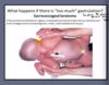
What can low hCG or high hCG mean during early pregnancy?

A

Low hCG may predict a spontaneous abortion or may indicate an ectopic pregnancy

High hCG may indicate a multiple pregnancy, hydatidiform mole, or gestational trophoblastic neoplasia

12
Q

____ is marked benign enlargement of chorionic villi (trophoblast) and is characterized by grapelike vesicles in uterus and absence of embryo, high hCG level.

A

Hydatidiform Mole

Gestational trophoblastic neoplasia (choriocarcinoma)

Malignant tumor of trophoblast

Some times they metastasize to liver and other organs (prognosis poor)

8.5.6

13
Q

What is paternal imprinting during hydatiform mole?

A
14
Q

What are the 7 main events that occur during week 3 of embryo development?

A

Bi-laminar germ disc forms the primitive streak

Gastrulation forms tri-laminar embryo

Neurulation or neural induction takes place

Somites are formed

Left-right asymmetry is determined

Cardiovascular development takes place

Extraembryonic spaces (coelom) and primitive placenta (villi) further develop

15
Q

How do you know between cranial and caudal end?

A

Connecting stalk on caudal side

16
Q

How does the primitive streak form?

A

¨Some of the hypoblast cells near the future mouth proliferate and form anterior visceral endoderm (forms the buccopharyngeal membrane)

¨Soon after formation of anterior visceral endoderm epiblast cells near the tail end proliferate and form primitive streak

Cephalic end of primitive streak:

primitive node: slightly elevated surrounding a small pit: primitive pit

17
Q

Explain Gastrulation

A

¨Epiblast cells migrate towards primitive streak, detach and slip beneath it

¨Some of the slipped epiblast cells displace hypoblast and form endoderm, others come to lie between epiblast and endoderm to form mesoderm, remaining epiblast cells form the ectoderm

18
Q

True or False: During gastrulation all germ layers develop from hypoblast

A

False, epiblast

19
Q

True or False: At gastrulation, primitive endoderm is replaced by definitive or embryonic endoderm then mesoderm is formed

A

True

20
Q

Gastrulation Summary

A
21
Q

Intraembryonic mesoderm migrates all over the embryo in between ectoderm and endoderm except at ________ membrane and _______.

A

buccopharyngeal and cloacal (anal) membrane

22
Q

What are the major signaling centers during gastrulation (understand the structures and mesoderm)

A

8.5.6

23
Q

How is the neural plate formed?

A

8.5.6

24
Q

How is left-right asymmetry established during gastrulation?

A

FGF 8 is synthesized by the node and the primitive streak

¨FGF 8 induces expression of Nodal

¨Nodal is restricted to the left side by accumulation of serotonin (5 – HT)

¨SHH and LEFTY 1 prevent nodal expression crossing over to the right side

¨Nodal upregulates LEFTY 2 and PITX 2:

¤PITX2 is the master gene for left sidedness

¤Left sided organs express PITX2 on their left side

25
What is situs inversus?
## Footnote 8.5.6
26
What happens if there's not enough gastrulation?
Caudal Agenesis (sirenomelia)
27
What happens when there is too much gastrulation?
Sacrococcygeal teratoma
28
What are the key features of sirenomelia?
_hypoplasia and fusion of lower limbs,_ vertebral anomalies, renal agenesis, imperforate anus, and genital organ anomalies
29
\_\_\_\_\_\_\_ clusters of pluripotent cells proliferate and form tumors in the region
Sacrococcygeal teratoma Teratomas can also arise from primordial germ cells that fail to migrate to the gonadal ridge
30
High doses of \_\_\_\_\_\_l during the third week of gestation kills cells in the anterior midline of the germ disc and produce a deficiency of the midline in craniofacial features resulting in \_\_\_\_\_\_\_
alcohol holoprosencephaly
31
\_\_\_\_\_ is fused eyes \_\_\_\_\_ is a single nasal opening formed by fusion of lateral nasal processes
Synophthalmia Proboscis
32
The primitive node secretes substances e.g. noggin, chordin, and follistatin that allow a region of the ectoderm to develop into \_\_\_\_\_\_.
nerve tissue
33
Explain neurulation: folding of the neural plate ## Footnote 8.5.6
¨Neural plate --\> neural groove --\> eural folds ---\> neural tube ¨As neural folds close, neural crest _delaminates and migrates away_ ¨Closure happens _first in middle of the tube_ and then _zips rostrally and causally_ Separates itself from the surface ectoderm
34
Explain neurulation: folding and closure of the neural plate
Folding and closure of the neural tube occurs first in the cervical region The neural tube then “zips” up toward the head and toward the tail, leaving two openings which are the _anterior_ and _posterior neuropores_ The _anterior neuropore_ closes around _day 25_ The _posterior neuropore_ closes around _day 28_
35
Neural tube defects: \_\_\_\_\_\_ failure of neural tube closure \_\_\_\_\_\_ failure of the anterior neuropore closure \_\_\_\_\_\_ failure of posterior neuropore closure and/or vertebral development 8.5.6
Rachischisis (image) Anencephaly Spina bifida NTDs have many genetic and environmental causes, but the strongest correlation is _folic acid deficiency_
36
Failure of neuropores to close can cause neural tube defects (image) anterior neuropore: anencephaly posterior neuropore: spina bifida
## Footnote 8.5.6
37
Where is spina bifida most common?
lumbosacral region Causes defects in spinal cord and bones of the spine
38
What is the 4th germ layer and how is it formed?
At the time of neurulation, cells at the lateral most edge of the neural plate are induced to form _neural crest_ The neural crest cells _transform from epithelial cells into migratory mesenchymal cells_ that contribute to forming _MANY_ tissues in the body Neural crest cells are pluripotential
39
What are neural crest derivatives?
Dorsal root _ganglia_ Autonomic ganglia Adrenal medulla Schwann cells Pia-arachnoid sheath Skin melanocytes _Thyroid parafollicular cells_ _Craniofacial skeleton_ Teeth odontoblasts _Aorticopulmonary septum (spiral septum of heart)_
40
What are the 4 types of mesoderm formed from the primative node?
_¨Axial mesoderm:_ passes through the node and migrates along the midline: forms the _notochord_ _¨Paraxial mesoderm:_ passes just caudal to the node and migrates slightly laterally: _Forms vertebral bones and associated cartilage, skeletal muscle, and dermis of the back_ _¨Intermediate mesoderm:_ Between paraxial and lateral plate mesoderm: Forms the gonads, kidneys and the duct system of the male reproductive system _Lateral plate mesoderm:_ passes more caudal and migrates more laterally: _forms limb bones, circulatory system and body cavity linings_
41
Subdivisions of the 4 mesoderm
Axial mesoderm/Chorda mesoderm ¤notochord Paraxial mesoderm ¤head mesenchyme ¤somites sclerotome myotome dermatome Intermediate mesoderm ¤urogenital organs Lateral plate mesoderm ¤splanchnic (viscera) ¤somatic (body wall) ¤extraembryonic
42
Summary of Mesoderm
43
What happens in the paraxial mesoderm?
¨Undergoes segmentation: Forms _somitomeres_ First appear in cephalic region and formation proceeds caudally ¨From occipital region caudally somitomeres organize into _somites_ ¨F_irst somite arises on the 20th day_
44
True or False: New somites appear in craniocaudal sequence approximately 3 pairs per day until, at the end of 5th week, 42- 44 pairs are present
true 8.5.6
45
What are the correct somite numbers?
Upper 4 - occipital somites (supplied by XII cranial nerve) 8 cervical 2 thoracic 5 lumbar 5 sacral 8-10 coccygeal
46
Where does somitogenesis begin?
Somitogenesis begins w/ 8th pair of somitomeres
47
How does the separation of somites work?
FGF signaling from the node drives proliferation; Retinoic acid from adjacent mesoderm drives differentiation. Because the node is caudal to the forming somites, there is a head-to-tail gradient of differentiation Proliferating cells express a ligand (called ephrin B1). As cells differentiate, they begin to express an INCOMPATIBLE receptor (called eph A) that causes the differentiating cells to repel the proliferating cells, thus pinching off and forming a new somite
48
How do epithelial somites then transform back into mesenchyme?
¨Signaling from ectoderm induces dermomyotome ¨Signaling from notochord and neural tube induces sclerotome
49
How does dermomyotome form dermis and muscle?
¨Dorsomedial portion of dermomyotome influenced by notochord becomes muscle that cannot migrate very far (epaxial muscles of the back) ¨Ventrolateral portion of dermomyotome exposed to high levels of BMP from lateral plate mesoderm, becomes migratory muscle (goes into limbs also “hypaxial” muscles of the lateral and ventral body wall, e.g. “latissimus” and “abdominal muscles”
50
How many types of sclerotome are there and what do they develop into?
Dorsal sclerotome: ¤Dorsal arch and spinous processes of vertebrae\* Medial sclerotome: ¤Meninges\* Central sclerotome: ¤Pedicles & transverse processes of vertebrae, proximal portions of ribs Ventral sclerotome: ¤Vertebral bodies and annulus fibrosus of intervertebral disks Lateral sclerotome: ¤Distal portions of ribs
51
summary of somite derivatives
¨Somite (Paraxial mesoderm) forms: ¤Sclerotome ¤Dermomyotome Sclerotome forms: meninges, vertebrae and ribs Dermomyotome forms: Dermis of the back of the body (from dermatome) Muscles (from myotome)
52
Intermediate mesoderm develops into the \_\_\_\_\_\_
urogenital system ## Footnote 8.5.6
53
Lateral plate mesoderm divides into ______ and \_\_\_\_\_\_mesoderm
somatic and splanchnic ## Footnote Somatic mesoderm: Lines body wall (somatic mesoderm + ectoderm = somatopleure) Splanchnic mesoderm: Covers endoderm (splanchnic mesoderm + endoderm = splanchnopleure) 8.5.6
54
Somatic (aka parietal) mesoderm stays with epidermis Splanchnic (aka visceral) mesoderm stays with endoderm
55
Blood and blood vessels develop from extraembryonic and lateral plate mesoderm
**¨Vasculogenesis:** blood vessels arise from “hemangioblasts” that develop into blood cells AND vascular tubes **¨Angiogenesis:** growth of new blood vessels from existing ones ¨Vessels from extraembryonic mesoderm go out to placenta and eventually hook up with blood vessels (and the heart) in the embryo that arise from lateral plate mesoderm to establish circulation. ¨2 major phases of hematopoiesis: **¤Embryonic** (weeks 1-4) : blood cells arise from yolk sac mesoderm **Definitive** (week 4-term): _blood cells arise from lateral plate mesoderm in the **AGM** (aorta-gonad-mesonephros region) that go on to seed the spleen, liver, and then bone marrow with hematopoietic stem cells_
56
Folding of embryo
§Occurs §Cephalocaudally §Laterally
57
How does lateral folding work?
¨Right and left somatic layer of lateral plate mesoderm fuse together ventrally and form rounded body form ¨Yolk sac gets incorporated into body of embryo- primitive gut tube ¨Communication between yolk sac and gut tube - Vitelline duct
58
What are the simaltaneuous events that happen during the closure of the body wall
Purse string-like closure of the body wall Growth of the amnion Regression of yolk sac
59
What happens if the body wall does not close properly?
**Ectopia cordis:** failure of the thoracic body wall to close
60
\_\_\_\_\_ is the failure of the abdominal body wall to close \_\_\_\_\_\_ is failure of the pelvic body wall to close \_\_\_\_\_\_ is failure of most of pelvic body wall to close
Gastroschisis Bladder Exstrophy Cloacal Exstrophy
61
Summary of Third Week
¨Primitive streak ¨Gastrulation à ectoderm, intraembryonic mesoderm and endoderm ¨Notochord formation ¨Neural tube and neural crest formation ¨Paraxial mesoderm à somites ¨Intermediate mesoderm ¨Lateral plate mesoderm cavitation and intraembryonic coelom formation ¨Angiogenesis and primitive heart tube formation ¨Secondary villi à tertiary villi à placental circulation begins
62