Embryos - Pregnancy Flashcards

1
Q

how does the uterus change during pregnancy

A
  • myometrium grows markedly
  • muscle fibres hypertrophy and increase in number
  • 3 LAYERS OF MUSCLE

outer longitudinal

middle interlacing

inner circular

  • CT becomes more vascular
2
Q

uterus and pelvic floor changes

what is the upper uterine segment attached to

A

peritoneum is intimately attached to upper uterine segment

loose and mobile all over the segment

uterus supports hypertrophy

broad ligaments show hypertrophy of all their content

levatores anii muscles hypertrophy and become softer ⇒ pelvic floor becomes progressively more distensible, thereby facilitating passage of the foetus

3
Q

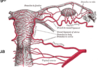
change in uterine blood supply

A

blood supply increases

uterine and ovarian arteries become large and very tortuous

PROTECTIVE FUNCTION:

lymphatics, like BVs, increase in size and number

large lymph spaces beneath the decidua and a well developed plexus under the enveloping peritoneum

4
Q

what happens from 2nd month onwards

describe blood supply by 9th month

A

hypertrophy of BVs and lymphatics produces progressive softening of whole body

by 9th month, the whole of uterus and outer pelvic viscera are so engorged with the blood and lymph that the outlines of the various organs become vague and difficult to define

5
Q

size and position of uterus

how does the position of uterus change throughout pregnancy

A

non-pregnant uterus = 2.5x5.7.5cm

full term = 23x25x30cm

uterus lies in true pelvis at 1st but by week 12 the fundus is level with the top of the symphysis pubis

by week 16 it lies mid way between the symphysis pubis and the umbilicus

6
Q

What might a woman experience towards the end of a pregnancy

A

lightening as the baby moves down

7
Q

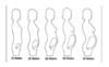
position of uterus @ 20 weeks and 24 weeks

change in position throughout pregnancy

A

20 weeks - below umbilicus

24 weeks - just above it

⇒ fundus rises 2 fingerbreadths every 4 weeks until 36 weeks when it lies @ xiphisternum

between 36 and 40 weeks it drops by 1 fingerbreadth per week and @ week 40 it lies at the same level that it had reached @ week 32

8
Q

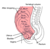
what causes the lightening in the last month

A

due to descent of foetal head into cavity of true pelvis

although the woman may feel more comfortable and may breathe more easily after lightening has occurred, she may notice frequency of micturition due to lack of space in the pelvis

9
Q

role of cervix

A

passive role

cervical blood vessels and lymphatics hypertrophy thereby causing progressive softening which may be detected very early in pregnancy

connective and muscular tissues, although they both become more vascular and softer, they do not undergo hyperplasia

10
Q

change in cervical mucosa

A

hypertrophies markedly until it constitutes nearly half of cervix @ full term

eventually, complex of glands resembles a honeycomb full of sticky tenacious mucus

when this protective mucus plug is expelled at onset of labour, it carries most of honeycombed mucosa with it

external os comes to have anterior and posterior lip, especially in multiparae

deep purple - engorged with blood

11
Q

how does the isthmus and lower uterine segment change

A

approx upper 1/3 of cervix = isthmus

unaffected in 1st month of pregnancy

dilates and is taken up into body of uterus to form the lower uterine segment

the foetal membranes are less firmly blended with the mucosa in the isthmus than elsewhere

the endometrium lining the lower segment does not undergo a full decidual change

12
Q

changes in vagina

A

similar to uterus

blood supply increases enormously - deep violet colour

hypertrophy of wall increases both length and width of vaginal canal

13
Q

changes in vulva

A

undergoes similar changes - increased blood and lymphatic supply

progressive softening

14
Q

changes in breasts

what happens at week 8

A

during the 1st 6 months - duct system proliferates

during the last 3 months - alveoli proliferate

also in alveoli, there is hypertrophy of BVs and lymphatics which supply them

WEEK 8 - Montgomery’s tubercles (mouths of enlarged sebaceous glands) become prominent in areola

WEEK 12 - darkening of primary areola occurs

WEEK 16 - a paler, secondary areola forms (more noticeable in dark-haired women)

15
Q

abdominal viscera changes

what is a common complication of pregnancy

A

stomach is displaced upwards during the 2nd half of pregnancy

diaphragmatic herniation is a common complication of pregnancy

16
Q

change in pelvis during pregnancy

A

symphyseal, sacroiliac and sacrococcygeal joint capsules soften and relax

reaches a maximum about week 28 and may cause sacroiliac back ache

may be accompanied by pain and tenderness in the symphysis

17
Q

changes in skin

A

deposition of melanin occurs in certain areas in the body - particularly dark haired women

in midline of abdominal wall - linea nigra

18
Q

chloasma uterinum

A

melanin deposition on forehead and cheeks

19
Q

moulding

A

fetal cranium is relatively deformable

bones of calvaria are thin and elastic and can alter their shape to some extent

they are attached to 1 another by relatively loose fibrous sutures

they can override one another somewhat in response to compression forces as the head is squeezed down through the pelvis

this is limited

must be sufficient prior congruity to permit first engagement and then passage of foetus through the pelvic cavity

20
Q

boundaries of pelvic inlet

angle it makes with pelvic floor

A

heart-shaped - bounded posteriorly by sacral promontory, laterally by iliopectineal line and anteriorly by symphysis pubis

plane of pelvic inlet makes an angle of 60° with that of pelvic floor

21
Q

true conjugate diameter

A

measured from top of symphysis pubis to sacral promontory and averages about 4.5 inches

22
Q

oblique diameter of pelvis

A

measured from sacroiliac joint to obturator foramen of opposite side and averages 4.75 inches

23
Q

transverse diameter of pelvis

A

widest measurement from side to side and averages 5.25 inches

24
Q

boundaries of pelvic cavity

measurement of diameters

A

bounded anterioly by symphysis pubis and posteriorly by sacrum and coccyx

diameter is usually taken at the level of junction of 2nd and 3rd sacral vertebrae posteriorly and middle of symphysis anteriorly

anteroposterior, oblique and transverse = 12cm

25
Q

pelvic outlet boundaries

A

outlet is bounded by pubic arch anteriorly, by ischial tuberosities and sacroiliac ligaments laterally and tip of coccyx posteriorly

26
Q

anteroposterior diameter of pelvic outlet

A

measured from lower border of symphysis pubis to the sacroiliac joint

5.25 inches

27
Q

transverse diameter of pelvic outlet

A

bituberous

taken between the lower borders of ischial tuberosities

4.25 inches

28
Q

subpubic angle - pelvic outlet

A

bounded by the pubic rami and symphysis

86°

29
Q

how is the inlet divided

A

into forepelvis and hindpelvis by the widest transverse diameter

walls of hindpelvis include that portion of the ilium overlying the sacroiliac notch - one of the most variable sections in the pelvis and this region is most affected by sexual and evolutionary differences

30
Q

how is the outlet divided

A

anterior and posterior segments by intertuberous diameter

31
Q

what influences the capacity of anterior segment (of pelvic outlet)

A

subpubic arch

either wide, moderate or narrow

its shape depends on the curve of the inferior pubic rami and is usually well curved in the female pelvis and straight-edged in male pelvis

side walls

may incline toward or away from one another or may be parallel as they pass downward

depth of pelvis

taken from iliopectineal line along the back of the obturator foramen to ischial tuberosity

32
Q

what influences the capacity of posterior segment of pelvic outlet

A

width of greater sciatic notch

sacral curve and inclination

the greater the upward and backward tilt of the lower end of the sacrum, the more room there is in the lower pelvis for passage of the foetal head

the degree to which the ischial spines project inward

33
Q

anatomical changes during normal labour - primigravida (first pregnancy)

A

head normally becomes engaged in pelvic inlet by week 37 or 38 of pregnancy

34
Q

anatomical changes during normal labour - multipara

A

engagement may not occur until the membranes rupture at the end of 1st stage of labour

35
Q

first stage of labour

A

rhythmic uterine contractions increase markedly in strength, freq and duration

typically experienced as pain, beginning in sacral region and passing around to the front of the abdomen, rising to a climax and then fading way

Associated with the success of contraction and retraction of the upper uterine segment, the lower uterine segment becomes progressively thinner and the cervix dilates

This leads to detachment of the mucosa lining the lower uterine segment with rupture of the small blood vessels attaching it to the uterine wall

Forewaters are formed

SHOW = the blood that has been shed mixes with the mucosa of the cervical plug, which separates at the same time, to form this blood-stained mucous discharge

36
Q

Effacement

A

cervical dilation

internal os of the cervix and the cervical canal are gradually “taken up” - merge with the cavity of the lower uterine segment

this process is completed by dilation of external os

37
Q

where are the pacemakers and what do they do

A

one on each side at the uterine end of each uterine tube

drive uterine contractions

38
Q

sequence of uterine contractions

A

Increasing strength

maximum

quick decline

period of rest

some of the shortening of a muscular contraction is permanently maintained

39
Q

retraction

A

progressive process

occurs throughout upper uterine segment but mainly at the fundus

with each contraction, traction is applied to the relatively passive lower segments and through it to the cervix

at the same time the forewaters and the presenting part are forced downwards

40
Q

retraction and the upper segment

A

retraction causes the upper segment to progressively thicken and the lower segment to stretch

41
Q

Bandi’s ring

A

junction of upper segment (which is progressively thickening)

and the lower segment (which is stretching)

42
Q

how is the birth canal formed

A

stretching and expansion of lower segment

effacement and dilation of cervix

all converts the cavities of the uterus, cervix and vagina into a single unit

43
Q

when is formation of birth canal complete

A

by the end of the 1st stage

a low resistance pathway down which the foetus may be driven by the upper uterine segment

44
Q

what contributes to membrane rupture

A

membranes begin to degenerate

increasing pressure exerted by retraction of upper segment

removal of support from below as a result of cervical dilation

45
Q

difference between effacement with primigravida vs multigravida

A
46
Q

2nd stage of labour

A

expulsion of foetus from full dilation of foetus until the child has been delivered

force is provided by the contraction and retraction of the upper uterine segment

After the membrane ruptures, uterine contractions become stronger, more frequent and more sustained

As the liquid drains away, the force of the uterine contractions is applied directly to the fetal breech

from the breech the force is transmitted to the foetal spinal column and from there to the foetal head

47
Q

change in position of uterus during 2nd stage

A

uterus rears forward, straightening out the curve of the foetal spine, resulting in elongation of the foetus which is an additional aid to its descent

48
Q

what happens to the anterior pelvic structures in the 2nd stage of labour

A

e.g. urethra, bladder and anterior vaginal wall

drawn upward out of the path of the descending foetus by contraction and retraction of the upper uterine segment -transmitted by the elastic tissues of the lower segment and intervening connective tissue

movement is facilitated by extensive softening of pelvic tissues late in pregnancy

49
Q

posterior pelvic structures in the 2nd stage of labour

A

posterior vaginal wall, rectum, anal canal, levator ani muscles

forced downward and backward

50
Q

foetal head during descent

A

rotates

51
Q

3rd stage of labour

A

separation and expulsion of placenta and membranes

52
Q

transverse caesarean incision

A
53
Q

indications for caesarean section

A
54
Q

ectopic pregnancies

A

implantation does not occur in upper part of uterine wall (which is usual)

55
Q

epithelium of foetal lungs @ 24 weeks

A

cuboidal

gases cannot diffuse across

56
Q

cells that produce surfactant

A

type I pneumocytes

type II pneumocytes