Epithelial Tissue Flashcards
(39 cards)
Describe tight junctions
impermeable; acts as a barrier on the apical surface of the cell; decreases permeability occludins and claudins
Describe gap junctions
communicating junctions fluid filled and connect the cells in order to communicate connexin aggregates
What are the 3 types of anchoring junctions?
- adherens 2. Desmosomes 3. Hemidesmosomes
Describe adherens
lateral adhesions: cadherins+actin
Describe desmosomes
lateral adhesions; cadherins+ intermediate filaments
Describe hemidesmosomes
basal adhesions: integrins and intermediate filaments that anchor to the basal lamina
What is the clinical relevance off tight junctions?
Bacteria that causes food poisoning target the tight junctions that are present in the GI tract and decreases the fluid flow into the intestines helicopter pylori: causes gastric ulcers and binds to the TJs in the stomach
Describe pemphigus vulgaris
Autoimmune disease in which there is an abnormal desmosome function decreases cell to cell adhesion and blisters of the oral mucosa
Describe the basement membrane
Adjacent to the basal domain selective barrier
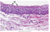
What is surrounding the cell (dark magenta)

The basement membrane
What are the three types of apical specializations of a cell?
Microvilli, stereocilia, and Cilia
Describe microvilli
Cytoplasmic
Have an actin core
Absorption
Increase the surface area of the cell for increased absorption
Describe stereocilia
Long microvilli, less mobile
actin core
increased surface area
ARE restricted to epididymis and hair cells of the inner ear
Describe cilia
Long and highly motile
beat in a wave like fashion to propel substances
Where is simple squamous located? What is the function?
Location: line blood vessels, serous membranes, alveoli, and loopof Henle in the kidney
Function: exchange, barrer, lubrication
Describe simple cuboidal
- Location
- Function
- Location: kidney, tubules, glands, and associated ducts, terminal branches, and covering of the ovary
- Function: absorption, barrier, secretion
Describe simple columnar
- Location
- Function
- Location: auditory tubes, uterus, oviducts, stomach, GI, gallbladder
- Function: absorption and secretion
Describe pseudostratified columnar ciliated
- Location
- Function
- Location: lining of the nasal caity, pharynx, trachea, bronchi
- Function: absorption and secretion, debris and particle movement
Describe urothelium
- Location
- Function
- Location: urinary bladder, uterus, urethra
- Function: barrier, distensible property
Describe nonkeratinized stratified squamous
- Location
- Function
- Location: oral cavity, portions of the pharynx, esophagus, anus, vagina, and cornea
Function: barrier and protection
Describe keratinized stratified squamous
- Location
- Function
- Location: epidermis
- barrier and protection
Describe stratified cuboidal
- Location
- Function
- Location: sweat glands and ducts, ovarian follicles, salivary glands
- Function: barrier and passageway
Describe a serous membrane
epithelial tissue that lines the internal body cavities
forms a smooth transparent, two layered membrane that is lubricated by serum
mesothelium: simple squamous that comprises a part of the serous membrane
Describe compound glands and differentiate between
- compound tubular
- compound acinar
- compound tubuloacinar