Exam 1 Flashcards
(221 cards)
Overview of the Nervous System
- Sensory receptors (the afferent system) and motor neurons/effectors are part of the PNS
- Sensory info feeds into the CNS, where info is processed and sent to the effectors
Cells of the Nervous system
-Neurons- main cell responsible for integration and relay of messages -Glia- support neurons
Neurons
-Cell body and dendrites receive input -Axon hillock= summation of APs, integrative -Axon= conductive -Terminal branches= output -Metabolically compartmentalized- proteins made in cell body and dendrites only *Specialized cells that conduct APs over long distances (quickly)
How do neurons vary?
-Number of dendrites -Branching pattern of dendrites -One axon, but vary in # of collaterals
What are the two types of neurons?
-Projection neurons (sensory, motor, tract) -10% pop -Take info from one place to another (large distances) -Largest and best studied -Local Interneurons -90% -Unmyelinated -Modify info within local/small area -Small, difficult to study
Pseudounipolar cells
-One cell body, axon that starts as one and then branches into 2 collaterals -Ex: sensory neurons
Multipolar cells
-multiple dendrites and an axon -Ex: motor neuron– synapses to muscle
Local Interneuron
-no myelin -Multipolar
Nissl Stain
-Shows ribosomes bound to ER and nucleolus (site of RNA synthesis 4 ribosomes -Doesn’t stain axon -Stains proximal dendrites and cell body
Neuronal Cytoskeleton
-Microtubule- Largest, hollow tube, support axons, laid end to end -Neurofilament- stable, middle size -Microfilament- actin, found in parts of neurons that rapidly change (dendrites)
Axoplasmic Transportation
-How materials are moved within the cell -Use molecular motors and microtubules -Various rates of transportation -Anterograde- cell body to synapse -Retrograde- synapse to cell body
Features of the synapse
-Synaptic vesicles/secretory granules present at presynapse–> Hold NTMs -Postsynaptic density- proteins connecting the two synapses -Glia surround the synapse -Mitochondria at synaptic button produce ATP
Dendritic Spines
-Dendrites have numerous spines to maximize surface area to receive synapses -Neurons receive thousands of synapses which summate to create an AP
Glial Cells
CNS- -Ependymal cells -Microglia -Astrocytes -Oligodendrocytes PNS -Schwann cells -Satellite cells
Ependymal cells
-Line the ventricular system -Fluid-filled part of CNS -Cilia face inward to keep CSF moving
Astrocytes
-Most numerous -Surround neurons -Protoplasmic in gray matter -Fibrillar in white matter -Control extracellular environment (K+) -Take up NTMs -Break down glucose and pass it down to neurons
Oligodendrocytes
-Myelinate in CNS only, more than one axon -Each axon requires more than one oligodendrocyte
Microglia
-Derived from immune system, not nervous system -Phagocytes–> clean up cells -Respond to Injury -Found in resting state
PNS Glia
-Schwann cells myelinate only 1 axon in PNS —-Need many schwann cells to myelinated the same axon -Satellite cells- Support cells around neurons in ganglia
Grey vs White Matter
-G: Little myelin, mostly cell bodies and dendrites -W: a lot of myelin and oligodendrocytes
Early Nervous System Development
-Develops from ectoderm (outermost germ layer) -Begins to form at day 19- ectoderm worlds due to thickening of the neural plate -Neural groove–> neural tube–> CNS cells -Neural folds–> neural crest cells–> PNS
Neural Crest Cells
-Migrate to diff places and make ganglia -Become: —Sensory ganglia (dorsal root ganglia) —Autonomic ganglia (sympathetic, para-) —Enteric ganglia of the digestive system —Arachnoid and Pia covering (CNS) —Schwann cells —Adrenal medulla
Ganglia
-A collection of nerve cell bodies in the PNS -Formed from neural crest cells
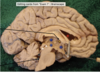
Neural Tube
-Cells become CNS structures —Cerebrum- cerebral cortex and deep nuclei —Diencephalon, Midbrain, Pons, Cerebellum, Medulla —Spinal cord —Glial Cells of CNS