Exam 1 Spring Flashcards

(175 cards)

1
Q

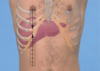
Quadrants and Regions of abdomen

A
2
Q

Visceral Pain of abdomen

A

hollow organs: distention/forceful contraction

solid organs: stretching capsule

desc: gnawing, burning, cramping or aching
assoc: sweating pallor, nausea, vomit, restlessness

3
Q

Parietal Pain of abdomen

A

orig: parietal pertoneum –> inflam

steady aching pain, more severe & precise localized than visc

worse with mvmt, cough

assoc with rebound tenderness

4
Q

Rebound Tenderness

A

Blumberg’s Sign

Pain introduced or increased by quick withdrawal of pressure

Suggests peritoneal inflammation

“Which hurts more when I press or let go?

5
Q

Referred Pain of abdomen

A

usually well localized

refer to chest, spine, pelvis

6
Q

Vomitus

A

ask for:

  • quant
  • odor
  • color
    • bile: yellow-green
    • blood: hematemesis:
      • COFFEE GROUND : blood altered by gastric acids
      • bright red
7
Q

diarrhea

A

Increased water content of stool , stool volume >200 grams in 24 hours

8
Q

Melena

A

Presence of black or tarry stool

upper GI bleed or from small bowel/right colon

9
Q

Hematochezia

A

bright blood in stool

lower GI tract bleed or brisk upper GI bleed

10
Q

Jaundice

A

Yellowish discoloration of the skin and sclera

From increased levels of bilirubin (a bile pigment derived mainly from the breakdown of hemoglobin)

Increased bilirubin is most suggestive of a hemoglobin problem (hemolysis) or a problem within the hepatobiliary system

Can range from benign (neonatal physiologic jaundice) to suggestive of life threatening disease (pancreatic cancer)

11
Q

•Supra Pubic pain

A

–Can be caused by bladder or pelvic

–Bladder infection

–Urinary Retention

12
Q

Order of the Physical Examination : abdomen

A

ØInspection

ØAuscultation

–Must precede percussion and palpation

ØPercussion

ØPalpation

ØSpecial tests

13
Q

abdominal profiles

A
14
Q
A

Abdominal Striae in Cushing’s Syndrome

15
Q
A

Caput Medusae seen in Portal Hypertension

16
Q
A

Sister Mary Joseph Nodule seen in Metastatic Disease

17
Q
A

CULLEN’S SIGN

– Periumbilical ecchymosis

– Typically occurs in the presence of hemoperitoneum, hemorrhagic pancreatitis, or uterine tube rupture in ectopic pregnancy.

–Physical findings: Bluish discoloration or ecchymosis around the umbilicus; abdominal tenderness may also be present

18
Q
A

GREY TURNER’S SIGN

–Flank ecchymosis

–Typically occurs in the presence of retro-peritoneal bleeding

–Hemorrhagic Pancreatitis

19
Q

Auscultation of Bowel Sounds

A

Perform before palpation and percussion.. Why?

  • Provides information about bowel motility

Use the diaphragm of your stethoscope

Note the frequency and the character of the bowel sounds

20
Q

Types of Bowel Sounds: normal

A
  • sounds occur 5 to 34 per minute in frequency and consist of gurgling and clicks in character
21
Q

Types of Bowel Sounds: borborygmi

A

•are loud, easily audible sounds. Prolonged gurgles of hyperperistalsis. They are normal, too.

22
Q

Types of Bowel Sounds: High pitched

A

•tinkling (raindrops in a barrel) sounds are a sign of early intestinal obstruction.

23
Q

Types of Bowel Sounds: Increased

A

•diarrhea or early intestinal obstruction

24
Q

Types of Bowel Sounds: Decreased

A

•(none for a minute) are a sign of decreased gut activity. Gut sounds may be markedly decreased after abdominal surgery; abdominal infection (peritonitis) or injury.

25
ØTypes of Bowel Sounds: absent
(no sounds for 2 minutes) caused by * longer-lasting intestinal obstruction * intestinal perforation * intestinal (mesenteric) ischemia/infarction
26
Auscultation of Vascular Sounds
pathology bruit: abdominal aorta, renal arteries , femoral arteries friction rub: Over the spleen and liver venous hum: between xiphisternum and the umbilicus
27
Auscultation- Bruits
ØTechnique: Use the bell\* of your stethoscope to listen for bruits in each of the areas shown in the picture: ØListen for: * Aortic bruits – in the epigastrium. They may be a sign of abdominal aortic aneurysm * Renal artery bruits in each upper quadrant. They may be a sign of renal artery stenosis, which is a potentially treatable cause of hypertension * Iliac/femoral bruits in the lower quadrants. They may be a sign of peripheral atherosclerosis.
28
oPercussionf abdomen
Assess the distribution of tympany and dullness * tympany: normal over gastric air bubble/gas filled portions of intestines * excess: gas during obstruction * dullness: normal over solid organs * unexpected: enlarged organs, mass technique: * Epigastric area * Gastric air bubble * Liver borders to estimate span * Spleen size
29
Palpation: what if pt complains of pain?
palp that area last
30
Palpation Technique- Light Palpation
Use one hand, fingers together and flat on the abdominal wall, pads of fingers palp during exhale = facil M relax * mouth breathe * voluntary guarding usu dec with these maneuvers
31
Deep Palpation of abdomen
use both hands: palmar surf
32
You ask your patient to tell you “ which hurts more when I press or let go” and then you proceed to firmly press your fingers down into her abdomen and then withdraw your hand quickly. By doing this technique you have assessed for the presence of: a) voluntary guarding b) Blumberg’s sign c) rigidity d) Cullen’s sign
Blumberg’s sign
33
Percussion- Liver Span
Measure the vertical span of the liver in the right midclavicular line * lower border: Start at a level below the umbilicus in the right lower quadrant and percuss upward toward the liver * upper border: nipple line down from lung resonance (have pt displace breast) normal: * greater in men & tall people * midsternal: 4-8cm * midclav: 6-12 cm
34
Liver Palpation
Use your left (posterior hand) to bring the liver anteriorly to facilitate palpation by your right hand Ask the patient to take a deep breath During inhalation the liver will move inferiorly (about 3cm below the costal margin in midclavicular line)and become easier to palpate cannot feel it move your palpating hand closer to the costal margin and try again
35
Hooking Technique
alternative method to palpation of the liver
36
Splenic Percussion
ØTwo Techniques: 1. 6th rib: * left lower anterior chest wall roughly from the border of cardiac dullness --\> anterior axillary line and down to the costal margin, Traube’s space. * If tympany is prominent, especially laterally, splenomegaly is not likely. 2. Test for the Splenic Percussion Sign: lowest interspace in the left anterior axillary line: usually tympanitic. * ask the patient to take a deep breath, and percuss again. * spleen size is normal, the percussion note usually remains tympanitic
37
Spleen Palpation
pt supine: Use your left (posterior hand) to bring the spleen anteriorly Ask the patient to take a deep breath * During inhalation the spleen will move inferiorly and become easier to palpate Repeat with the patient lying on their right side with their hips and knees partially flexed §~In 5% of adults a spleen tip is palpable
38
Kidney Palpation
With patient supine- Palpate the right kidney Use your left (posterior hand) to bring the kidney anteriorly Ask the patient to take a deep breath * During inhalation the kidney will move inferiorly * normal right kidney may be palpable * normal left kid = rarely palp
39
Costovertebral Angle Tenderness
location: lower border of 12th rib and TP of upper lumbar vert •Palpation first –Use fingertips –May not be enough to elicit tenderness •Fist Percussion –Place ball of one hand in the CVA and strike it with the ulnar surface of your fist (of the other hand) Use enough force to cause a perceptible but painless jar or thud common in UTIs (ascending), kidney stones * 11/12th rib
40
Bladder Palpation
* Cannot normally be palpated * Check for tenderness * Suprapubic tenderness is present in a bladder infections •Bladder can be palpable if it is distended above the pubic symphysis * dome feels smooth and round •Use percussion to determine how high the bladder rises above the public symphysis –If filled with urine, will be dull to percussion –If empty will be tympanic to percussion
41
Palpation of the Aorta
Press firmly and deeply in the upper abdomen, Slightly to the left of midline One hand on each side of the aorta Feel for the aortic pulsations Assess the width –\>3cm is suggestive of an AAA
42
Ascites
percussion gives dull note (fluid sinks with gravity and gas
43
ØTest for shifting dullness: ascites
* turn onto one side * In a person without ascites, the border between tympany and dullness usually stay relatively constant.
44
Test for a fluid wave: ascites
ØTest for a fluid wave. •Ask an assistant to press the edges of both hands firmly down the midline of the abdomen. * stop the transmission of a wave through fat. •While you tap one flank sharply with your fingertips, feel on the opposite flank for an impulse transmitted through the fluid. An easily palpable impulse suggests ascites.
45
The presence of these three make the diagnosis of ascites highly likely:
§A positive fluid wave § shifting dullness § Peripheral edema
46
Appendicitis
§Inflammation of the appendix §Acute appendicitis often presents as “acute abdomen” §Early diagnosis is critical §Most often, the inflamed appendix will be surgically removed so it does not rupture disease progress progresses, the parietal peritoneum becomes affected by inflammation and the pain migrates and localizes to RLQ
47
Assessing for Signs of Appendicitis
* Ask patient to show you where pain began and where it is now * Ask patient to cough to see where pain occurs * Search for the area of local tenderness … Classically at Mc Burney’s Point * Assess the Abdomen for Peritoneal signs (guarding rigidity and rebound tenderness) * Asses for a Rovsing’s Sign * Assess for a Psoas Sign * Assess for an Obturator Sign * Perform Rectal and Gynecologic exam
48
McBurney’s Point-
§lies two inches from from the Anterior superior iliac spine to navel
49
ROVSING SIGN-
§pressure (via palpation) on LEFT lower quadrant moves the parietal peritoneum, (including the RLQ, over inflamed appendix) resulting in the RLQ pain Referred rebound tenderness-
50
Appendicitis- Psoas Sign
•Pain with resisted flexion on the right side suggests inflammation of the right psoas muscle by the overlying inflamed appendix
51
Appendicitis- Obturator Sign
pain with passive internal rotation of flexed thigh lower leg lat while applying resistance to lat side of knee
52
You are concerned that your patient has acute appendicitis. You press on her left lower quadrant and she states she feels pain in her right lower quadrant. You have just determined that the patient has a positive: a) Obturator sign b) Rovsing’s sign c) Turner’s sign d) Murphy’s Point Tenderness e) Psoas Sign
Rovsing’s sign
53
Cholecystitis
54
Murphy’s Sign
Cholecystitis ## Footnote §elicited by firmly placing a hand at the costal margin in the right upper abdominal quadrant and asking the patient to inhale deeply §If the gallbladder is inflamed, the patient will experience pain and catch their breath as the gallbladder descends
55
Ventral Hernia
Are hernias in the abdominal wall exclusive of groin hernias. Protrusions of the intestines through the abdominal wall * may be observed as a budge or felt as a mass Special Technique: * ask the patient to raise both head and shoulders off the table. The bulge of a hernia will usually appear with this action. * intra-ab mass will bve obscured by overlying ab M
56
Assessing for Peritonitis
Ask patient to cough and identify where pain is reproduced Palpate gently first with one finger then with your hand Check for rigidity, rebound and percussion tenderness If positive--- suggests peritonitis Peritonitis Signals an Acute Abdomen
57
risk factors for AAA
over 65 years old smoking hx male 1st degree relative with hx
58
A 60 year old male presents with passing bright red blood in his stool for the past month. Your patient is having: a) melena b) hematochezia c) hematemesis d) hemoptysis
hematochezia
59
A 30 year old female with no past medical history presents with stomach pain and vomiting. When asked to describe the vomit she states that is looks like coffee grounds. You suspect she has: a) hemorrhoids b) peptic ulcer disease c) small bowel obstruction d) pregnancy ●
b)peptic ulcer disease
60
A 45 year old male presents with frequent heartburn associated with nausea and vomiting. He is not a new patient to the office. In addition to a thorough examination of his abdomen which other parts of the physical exam will you perform at this visit?
The CORE Physical Exam The basics-the fundamentals that are always\* done: * General Survey * HEENNT * Cardiac * Respiratory * Abdomen/GI * Musculoskeletal
61
The proper sequence of the abdomen examination is: ● a) Inspection, percussion, palpation, auscultation, special testing b) Inspection, palpation, percussion, auscultation, special testing c) Inspection, auscultation, percussion, palpation, special testing d) Auscultation, inspection, percussion, palpation, special testing
62
Cullen’s Sign
63
A 25 year old male presents with diarrhea for the past 12 hours. On auscultation of his abdomen you hear: a) decreased (hypoactive) bowel sounds b) normal bowel sounds c) increased (hyperactive) bowel sounds d) absent bowel sounds
increased (hyperactive) bowel sounds
64
Diverticulitis
65
A 35 year old female presents with pain in her “stomach ”. She describes the pain as a steady aching pain which is exacerbated when she moves. To further assess her pain you ask her “ what hurts more when I press in or when I let go” The patient tells you that the pain is worse when you withdraw or let go. She most likely has: a) Visceral pain b) Parietal pain c) Referred pain
a)Parietal pain
66
Abdominal pain worse as you withdraw your hand on palpation is known as:
Rebound Tenderness
67
A 40 year old female presents with “pain in my right side” as she points to the right upper quadrant and epigastric region. The patient admits to nausea and her pain worsens after a meal. This patient most likely will have referred pain to the right: a) Sternal region b) Subscapular region c) Iliac region d) Groin region •
a)Subscapular region
68
To further assess the cause of her pain you firmly place your hand at the costal margin in the right upper abdominal quadrant and ask the patient to inhale deeply. Upon palpation of this area the patient experiences pain and catches her breath. This is a positive: a) Murphy’s sign b) Rovsing’s sign c) Obturator sign d) Psoas sign
Murphy’s sign A sudden increased in tenderness with a sudden stop of inspiratory effort indicates: cholescystitis
69
During the abdominal exam, you palpate deep to the left costal margin during inspiration. At this site you most likely will palpate the: a) Liver edge b) Spleen edge c) Appendix d) Gallbladder e) Kidney ●
a)Spleen edge
70
The normal liver span is approximately : a) 4-12 cm in the midsternal line b) 6-8 cm in the midclavicular line c) 4-8 cm in the midclavicular line d) 6-12 cm in the midclavicular line
6-12 cm in the midclavicular line
71
Which part of the abdominal exam is important in a relatively healthy 68 year old male with a 50 year pack history of tobacco? a) Assessing for a ventral hernia b) Assessing size of the abdominal aorta c) Assessing for hepatomegaly d) Assessing for splenic enlargement
a)Assessing size of the abdominal aorta
72
Risk factors for AAA - Abdominal Aortic Aneurysm
* \> 65 years old * Smoking history * Male * First degree relative with history •
73
During your 3rd year rotation you are assigned to follow up on a patient admitted with abdominal pain. You review the chart and note that the resident documented in the abdomen portion of the physical exam that the patient has a positive fluid wave and shifting dullness. This finding indicates that the patient has:
Ascites
74
A 25 year old female presents with urinary frequency and burning. On physical examination her vital signs are: Temp 101F, BP 110/80mm Hg , HR 80 bpm and RR 16 bpm. There is tenderness on suprapubic palpation but no distention of the urinary bladder. You should further assess her for: a) Splenomegaly b) CVA tenderness c) Ascites d) Periumbilical tenderness
CVA tenderness: costovert angle
75
breast health hx concerning symptoms
lump pain/discomfot nipple discharge skin changes
76
breast ca risks: modifiable
post-menopause obesity HRT alcohol physical inactivity breast feeding contraceptives
77
breast ca risks: NON-Modifiable (5)
age: * \> 50 * 1st full-term preg: late or none * menarche: early \< 12y/o * menopause: late \> 55 prev hx: * fam breast/ovarian - 1st degree relative * breast ca * chest wall rad atyp hyperplasia breast density probably: hx not breastfeeding
78
ORDER of Breast exam
1.INSPECTION (axilla and breast) with patient’s: * arms at sides * arms over head * hands on hips * then leaning forward 2. PALPATION: * Axilla (seated) * Breast (supine) * Nipple 3. If you palpate a mass, go back & recheck for nodes.
79
AXILLAE EXAM
pt seated: rash, infection, color * L ax: pt relaxes with arm down * supp with L hand * cup R hand --\> reach towards apex --\> try feel nodes
80
If central axillary nodes enlarged, feel for
–Pectoral nodes: grasp anterior axillary fold & palpate inner border of pectoralis –Subscapular nodes: from behind patient feel inside posterior axillary fold –Lateral nodes: feel along upper humerus from high in axilla –Remember Infraclavicular and Supraclavicular nodes
81
Lymphatic flow –female breast
central = deep in ax 1. PECTORAL NODES=ANTERIOR: along lower border of Pectoralis Major inside AAF Drain: anterior chest wall & much of breast 2. SUBSCAPULAR NODES = POSTERIOR: along lateral border of scapula; palpated deep in PAF Drain posterior chest wall and part of arm. 3. LATERAL –along upper humerus. Drain most of arm
82
Fibroadenoma
* Ages 15-25 * Single, occasionally multiple * Round, disc-like, or lobular * Firm and rubbery * Well delineated, mobile * Nontender * No retraction signs
83
Patient education: teach Breast Self Examination
5-7 days after onset of menses 1. supine: pillow under R shoulder, R arm behind head 2. palp with 3 middle fingers of L hand in vert strip pattern 3. exam L 4. soapy shower: ! arm behind head --\> palp 5. mirror: arms @ sides, hands on hips
84
PALPATION CBE: clinical breast exam
front opening gown: * full exposure for inspection * drape 1 while palp other: 3 min each vert strip pattern * circ motion with pads of 3 fing: vary light --\> med --\> deep * may need lub to dec noise/skin friction supine: clav --\> inframmam fold, mid stern --\> midax: started at tail of breast lateral: pt on opp hip with hand on forehead: ax --\> bra line --\> clav --\> end at nip medial: pt shoulders flat with hand @ neck and elbow even with shoulder: nip --\> bra line --\> clav --\> midsternum
85
Nodules: look for...
1. Location, size (cm), shape 2. Consistency 3. Delimitation-well circumscribed 4. Tenderness 5. Mobility
86
Assessment of Nipple Discharge
* Compress circumferentially on areola to determine origin. * Note # of ducts nipple discharge is from * NOTE: color, consistency, quantity, location
87
FEMALE GENITALIA exam, Lithotomy position
88
Obtain urine specimen ______ GYN exam
before
89
GYN exam order
1.External inspection & palpation * external genitalia * pubic area * Bartholins glands 2.Speculum exam: * cervix * Obtain specimens (Pap smear then cultures) 3. Internal inspection: vaginal walls, check for muscle tone 4. Bimanual exam: palpation of cervix, uterus, adnexa 5. Rectovaginal exam 6. Rectal exam- guaiac/hemoccult (if applicable)
90
Anatomy
ectocerv: visible * red columnar epith around os * ext os = vis opening of cervix: round/slit * pink shiny sq contin with vag opening endocerv: * columnar: thick, red * sq-columnar jx •@ Puberty- columnar encircling Os= (ectropion )is replaced by squamous epithelium. Later risk dysplasia! PAP smear.
91
Lymphatics
•Inguinal nodes receive drainage from vulva and lower vagina • •Pelvic and abdominal nodes drain upper vagina and internal organs • •Only Inguinal Nodes are accessible to examination
92
Health Promotion and Counseling
1. Cervical cancer screening: Pap smear & HPV 2. Family planning 3. STIs and HIV 4. Menopause
93
•For best results with cervical cancer screening
not on period avoid sex, douches, tampons, contraceptive forms/creams, vag suppositories for 48hrs prior to exam
94
Risks for cervical cancer
viral: HPV 16, 18 behavioral: early sex, mult partners, STIs, no PAP, age, nutrition, smoking, immune status
95
Maximize Comfort for cerv exam
empty bladdar first: urine specimen supine: head/shoulders elev, arms on side/chest drape mid-ab to knees, depress in center for eye-contact assist into lithotomy pos * side to edge of exam table with butt just past tbl edge * stirrups: thighs flex, abducted, hips ext rotated no sudden moves --\> may touch inner thigh first warm speculum if not plastic
96
•Examiner _____ for speculum exam & ____ for bimanual exam
sits stands
97
lubricant
water only gel may interfere with cultures and cytological studies
98
gloves while inspecting external genitalia
both hands: double on dom hand
99
palp of external genitalia
bartholin's (only if hx or inspection suspects a problem) * index into introitus post/lat with thumb outside post labia maj palp for lump inpect for discharge culture
100
Speculum insertion
1. water lub 2. hold 45 deg: entry angle 3. intro closed speculum oveer finger --\> vag 4. direct post/downward: slide down post vag wall 5. remove finger 6. rotate to horiz pos 7. open blades: warn of clicking noise 8. look into vag: rotate until vis cervix 9. pos ends until they cup cervix 10. "lock" blades into place
101
pap smear
plastic scraper * longer end into os --\> press/turn/scrape in full clockwise circle * include txformation/sq-colum jx * smear --\> glass slide endocerv brush * cone shapred brush --\> cerv ox * roll in circle b'w thumb/index * smear onto glass in paint/rolling motion liquid base test * less false neg, no slides/ fixative * use broom --\> obtain specimen --\> detach tip of broom into container preg = cotton-tip applicator & saline
102
CMT
pain with cerv mvmt: cerv motion tenderness Chandelier's sign * + adenexal tenderness --\> PID
103
blueish hue to the cervix or vaginal walls
–Chadwick’s Sign of early pregnancy
104
palp uterus
ext hand b/w pubic symp & umb --\> press down and in int hand: elev cervix --\> slide to anterior fornix & palp body of uterus b'w 2 hands internal pelvic fingers feels anterior uterus ab hand feels part of posterior uterus if unable: pt's uterus may be tipped post --\> slide fingers to posterior fornix
105
palp ovaries
standing ab hand on RLQ: press in/down --\> push adnexa to pelvic hand pelvic hand in R lat fornex
106
Assess Pelvic Muscle Strength
107
Rectovaginal Examination
re-glove by removing glove of dom hand (remember it was dbl gloved) most useful= feel post of retroverted uterus 1. explain 2. index = vag, middle = anus * ask pt to strain down to relax anal sphincter as you insert finger
108
URETHRAL EXAM
perform for: * urethritis * inflam paraurethral glands 1. index finger --\> vag --\> milk outwards urethral culture for GC, chlamydia
109
Breast masses: characteristics : benign
* +/- skin changes * Smooth, soft ,firm and mobile * Well defined * But, May still be cancer
110
Breast masses: characteristics : malignant
* Hard * Immobile * Fixed to skin or soft tissue * Irregular margins * Skin changes
111
anorectal jxn
pectinate/dentate line * serrated line boundary b/w * somatic/visc N supp * vasc * lymph * embryo * histology anal columns (Morgagni) * each = A + V * from retum --\> anorectal jxn * incr venous P --\> vein = hemorrhoid
112
prostate
secr thin, milky alka fl = spern viability
113
peritoneal reflection
covers only upper 2/3 males = rectovesical pouch females: rectouterine pouch
114
valves of Houston
3 semilunar txverse folds cross 1/2 circum of rectal lumen lowest can be palp: do not mistake for intrarectal mass
115
technique for rectal exam
drape pos: L lat
116
Pilonidal Cyst or Sinus
* Common * Congenital * Sinus tract opening * Generally asymptomatic * Slight drainage
117
anal fistula
* Inflammatory tract or tube * Openings at skin and anus or rectum
118
Hemorrhoids
* Chronic increased venous pressure, forming a hemorrhoid * Internal Hemorrhoids: –Occurs above dentate line –Painless rectal bleeding •External Hemorrhoids: * –Occurs below dentate line * –Painful swelling of that area
119
Pruritus Ani causes = 7
* May be due to pinworms, especially in younger patients * Other causes: –Hemorrhoids –Excessive skin tags –Fecal soilage or incontinence –Anal fistulae (abnormal passageways between the bowel and an organ or skin surface) –Anal warts –Irritation –Fungal infection
120
Enterobius vermicularis
pinworm: egg deposit on perianal folds itching = most comm symptom reinfection common --\> can infected entire household
121
anal fissure
* Painful * Oval ulceration * Sentinel tag * Associated with prior abscess, proctitis, Crohn’s disease * Anal intercourse places men and women at risk for perianal and rectal abrasions/ lesions and transmission of HIV and other STI’s
122
Condyloma Acuminata
* Ano-genital warts * Human Papillomavirus * Symptoms range from asymptomatic to pruritus, bleeding, burning, tenderness * Distinguish from condyloma lata
123
Condyloma lata
* Secondary syphillis * Flat and velvety * Distinguish from condyloma acuminata
124
Rectal prolapse
projection of pink mucosa
125
Rectal Polyps
* Common * Variable size and number * May be pedunculated (on a stalk) or sessile (flat) * May be difficult to palpate
126
Rectal Cancer
* Irregular border * Firm, nodular, rolled edge * Central Ulceration
127
Rectal Shelf
* Peritoneal metastasis to peritoneal reflection anterior to rectum * Source can vary * Firm or hard * Felt with tip of examining finger
128
Fecal Occult Blood Testing
hidden blood may indicate bleeding in GIT, colon ca, polyps * but don't always bleed one neg stool sample does not rule out ca * false-pos if person ate red meat w/in 3 days of test
129
United States Preventive Services Task Force (USPSTF) Colorectal Cancer Screening Guidelines
Screening Test Intervals: * Annual screening with high-sensitivity fecal occult blood testing * Sigmoidoscopy every 5 years, with high-sensitivity fecal occult blood testing every 3 years * Screening colonoscopy every 10 years DRE: digital rectal exam * no recomm as stand-alone for colorectal ca * reach = lim * stool sample during: can miss\>90% colon abnorm, inclu most ca
130
Normal Anal / Rectal Exam (5) male and female
No sacrococcygeal, anorectal lesions or fissures. External sphincter tone intact. Rectal vault without masses. Stool brown Fecal Occult Blood Test negative. Male: Prostate smooth and non-tender with palpable median sulcus. Female: Uterine cervix non-tender.
131
A 24 year old female with a history of hypothyroidism presents with pain with defecation and occasional blood on the toilet paper. She was well until last week when she had food poisoning associated with nausea, vomiting and diarrhea. She had runny stools but no black or bloody stools. Ever since her illness, she has severe pain with bowel movements. She now tries to put off defecation as long as possible and is having constipation. She denies any further diarrhea or leakage of stool. There is no family history of colon cancer. She has had no weight changes or night sweats. On examination, Vital Signs: T 98.6 F, BP 115/70 mm Hg, pulse 80 bpm, RR 16bpm. Abdominal examination: +active bowel sounds, non-tender in all quadrants, and has no masses or hepatosplenomegaly. Inspection of the anus reveals inflammation with erythema. You are unable to insert your finger in the anal canal due to pain. At this point you want to assess her for an: a) Anorectal fistula b) External hemorrhoid c) Anal fissure d) Anorectal cancer
a)Anal fissure
132
A 65-year-old male presents to clinic for a routine examination. The following is the documentation of his prostate examination. Which finding would be of concern? a. Firm b. Heart-shaped c. 2.5 cm long d. Median sulcus palpable
Firm
133
DIPSS
male genitalia concerns: Discharge from penis Infection (Previous) Pain in scrotum Swelling in scrotum Sores or growths on penis
134
Phimosis-
•inability to retract the foreskin over the glans. -Can lead to infection, iatrogenic trauma, and SCC. Tx: circ or dorsal slit
135
Smegma-
cheesy & whitish secretion under foreskin
136
Paraphimosis
•Retracted foreskin cannot be returned to anatomical position. –Usually caused by healthcare personal. –If not addressed in timely manner can lead to impairment of circulation and result in gangrenous changes of the glans. –If reduction is not successful may need surgical dorsal slit.
137
Hypospadias
–displacement of urethral meatus to the underside
138
Balanitis
–inflammation of the glans
139
Balanosposthitis
–inflammation of glans and foreskin
140
how to palp penis
shaft b/w 1st and 2nd fingers
141
Anogenital warts
* Most common viral STI in the United States (HPV) * Asymptomatic or pruritus, bleeding, burning, tenderness, or pain * Can occasionally form large exophytic masses that can interfere with defecation & intercourse lesions: colored/pink, flattten papules to verrucous papilliform location: shaft in circumcised, glans in uncircumcised tx: chem/phys destruction, immunologic therapy, sx excision
142
Genital Herpes
HSV1 and HSV2 •Presents with painful ulcers, dysuria, fever, tender local lymphadenopathy, headache * maybe mild, subclinical, or entirely asymptomatic * small vesicles followed by painful ulcers on red base has latent state followed by reactivation and recurrent local disease. •Average incubation period after exposure is four days (2-12 days)
143
Syphilis
txmitted via direct contact: early lesions (primary/secondary) = very infectious incub period = 2-3 weeks papule (usu painless) @ inoc site --\> ulcer --\> 1-2 ulcer with raised, indurated margin (classic) chancres heal spont w/in 3-6 wks even without tx tx: long-acting pcn
144
Hypospadias
congenital displac urethral meatus to inferior surf of penis one of most comm congen anomalies can have addn congen of genital organs: undesc testes, intersex * no circumcision
145
•Peyronie’s disease:
acquired, localized fibrotic d/o of the tunica albuginea resulting in penile deformity, pain, and in some men, erectile dysfunction . –Pt c/o crooked painful erections –Palpable nontender hard plaques beneath skin, dorsum of penis –can resolve spontaneously minority of cases –Tx: observation, medical, or surgical therapy, depending
146
Penile Fracture
–Rupture of the tunical albunginea of the corpus cavernosum due to external trauma of the erect penis. –On examination must rule out associated urethral injury.
147
Carcinoma of the Penis
–rare in developed countries (up to 20 % of cancers in men in some parts of Africa, Asia, and South America) indurated, nontender nod/ulcer: biopsy mandatory if no response to conserv therapy may be masked by foreskin seen in uncircum inc risk with HPV, HIV, smoking, PUVA exposure, AA 95% sq cell carcinoma
148
* The pt is sexually active with only 1 female partner in the last yr. He has had 1 other female partner prior. He became sex active at the age of 15. * He denies any penile discharge, but is concerned about lesions on his penis. He is anxious and embarrassed. The pts states that these lesions appeared about 7 years earlier. * He was previously dx with condyloma accuminatum. The lesions did not respond to tx with various topical wart preparations. * What do you think this patients diagnosis is?
Pearly Penile Papules * This normal variant is seen more commonly in AA and circ men. * Asymp acral angiofibromas, typically dist circumferentially on the corona & sulcus of the glans * More frequently after puberty * Important: –Be Familiar with normal anatomical variants which may resemble various dermatologic conditions. –This helps to relieve anxiety in pts & prevent unnecessary treatments.
149
Epididymis
sup/post surf of testicle: feels cordlike/nodular
150
Transillumination of the Scrotum
scrotal swell = red glow = serous, hydrocele no glow = blood, testis, tumor, most hernias
151
Hydrocele
* Nontender * fluid-filled mass in the tunica vaginalis * fingers can get above mass within the scrotum •
152
Scrotal Edema
* Pitting, taut skin * May be seen in: –CHF –Nephrotic syndrome
153
Epidermoid Cysts
•Firm, yellowish, nontender skin nodules on scrotum
154
Cryptorchidism
testi = absent/undescended * inguinal canal, abdomen = undev scrotum * most common = just outside ext ring (suprascrotal) inc rist for tes ca if not corrected * infertility * tes torsion * inguinal hernia most descend spont by 6 mo: if not = sx asap
155
Acute Orchitis
* Inflamed, painful, tender & swollen testicle * May be difficult to distinguish from epididymis * Possibly red scrotum * Usually Unilateral * Mumps, viral
156
Small Testis
tes length: = 3.5 cm can be due to * –Atrophy in cirrhosis –Myotonic dystrophy –Estrogen use –Hypopituitarism –or may follow orchitis: klinefelters (= 2 cm. small firm testes)
157
Testicular Tumors risk fac (7)
MOST COMMON neoplasm in men Age 15- 35 * early painless nod --\> tumor replacing testicle * tes feels heavier than usual Risk factors: * Cryptorchidism * Carcinoma of contralateral testicle * Mumps orchitis * Childhood hydrocele * Tumor markers-AFP, bHC * Lymphatic spread to chest & brain Dx-ultrasound & bx
158
Testicular Self Examination TSE age range
15-35
159
Epididymitis
* Acute and subacute forms * May have red scrotum * May have inflamed vas deferens * Mainly adults * Commonly seen in conjunction with acute prostatitis
160
Acute Epididymitis
* Severe swelling and exquisite pain in the involved side * Often accompanied by high fever, rigors, and irritative voiding symptoms (frequency, urgency, dysuria) secondary to UTI * May be difficult to distinguish from testis
161
Varicocele
* “Bag of worms” * Varicose veins of spermatic cord * Usually left side * Possible infertility * Collapses when scrotum elevated * Patient Standing for varicocele exam best position
162
Tuberculosis of the Epididymis
–Chronic inflammation of Tb produces firm enlargement of epididymis –Sometimes tender –Thickening or beading of vas deferens
163
spermatocele/ epididymal cyst
s = \>2cm ec = \< cm painless, movable cyst above testis: txillumin
164
Testicular Torsion or Torsion of the Spermatic Cord
Twisting of the testicle on its spermatic cord * affect bv --\> necrosis * painful * tender * swollon * retracted up * red/edema scrtum pain = constant, severe no reflex
165
Inguinal Hernia - Palpation
pt standing: invag scrotal skin with index --\> follow spermatic cord, above inguinal lig --\> ext inguinal fing --\> enter ring --\> ask pt to strain/cough --\> hernia will touch finger
166
Indirect Inguinal Hernia
\*Most common groin hernia in men and women * defective obliteration of the fetal processus vaginalis •Origin: lateral to the inferior epigastric artery * Comes down inguinal Canal & touches fingertip, often into scrotum * Often felt as a bulge near the internal inguinal ring most = * congenital * on the right, the side that descends last
167
Direct Inguinal Hernia
Less common; usually men \> 40 y/o Hesselbach's triangle * inguinal ligament inferiorly, the inferior epigastric vessels laterally, and the rectus abdominus muscle medially weakness in the floor of the canal Origin: above Inguinal Ligament near pubic tubercle (near EIR) Bulges anteriorly & pushes finger forward Can be palpated as a bulge near the external inguinal ring Rarely enters scrotum
168
Femoral Hernia
palp anterior thigh over fem canal: ask pt to strain/cough never enters canal/scrotum women \> men: least comon hernia
169
most common methods for prostate ca screening
PSA: glycoprotein from prostate DRE * many false positives
170
Anatomy of the Prostate
* 2 LATERAL LOBES against anterior rectal wall. * It is palpable as a rounded or heart-shaped structure 2.5 cm long * Lobes separated by MEDIAN SULCUS (groove) * 3rd (median) lobe anterior to urethra, that is not palpable * Seminal vesicles lie above prostate, NOT normally palpable * Prostate increases in size fivefold from puberty to age 20 * 5th decade: prostatic hyperplasia causes further increase in size •
171
Examination Findings Prostate
normal: * round/heart shaped * 2.5 cm * palp median sulcus & 2 lat lobes BPH: * enlg symm * smooth, firm, elastic * protrues into rectal lumen * medial sulcus may be obliterated
172
prostate ca
hard area: feels like nodule --\> enlg --\> irreg & ext beyond gland median sulcus may be obscured symp: urin urgency, nocturia, freq/hesitancy with pee gen cmpt: higher with increased age and fam hx * usu 50-90 --\> lymph spread to bone **new onset ED = suspicion for prostate gland path**
173
Prostatitis
Acute bacterial: tender, swollen, firm, warm prostate, fever * mostly gram (--) infections elev PSA
174
Benign Prostatic Hyperplasia BPH
Chronic enlargement of prostate Seen in 1/3 of all men by age 65 Irritative s/s: urgency, frequency, nocturia obstructive s/s: decreased stream, incomplete emptying straining or both Can cause urinary obstruction and chronic UTI
175