Exam 1 (X-Ray images B) Flashcards

(89 cards)

1
Q

Thoracic and Lumbar fractures FACTS:

A

most (90%) occur between T-11 and L-2

FX of mid to upper thoracic uncommon multiple (often contiguous)

fx common compression fractures (MC)

2
Q

Biomechanical region of T1-T8

A

relatively rigid Ribcage

Kyphosis

Flexion injurt pattern predominates

3
Q

Biomechanical region of T9-L2

A

transition: immobile-mobile
transition: kyphosis- lordosis

MOST injuries occur here

4
Q

Biomechanical region og L3-sacrum

A

mobile, lordosis

axial load injuries predominate

5
Q

Thoracic and Lumbar compression fractures

A

MC fracture of thr thoracic and lumbars

flexion mechanism

osteoporotic compression fx maybe no trauma

6
Q

Thoracic and Lumbar compresssion fractures CONT.

A

anterior wedging ( decreased ant. height, depression of the superior endplate, posterior body height maintained, may see step defect and zone of condensation

7
Q

What is this diagnosis

A

Compression fracture

8
Q

If compression fractures are random, what should you think first?

A

PATHOLOGY. Not normal for trauma

9
Q

What is a step defect and where does it occur?

A

It is seen on lateral projection, failure of anteriior superior cortex of vertebral body, superior endplate shift compresses and forward anterior cortex fails and creates step

10
Q

What is zone of impaction aka (Zone or Line of condensation)?

A

radiographically represents as a thick, dense white band just below the compressed endplate

11
Q

Zone of impaction and step defect represent NEW or OLD fracture?

A

NEW!!!!! It is acute

12
Q

What is this finding?

A

Zone of impaction

13
Q

What are 3 pathologies you should think of when a patient has compression fractures without trauma?

A
  1. Osteoporisis, 2. Metastatic Cancer, 3. Multiple Myeloma
14
Q

Osteroporotic compression fractures FACTS

A

More common after age 50, MC in Females, MC in dorsal and thoracolumbar spine, may increase kyphosis (dowager’s hump), initialy reabsorption of horizontal trabeculae, accentuated vertical striations

15
Q

Osteoporotic compression fractrures

A

Decreased anterior body height, New vs old diffuclt (old films), if multiple = contiguous, discontinues means CONCERN and needs special imaging

16
Q

Name this diagnosis?

A

Osteoporosis

17
Q

Name the diagnosis

A

Osteoporosis caused compression fracture

18
Q

Difference between Pathlogical fracture and osteoporosis

A

Pathlogical fractures decrease height of the anterior, osteoporosis, metaststis, or multiple myeloma, proper work up needed

19
Q

BURST fractures ( bursting compression fractures)

A

axial compression mechanism, vertebral body “explodes”, may see vertical cleft on AP, up to 50% cause of cord injury

20
Q

Bursting fractures continued…

A

may have posterior body convexity, retropulsion of the posterior fragments, CT exam is warranted, widened intrapedicular distance (neural arch FX)

21
Q

Name this diagnosis

A

Bursting fracture in cervicals…. try to name segment!

22
Q

Signs of Bursting fractures

A

Decreased height and posterior body convextiy and increased pedicle distance

23
Q

Name the finding

A

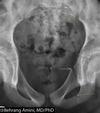
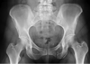
increased PEDICLE distance

24
Q

Chance Fractures AKA Lap Belt Fractures

A

Horizontal splitting of the arch and body, flexion distraction mechanism, seatbelt acts liek a FULCRUM, MC in L1-L3, “empty vertebra” sign, commonly associated with compression fx

25
Name this diagnosis
CHANCE fractures
26
Name this diagnosis
Chance fracture If you can be specific by naming level
27
Transverse process fractures
2nd MC lumbar FX, direct trauma or an avulsion, most often at L2 and L3, vertical to oblique in orientation (perpedicular to ground), often unilateral and often multilevel
28
Transverse process fractures
may or may not be displaced, often obscured by gas and fecal material, POTENTIAL renal damage, need a urinalysis for potential hematuria
29
Name this diagnosis
Transverse process fractures at L2 and L3
30
Name this diagnosis
Transverse process fracture (Look for hemorrhage)...
31
Spondylolisthesis
spondylolysis= interuption of the pars, spondylolisthesis= anterior displacement, prespndylolithesis= spondylolysis without the anterior, pseudospondylolisthesis= anteriorly without pars defects
32
Spondylolisthesis
1st described in 1782 by beligian obstetrition (DON'T MEMORIZE), 5-7% prevalence in white population, 40% prevalence among alaska eskimos
33
NAME this diagnosis
left= Spondylolysis because of no displacement Right= anterior displacement is spondylolisthesis
34
Spondylolisthesis (clinical)
May or may not have pain, if painful may or may not be due to spondylo, displaced RARE after 18 (usually happens before and during puberty
35
Spondylolisthesis
Conservative management, some require more aggressive management, onset: isthmix= (child) degenerative= late (2nd MC reason)
36
Name this Diagnosis
Spondylolisthesis
37
Spondylolisthesis Classifications
Type 1: Dysplastic (Congenital) RARE, Type II: Spodylolytic (isthmic) MC, Type III: Degenerative 2nd MC, Type IV: Tramatic RARE, Type V: Pathological RARE, Type VI: Post Surgical RARE
38
Where is Spondylolisthesis most commonly located?
L5
39
CONGENITAL spondylolisthesis
Rare, malformation of sacrum and L-5, congenital predisposition, such as a congentially thin pars, NEVER present as birth
40
Spodylolisthesis (Spondylytic)
Isthmic, stress FX, elongated pars or acute fx (rare), biomechanical stress, MC type for the younger age onset, MC at L5
41
Spondylolithesis (degenerative)
Due to degeneration of the posterior joints, Pars in tact (pseudospondylo), most commmon at L-4, more common in females, Most commmon type in the older age onset
42
Name this diagnosis
Degenerative Spondylolisthesis
43
Spondylolisthesis (Traumatic)
Acute one time trauma is rare, MC type would be HANGMAN'S fracture of C2
44
Name the diagnosis
traumatic (Hangman's)
45
Spondylolisthesis (Pathologic)
Gernalized or localized bony disorders, metastasis, Paget's, Osterpetrosis, RARE
46
POst surgical Spondylolithesis (Latragenic Spondylolithesis)
Stress Fx at the level above or below anthrodes, or at the level at laminectomy, RARE
47
Spondylolisthesis (RADIOGRAPHIC)
Best detected by George's line, graded by Meyerding's method and / or %, may or may not need obliques, trapezoidal L-5
48
Why don't spondylolisthesis have huge effects?
During puberty a big change young enough can be accomadated
49
Spondylolisthesis (Radiographic)
Inverted Napoleon hat, Bow line of Brailsford, Gendarmes cap, may or may not jave pars defects
50
Name this finding?
Inverted Napoleon hat
51
What is uniquw with Type 2 spondylolisthesis
Scotty dog pedicle fractures
52
What is unique radiographically with Type 3 spondylolisthesis?
On the film, you would see degeneration of the facet joints. It looks brighter than rest of the film
53
Sacral fractures
Direct trauma or results of fall on buttocks, associated with other pelvic fractures, most are horizontal near 3rd and 4th segs, examine sacral foramen CAREFULLY
54
Sacral fractures continued....
Cortical offset on the lateral, potential angulation (compare old films), vertical fractures can occur from indirect trauma, re-exam may be needed, obscured by bowel gas and fecal material
55
Name the classification
Denis classification
56
Name diagnosis
Sacral fracture
57
Coccygeal fracture
Most are horizontal in orientation, direct trauma, best last seen on the lateral, potential angulation (although may be normal), Correlate clinically
58
Pelvic fractures are stable if they have a _____ break of the ring. Unstable fractures usually occur with _______ ring breaks
Single ring break = Stable Double ring break = Unstable
59
T/F 2/3 of pelvic fractures are unstable
FALSE 2/3 are stable
60
Unstable fractures usually result from ___________ and has considerable ___________ injury
Severe trauma Soft tissue
61
Pathology -What mechanism of injury is most commonly associated with this?
Duverney Fracture - a.k.a Iliac wing fracture - Direct lateral force -
62
T/F Duverney Fractures are stable
True -Single break of the ring = stable
63
Pathology
Duverney Fracture
64
Pathology | (lowest 2 arrows)
Ischiopubic Rami Fractures (in this case, superior and inferior fractures) -Medial portion of the inferior rami is most common
65
Most common stable fracture of the pelvis
Ischiopubic Rami Fracture
66
Radiographic findings -What is the Dx?
Cortical offset of the ramus that is absent on the opposite side -Inferior Ischiopubic Rami Fracture
67
Pathology
Superior and Inferior Ischiopubic Rami Fractures
68
Pathology
Malgaigne Fracture -Ipsilateral double vertical shearing fracture of superior and inferior pubic rami with fracture or dislocation about ipsilateral SI joint
69
Most common unstable pelvic fracture
Malgaigne Fracture
70
Pathology
Malgaigne Fracture - Superior and inferior ischiopubic rami fractures - Widened/dislocated/fractured SI joint ipsilaterally
71
What patient profile would lead you to consider a possible avulsion fracture of the pelvis?
Adolescent with athletic participation in sports that involve sprinting, long jumping, hurdling, gymnastics, etc. (Muscular traction leads to the avulsion)
72
Location and muscle associated with avulsion fractures of the pelvis
- ASIS = Sartorius - AIIS = Rectus Femoris - Ischial Tuberosity = Hamstrings (most common)
73
Pathology
Avulsion Fracture of the Left ASIS -Curvillnear ossific density near the affected side ("Finger nail clip")
74
Most common avulsion of the pelvis
Ischial Tuberosity avulsion
75
Pathology
Avulsion Fracture of the Lesser Trochanter of the Femur -Less common
76
Name the finding
Healing avulsion fracture of the pelvis NOT TUMOR
77
Bucket- Handle Fracture
- Contralateral double vertical fx - Superior and inferior pubic rami (BE SPECIFIC ON TEST) - Fx or dislocation about contralateral SI - UNSTABLE
78
Is a Bucket-Handle fracture stable?
NO.
79
Name the Diagnosis
Bucket- Handle fracture on pubic Superior rami and contralateral SI
80
What is the difference between Bucket-Handle fracture to Straddle fracture
Staddle fractrue is BILATERAL superior and inferior rami FX -Substantial soft tissue injury
81
T/F Staddle fractures are stable
FALSE unstable
82
Name Diagnosis
straddle fracture
83
Sprung Pelvis AKA
"Open Book" Diastasis of pubic symphisis Diastasis od one or both SI joints
84
T/F Sprung Pelvis is UNSTABLE
TRUE
85
Name Diagnosis
Sprung Pelvis
86
T/F Complex Pelvic Fractures are STABLE
F UNSTABLE
87
What causes complex pelvic fractures
Usually from a severe injury (Car accident)
88
Complex pelvic fractures are difficult to _____________ and are complex ____________ fractures
- Classify - Multiple
89
Name Diagnosis
Complex pelvic fracture