Exam 3 Flashcards
(35 cards)

Where were the Osteoclasts, Osteocytes and Osteoblasts located?
Identify by the structures in this photo.

Osteons
Haversian Canals
Lamellae

Chondroblasts

Chondrocytes
Top Arrow?
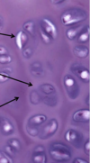
Capsular Matrix w/ Type IV Collagen
Middle Arrow?

Territorial Matrix secreted by isogenous groups
Bottom Arrow?

Interterritorial Matrix
What does the darker staining indicate?

Higher concentration of proteoglycans
What type of cartilage is this?

Elastic
What type of cartilage is this?

Fibrocartilage
What type of cartilage is this?

Hyaline
What are the 5 zones of Endochondral Ossification are indicated in this image?

a) Zone of reserve cartilage
b) Zone of proliferation
c) Zone of Hypertrophy
d) Zone of calcification
e) Zone of ossification

Zone of Reserve Cartilage

Zone of Proliferation

Zone of Hypertrophy

Zone of Calcification

Zone of Ossification
Top Arrow

Osteon = concentric rings of lamellae
Middle Arrow

Haversian (central) canal
Bottom Arrow

Volkmann’s (perforating) canal

Canaliculi

Meninges = Dense Irregular Connective Tissue

Schwann Cells

Satellite Cells


