External Eye Disorders Flashcards
Objectives (26 cards)
1
Q
PANCE External Eye Conditions
A
- Hordeolum
- Hyphema
- Blepharitis
- Chalazion
- Conjunctivitis
- Orbital cellulitis
- Pterygium/Pinguecula
- Cataracts
- Corneal abrasion
- Dacryocystitis
- Ectropion
- Entropion
- Foreign bodies
2
Q
Differentiate between
A
- Benign eyelid vs. conjunctival conditions
- Sight threatening vs malignant eyelid/conjunctival conditions
3
Q
Benign Eyelid Lesions
A
- HIDROCYSTOMA
- HORDEOLUM
- CHALAZION
- BLEPHARITIS
- XANTHALASMA
- KERATOACANTHOMA
- CAPILLARY HEMANGIOMA
- BENIGN EYELID NEVUS
4
Q
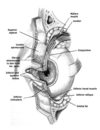
Meibomian Glands
A
- Modified sebaceous glands located within tarsal plates of eyelids
- responsible for secretion of oily layer of tear film
- critical for normal ocular surface lubrication
- Preventing tear evaporation, facilitating spread of tears over ocular surface
5
Q
HORDEOLUM
A
- Acute, purulent,inflammation of eyelid
- External
- infection of lash follicle/glands of Zeis /Moll – points to skin
- Internal
- infection of meibomian gland- large swelling on conjunctival surface of lid
- External
- S/S
- Red, swollen, tender nodule with central core of pus-upper/lower eyelid
- Tearing, photophobia, foreign body sensation
- Etiology
- s. aureus
- TX
- Warm compresses
- I&D
- topical abx

6
Q
CHALAZION
A
- Obstruction of meibomian glands within tarsal plates
- Internal hordeola may develop into chalazion
- Results in formation of granulomas
- Noninfectious
- S/S
- begin as localized eyelid swelling/erythema, then develops into a painless rubbery nodule
- Heaviness of eyelid; vision distortion
- Common in patients with blepharitis and rosacea
7
Q
Treatment of Chalazion
A
- Warm compresses/massage to soften/drain them
- Antibacterial ointment
- Steroid injections
- Surgical incision and removal
- DDX if persistent
- carcinoma
- BCC
8
Q
BLEPHARITIS
A
- Chronic inflammation of eyelids with intermittent acute exacerbations
- 2 types
- anterior
- posterior
- Typical patient
- female and younger
9
Q
Anterior BLEPHARITIS
A
- infection at base of eyelashes
- staphylococcal: colonization leads to scales/crust around lashes
- •Aka:”collarette”/scurf

10
Q
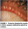
Posterior BLEPHARITIS
A
- More Common than anterior blepharitis
- inflammation of the inner eyelid, at level of meibomian glands
- Meibomian gland dysfunction causing plugging/hypertrophy of sebaceous glands
- Associated w/ rosacea/seborrheic dermatitis
11
Q
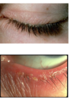
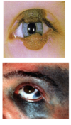
Clinical manifestations if blepharitis
A
- burning, gritty sensation
- Itching
- epiphora
- an overflow of tears onto the face
- sign that constitutes insufficient tear film drainage
- crusting of lashes in am
- flaking skin
- red rimming
- Phlyctenulosis
- a characteristic nodular growth occurring as an allergic response of the conjunctival and corneal epithelium
- in image

12
Q
blepharitis PE
A
- punctate epithelial erosions on cornea
- From hypersensitivity reaction to staph antigens and toxins
- Diffuse conjunctival injection.
- Foamy appearance of tear film
13
Q
BLEPHARITIS TREATMENT
A
- Lid Hygiene
- warm compresses
- lid massage
- lid washing – baby shampoo
- Treat with Antibiotic ointments
- erythromycin, bacitracin, or sulfacetamide
- Steroid/Antibiotic ointments
- Patient education and counseling.
- Maintaining a regimen to prevent future exacerbations.
14
Q
XANTHALASMA
A
- Cholesterol filled soft yellow plaques
- medial aspects of eyelids bilaterally
- nontender
- ETIOLOGY
- conditions that cause elevated blood lipids; deposition of cholesterol laden histiocytes
- Hyperlipidemia (50%), primary biliary cirrhosis
- Appear with elevated triglycerides
- *May have normal levels of cholesterol
- conditions that cause elevated blood lipids; deposition of cholesterol laden histiocytes
- TX
- Surgical Removal-cosmetic
- Correction of underlying cause

15
Q
HIDROCYSTOMA
A
- Benign fluid filled tumor
- S/S
- Solitary, dome shaped translucent nodule on eyelid –bluish gray
- Not confined to lid margins
- asymptomatic
- Etiology
- produced by the cystic proliferation of apocrine/eccrine glands
- High-frequency U/S confirms dx
- TX
- Excision to r/o BCC
- Removing all ectopic epithelium to prevent recurrence

16
Q
KERATOACANTHOMA
A
- Common skin tumor originating from neck of hair follicle
- Flesh-colored, dome shaped, symmetrical nodule, with smooth inflamed skin
- Central keratin/debris filled CRATER
- Etiology
- low-grade subtype of Squamous cell Carcinoma
- S/S
- Rapidly developing nodule reaches full size within 2 mo
- Resolves spontaneously
- If untreated, will starve itself from nourishment, slough off and heal with significant SCARRING
- Sun-exposed areas
- TX
- Biopsy/excision

17
Q
CAPILLARY HEMANGIOMA
A
- Superficial hemangioma of infancy
- Most Common congenital vascular tumor of eyelid and Most common tumors seen in pediatrics
- Characterized by a growth phase and involution phase.
- Presents during 1st 6 months of life
- 50% involuted by age 5, 90% age 9
- TX
- observe/resassurance/steroid injections/laser
- Propranolol oral solution- (Hemangeol) is FDA approved for treatment of infantile hemangiomas
- DDX
- benign vascular tumor
- vascular malformations

18
Q
Hemangiomas: before and after laser treatment
A
- ½ kids with hemangiomas experience residual changes such as scarring, atrophy, redundant skin, discoloration, and telangiectasias. Clinicians who evaluate children who have hemangiomas should be realistic with parents about the potential outcomes:
- Superficial, very raised hemangiomas, especially those that exhibit a sharp, ‘cliff-like’ border or pedunculated lesions are at risk for residual fibrofatty tissue, which may ultimately require surgical intervention.

19
Q
BENIGN EYELID NEVUS
A
- Well-demarcated, smooth, pigmented congenital lesions
- Nevus of Ota
- congenital disorder of melanocytes: discoloration of conjunctiva, skin, eyelids, periorbital tissues
- Uniform in color
- NO loss of lashes
- Junctional
- flat with cells at interface of epidermis and dermis: malignant potential
- Intradermal
- cells lie entirely in dermis
- More pigmented/elevated during puberty
- Risk of malignant transformation : 5 %
- TX
- Observe for changes
- Change in appearance
- excised
**Congenital dermal melanocytosis involving the areas innervated by the first and second divisions of the trigeminal nerve [most commonly in Asians and blacks- potential for malignancy- f/u yearly with ophthalmologist
20
Q
Malignant Eyelid Tumors
A
- Meibomian Gland Carcinoma
- Basal Cell Carcinoma
- Merkel Cell Carcinoma
- Kaposi Sarcoma
21
Q
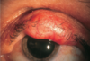
MEIBOMIAN GLAND CARCINOMA
A
- Sebaceous gland carcinoma
- Slow growing
- Highly malignant tumors in tarsal part of eyelid-arising from meibomian/zeis glands
- Upper eyelid
- Yellow appearance
- Loss of eyelashes
- Classically: firm, painless, indurated mass
- May be at site of previous chalazia/blepharitis treatment
- women>men
- TX
- wide surgical excision of tumor
- Biopsy the lymph nodes
- FOLLOW-UP!
22
Q
Kaposi Sarcoma of Eyelid
A
- Aggressive, Small, purplish highly vascular elevated mass w/ surrounding telangiectasias
- Often seen in AIDS pts
- ETIOLOGY
- lymphatic or viral: HIV
- Organ transplant recipients w/ immunosuppression
- S/S
- pain, photophobia ,epiphoria, dry eyes, heavy eyelids
- Indolent tumor, with growth
- Can cause significant disfigurement
- TX
- local radiation/surgical if disfiguring, otherwise manage HIV w/ reducing risk of opportunistic infection

23
Q
BASAL CELL CARCINOMA
A
- Most common malignant tumor of eyelid
- NONHEALING, BLEEDING ULCER
- Firm, small, pearly bordered nodule
- Telangiectasic vessels
- Loss of eyelashes
- Grows slowly with central ulceration
- Painless
- 1/2–2/3 involve the lower eyelid margin
- Locally invasive, rarely metastasize
- ETIOLOGY
- hx of UVB exposure, multifactorial
- Most Common on Left Eye in US, Right Eye in England
- DDX
- Actinic keratosis, malignant melanoma
- TX
- Mohs excision, cryotherapy
- FOLLOWUP!

24
Q
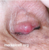
MERKEL CELL CARCINOMA
A
- Neuroendocrine carcinoma
- Rare , AGGRESSIVE, highly malignant skin cancer
- Firm, painless, solitary red nodule
- Most commonly found on sun-exposed areas of the head/neck/extremities
- Pts seek advice on rapidly growing bump or breaking down skin
- Pts >50 years old
- Etiology
- UV radiation, sun exposure
- merkel cell polyomavirus, 8/10 : (Jan 2008)
- TX
- surgical excision, sentinel LN biopsy
25
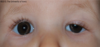
PTOSIS
* Drooping of upper eyelid that results from congenital or acquired abnormality of muscles that innervate eyelid
* low-lying upper eyelid margin: more obvious if eye is focused in primary gaze: secondary to abnormality of levator/muller muscle
26
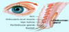
Important Muscles contributing to Ptosis
* Circumferential orbicularis oculi
* Innervated by facial (7th CN)
* close upper and lower eyelids.
* LEVATOR M: Oculomotor (3rd CN)
* opening of upper eyelid
* Müller's muscle, sympathetic NS
* arises from levator
* inserts into superior tarsal plate
* Function:
* over-elevation of the eyelid
* excited or fearful
* leads to mild ptosis with fatigue/inattention.