Final Exam- Important Concepts Flashcards
What are you assessing when you test the menace response? Why?
* the animal cannot see out of that eye, but that the lesion is not necessarily in the optic nerve or eye, but the lesion could be in the brain. If so, it is known as cortical blindness. PROSENCEPHALIC (forebrain) LESIONS.
** Menace response assess: CN II, retina, cerebellum, CN VII
Why? Because the menace pathway starts where vision left off– neurons in the occipital cortex–> motor cortex–>via internal capsule and crus cerebri to synapse with nuclei in the PONS–> contralateral cerebellar hemisphere (for coordination) –> facial nerve nucleus to drive blinking (orbicularis oris)
What are pathways that involve the optic nerve?
- Vision
- Control of eyeball movements (from the rostral colliculus, neurons project to the motor nuclei of CN III, IV, and VI of both sides) (Voluntary eyeball movement is different)
- Pupil Constriction (rostral colliculus left and right parasympathetic nuclei of the oculomotor nerve CN III)
- Control of turning of the head and neck (rostral colliculus neurons send axons which decussate and descend through th brain into the spinal cord forming the tectospinal tract– reflex turning of the head and neck towards sudden source of light or movement)
- Projections into the reticular activating formation (some neurons from the rostral colliculi project into the reticular activating formation to provide general arousal stimnuli to the cerebral cortex)
What are the 3 neurons in hearing?
SCM
Neuron 1: cell body in the spiral ganglion of CN VIII receiving impulses from the neuroepithelial cells in the spiral organ (of corti). Axon runs in CN VIII (vestibulocochlear)
Neuron 2: Located in the cochlear nuclei– many axons decussate at once forming the TRAPEZOID BODY.–> Lateral lemnicus
Neuron 3: Medial geniculate nucleus
What 3 neurons are in the taste pathway?
Neuron 1: Ganglia of CN VII (rostral 2/3 of tongue), IX (caudal 1/3), or X (tastebuds near epiglottis)
Neuron 2: Nucleus of the solitary tract
Neuron 3: Ventral group of thalamic nuclei–> cerebral cortex to the somaesthetic cortex
What 3 neurons are in smell?
Neuron 1: Olfactory neuroepithelial cell (constantly dividing and replenishing)–> axons pass through the cribiform plate and synapse with neuron 2 in the olfactory bulb
Neuron 2: Olfactory bulb–> olfactory peduncle–> divides into the medial and lateral olfactory tracts end in the Olfactory tubercle
Neuron 3: Olfactory tubercle–> axons to cerebral cortex via pyriform lobe
** No relay to the thalamus because most primitive part of the brain**
What is a seizure?
Epilepsy is idiopathic. But if they occur due to brain injury. Then injury can cause changes to inherent excitability of glutamatergic neurons. Excessive neuronal activity can cause the accumulation of potassium and glutamate if the astrocytes are dysfunctional. And the accumulation of potassium can cause depolarization of neighboring axon terminals. The accumulation of glutamate at excitatory synapses and activate post synaptic receptors.
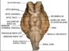
What’s the mnemonic for the cranial nerves?
On Old Olympus’s Towering Tops A Fair Voluptuous German Vaulted And Hopped
- Olfactory
- Optic
- Oculomotor
- Trochlear
- Trigeminal
- Abducens
- Facial
- Vestibulocochlear
- Glossopharyngeal
- Vagus
- Accessory
- Hypoglossal
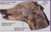
What are the most likely potential routes for the transport of bacteria into the CNS?
CN I (cribiform plate), II, and VII (travels with VIII through the internal acoustic meatus – susceptible to middle ear disease)
Which cranial nerves attach together and may be involved in one lesion?
CN V, VII, VIII
Innervation of the pharynx and larynx?
Pharynx- sensory- 9; musculature- 9 & 10
Larynx- sensory- 10 & 11; musculature- 10 & 11
Which is the only nerve that exits the brain dorsally? What does it do?
CN 4 (Trochlear)- exits through the orbital fissure in the dog to innervate the dorsal oblique muscle (inward eye rotation)
What part of the brain is the Abducens CN part of? What are the main functions? Dysfunction?
* Medulla
* Lateral eye movement (Lateral rectus muscle)
* Double vision; strabismus: eye deviation medially
What part of the brain is the Trochlear CN (IV) associated with? Main function? Dysfunction?
* Midbrain
* eye movement (dorsal oblique m.)
* Dorso-lateral strabismus


What part of the brain is the Vestibulocochlear CN part of? Function? Dysfunction?
* Medulla (Myelencephalon)
* Hearing and balance- horizontal and vertical eye movement
* Deafness, head tilt, nystagmus
Function of Glossopharyngeal? Dysfunction?
* Caudal 1/3 of Tongue and pharynx (sensory), carotid sinus, motor to stylopharyngeaus, PS to parotid and zygomatic salivary glands
Function of Facial CN? Dysfunction?
* Taste on rostral 2/3 of tongue, motor to muscles of facial expression, PS to mandibular, sublingual, palatine, nasal, lacrimal glands
* Paralysis of facial muscles (drooping of ear, lip, and eyelid), decreased lacrimation, decreased taste sensation
Function of Vagus? Dysfunction?
* Sensory to pharynx, larynx, and viscera, sensory to external ear canal, taste on root of tongue and epiglottis, PS to viscera
* dysphagia (difficulty in swallowing), respiratory noise (laryngeal paralysis)
Function of Accessory? Dysfunction?
* motor to trapezius and brachiocephalicus muscles
* Atrophy, dysfunction of trapezius, brachiocephalicus and sternocephalicus
Function of hypoglossal? Dysfunction?
* Motor to tongue muscles
* Paralysis and deviation of the tongue if unilateral lesion
Where does CN I exit from the skull?
Cribiform plate
Where does CN II exit from the skull?
Optic foramen
Where does CN III exit from the skull?
Orbital fissure in small animals, orbitorotundum in ruminants