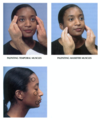
Flashcards for Midterm Exam

(155 cards)

1
Q

Labs and Diagnostic Testing: CBC, H&H

A

Consider:

  • Proposed surgery
  • Potential blood loss
  • Clinical indications: hematologic dx, chronic kidney/liver dz, anticoagulants, etc.
  • NICE guidelines: ASA-PS 3 or 4 intermediate procedures, all pts having major procedures
2
Q

Lab and Diagnostic Testing: Electrolytes

A

Clinical indications for electrolytes:

  1. chronic kidney dx
  2. cirrhosis
  3. certain meds
  4. DM
  5. dialysis
  6. type of surgery
3
Q

Lab and Diagnostic Testing: Glucose

A
  • Clinical indications for glucose: DM, steroid use, cirrhosis
4
Q

Lab and Diagnostic Testing: Renal Function

A
  • Assess tubular function & GFR
  • Clinical indications: HTN, DM, dehydration, renal dx, h/o transplant, etc.
  • NICE guidelines:
    • Routine: ASA 3 or 4 having intermediate procedure & ASA 2, 3, or 4 having major procedure
    • Risk for AKI: ASA 3 or 4 having minor procedure, ASA 2 for intermediate
5
Q

Lab and Diagnostic Testing: Liver Function

A
  • History of liver injury & exam findings
  • Clinical indications: hepatitis, jaundice, cirrhosis, portal HTN, bleeding disorders, etc.
6
Q

Lab and Diagnostic Testing: Coagulation Profile

A
  • Routine testing not indicated unless known or suspected coagulopathy
  • Clinical indications: bleeding disorder, anticoag meds, liver dx
  • NICE guidelines: ASA 3 or 4; intermediate, major, complex procedures; anticoag meds or chronic liver dz
7
Q

Lab and Diagnostic Testing: Urinalysis

A
  • No indication for routine use
  • Clinical indications: suspected UTI, unexplained fever or chills
8
Q

Lab and Diagnostic Testing: T&S, T&C

A
  • Decision usually guided by institution policy (surgical blood order schedule)- reduce unnecessary blood order$
  • Clinical indications: suspect blood transfusion
9
Q

Lab and Diagnostic Testing: ECG

A
  • No indication for routine use
  • Clinical indications: h/o IHD, HTN, DM, HF, CP, syncope, DOE, etc.
  • 2014 ESC/ ESA guidelines: risk factors for IHD, CVD, sig. arrhythmia, or symptomatic; intermediate/ high risk surgery
  • NICE guidelines: ASA 3 or 4 intermediate procedure; ASA 2, 3, or 4 major procedures
10
Q

Lab and Diagnostic Testing: Chest X-ray

A
  • No indication for routine use
  • Clinical indications: advanced COPD; bullous lung disease; suspected: pulmonary edema, pneumonia, mediastinal mass; suspicious findings on exam
  • Patients undergoing thoracic, upper abdominal, AAA surgery
11
Q

Lab and Diagnostic Testing: ECHO

A
  • No indication for routine use
  • Clinical indications: heart murmur and symptomatic, S&S of HF, unexplained dyspnea
  • Clinically stable, ventricular dysfunction not tested in previous year
12
Q

Lab and Diagnostic Testing: PFT’s

A
  • Considered for type and invasiveness of surgery (CABG, lung resection)
  • Clinical indications:
    1. severe asthma
    2. symptomatic COPD
    3. scoliosis
    4. restrictive lung dx
13
Q

Patients considered “Aspiration Risk”

A
  1. Age extremes <1 or >70
  2. Prematurity
  3. Pregnancy
  4. Ascites (ESLD)
  5. Neurologic dz
  6. Collagen vascular dz, metabolic disorders (DM, obesity, ESRD, hypothyroid)
  7. Hiatal Hernia/GERD/Esophageal surgery
  8. Mechanical obstruction (pyloric stenosis)
  9. Having eaten food or non-clear drinks
14
Q

Continue meds day of surgery except

A
  • ACEIs and ARBs
  • ASA: stop 3 days prior unless PCI, high-grade IHD, sig. CVD
  • P2Y12 inhibitors: stop 5-10 days prior unless drug-eluting stent (6-month dual antiplatelet therapy) & bare metal stents (1-month dual antiplatelet therapy) *** Discuss risk w/ surgeon/ cardiologist
  • Warfarin: stop 5 days prior
  • NSAIDS: 48 hrs prior
  • Short-acting insulin; none or ½ dose of long-acting insulin day of *** insulin pump- basal rate only
  • Non-insulin antidiabetic meds (metformin →lactic acidosis)
  • Topical
  • Diuretics
  • Sildenafil or similar
15
Q

Cardiovascular Assessment
Renal Failure

A
  1. ↑CO – compensation for ↓ O2 carrying capacity
  2. HTN – Na+ retention, RAAS activation
  3. LVH common
  4. CHF w/ pulmonary edema after limits of compensation reached
  5. Deposition of Ca++ - in conduction system & on heart valves
  6. Arrhythmias – electrolyte imbalances
  7. Uremic pericarditis – can be asymptomatic, chest pain, tamponade, usually 2 to inadequate dialysis
  8. Accelerated CAD, PVD
16
Q

Fluid Balance Assessment
Renal Failure

A

•Fluid overload VS intravascular depletion s/p dialysis/ aggressive diuretic therapy

  1. Body weight
  2. VS (orthostatic hypotension & tachycardia)
  3. Atrial filling pressures
17
Q

Pulmonary Assessment
Renal Failure

A
  • ↑ MV to compensate for metabolic acidosis
  • ↑ pulmonary extravascular water = interstitial edema = widened alveolar/arterial O2 gradient
  • “Butterfly wings” on CXR 2 to ↑ permeability of alveolar capillary membrane (edema even with normal PCWP)
18
Q

Endocrine Assessment

Renal Failure

A
  • Peripheral resistance to insulin = poor glucose tolerance
  • Hyperparathyroidism = prone to fractures
  • Abnormal lipid metabolism = accelerated atherosclerosis
  • Kidneys do not degrade hormones and proteins normally = increased circulating PTH, insulin, glucagon, GH, LH, PL
19
Q

Normal Na+

A

135-145 meq/L

20
Q

Normal K+

A

3.5-5 meq/L

21
Q

Normal Cl-

A

95-105 meq/L

22
Q

Normal HCO3-

A

Venous: 19-25 meq/L

Arterial: 22-26 meq/L

23
Q

Normal Ca++

A

4.5-5.5 meq/L

24
Q

Normal Phosphate

A

2.4-4.7 mg/dL

25
Normal Mg+
1.5-2.5 meq/L
26
Normal Serum Osmolality
280-300 mOsm
27
Labs/Test Abnormalities common w/ renal failure
1. Assess adequacy of dialysis 2. Metabolic acidosis with high AG 3. **Hyperk**alemia (hypok w/ dialysis) 4. **Hyperglyc**emia (if insulin resistant) 5. **Hypermag**nesemia 6. **Hypona**tremia (hyper is rare) 7. **Hypoca**lcemia (unclear etiology) 8. **Hypoalb**uminemia (esp. w/ dialysis)
28
Hyperkalemia EKG findings
Tall T waves ST depression Wide QRS
29
Hypokalemia ECG findings
Peaked or flattened T waves Prolonged PR & QT
30
Hypocalcemia ECG findings
Prolonged QT
31
Assessment of Muscle Strength: elbow flexion & extension
elbow flexion = C5, C6 elbow extension = C6, C7, C8
32
Assessment of Muscle Strength: Grip
C7, C8, T1
33
Assessment of Muscle Strength: Finger ABduction
C8, T1, ulnar nerve
34
Assessment of Muscle Strength: Opposition of thumb
C8, T1, median nerve
35
Assessment of Muscle Strength: hip flexion & adduction
L2-L4
36
Assessment of Muscle Strength: Hip ABduction
L4, L5, S1
37
Assessment of Muscle Strength: Hip extension
S1
38
Assessment of Muscle Strength: Knee extension
L2-L4
39
Assessment of Muscle Strength: Knee flexion
L4, L5, S1, S2
40
Assessment of Muscle Strength: Dorsiflexion
L4, L5
41
Assessment of Muscle Strength: Plantar flexion
S1
42
List of MAOI's
* Phenelzine * Isocarboxazid * Selegiline * Brofaromine * Befloxatone * Iproniazid * Moclobemide
43
Interferon B
Disease modifying therapy/ immunomodulator for MS Side Effects: flu-like symptoms, hepatotoxicity, myelosuppression, depression
44
Dimethyl Fumarate
Disease modifying therapy/ immunomodulator for MS Side Effects: GI discomfort, infections
45
Glatiramer Acetate
Disease modifying therapy/ immunomodulator for MS Side Effects: well tolerated
46
Natalizumab
Disease modifying therapy/ immunomodulator for MS Side Effects: Leukoencephalopathy, hepatotoxicity
47
Fingolomid
Disease modifying therapy/ immunomodulator for MS Side Effects: bradycardia, hepatoxicity
48
Teriflumonide
Disease modifying therapy/ immunomodulator for MS Side Effects: neutropenia, hepatotoxicity
49
Mitoxantrone
Immunosuppresant drug for MS Side Effects: severe myelosuppression and cardiac toxicity-- reduced EF--heart failure
50
Drugs that affect coagulation status (SLE)
1. Ibuprofen 2. Indomethacin 3. ASA 4. Cox-2 Inhibitors 5. DVT preventative therapy
51
Leads that correspond to inferior wall (RCA)
II, III, avF
52
Leads that correspond to lateral wall (circumflex branch of LCA)
I, avL, V5-V6
53
Leads that correspond to anterior wall (LCA)
V3-V4
54
Leads that correspond to septal wall (left descending coronary artery)
V1-V2
55
V1 placement
+ electrode placed directly over R atrium; corresponds to septal wall (LAD)
56
V2 placement
+ electrode placed just anterior to the AV node; corresponds to septal wall (LAD)
57
V3 & V4 placement
+ electrode placed over ventricular septum; corresponds to anterior wall (LAD)
58
V5 & V6 placement
+ electrodes placed over lateral surface of L ventricle; corresponds to lateral wall of heart (circumflex)
59
Causes of left axis deviation
* LBBB * Chronic HTN * Aortic stenosis * Aortic insufficiency * Mitral regurgitation
60
Causes of right axis deviation
1. PE 2. Pulm HTN 3. COPD 4. Cor pulmonale 5. Acute bronchospasm
61
Right ventricular hypertrophy
Large R wave in V1 and gets progressively smaller in V2, V3, V4
62
Left ventricular hypertrophy
Large S wave in V1 and larger R wave in V5 * Depth (in mm) of S in V1, plus height of R in V5 if \>35 mm = LVH
63
Ischemia
T wave inversion or ST segment depression
64
T wave inversion or ST segment depression
Ischemia
65
Injury
ST segment elevation (\>1 mm)
66
ST segment elevation (\>1 mm)
Injury
67
Infarction (old MI)
Q waves which are \>1 small box or 1/3 size of QRS
68
Q waves which are \>1 small box or 1/3 size of QRS
Infarction (old MI)
69
ASA I
Normal, healthy pt; no systemic dz
70
ASA II
Mild to moderate systemic dz, well-controlled, no functional limitations
71
ASA III
Severe systemic dz, functional limitation
72
ASA IV
Severe systemic dz that is a constant threat to life
73
ASA V
Moribund pt, not expected to survive (+/- surgery)
74
ASA VI
Pt declared brain dead; harvesting organs for donation
75
GI/Liver Assessment Renal Failure
* 10-30% pts develop **GI hemorrhage** * High incidence of **Hep B&C** (multiple transfusions) * **Anorexia, N/V** * **Ascites** w/ dialysis * **Hypersecretion** of gastric acid **+ delayed emptying** (autonomic neuropathy)
76
Renal Failure: Impact on Drugs
Effects altered due to: * **Anemia** * **Decreased** serum **protein** * **Fluid retention** = *change Vd for MR's* * **Electrolyte** abnormalities = *pts more prone to dig toxicity* * Abnormal **cell membrane** activity Drugs eliminated by the kidneys unchanged are **_CONTRAINDICATED_** * **_Gallamine, phenobarbital, LMWH_** \*\*\*LMWH is cleared by the kidneys and NOT removed during dialysis = prolonged duration and r/f bleeding!
77
Renal Function Tests for **GFR**
1. Blood Urea Nitrogen (10-20 mg/dL) 2. Plasma creatinine (0.7-1.5 mg/dL) * GFR can decreased 50% without rise in plasma creatinine. Not accurate indicator, especially in elderly 3. Creatinine clearance\*\* (110-150 mL/min)
78
* Chronic Kidney Disease (CKD) = * Chronic Renal Failure (CRF) = * ESRD = * Acute Kidney Injury (AKI) =
_Chronic Kidney Disease (CKD)_ = GFR \< 60 mL/min/1.73m2 for 3 months _Chronic Renal Failure (CRF)_ = GFR 15 mL/min/1.73m2 _ESRD_ = Loss of renal function for 3 months or more (Diabetes accounts for ½ of cases & HTN for ¼. Polycystic 10%-genetic autosomal dominant) _Acute Kidney Injury (AKI)_ = Sudden decreased function/UOP
79
What situations requires dialysis?
1. Oliguria 2. Fluid overload 3. Hyperkalemia 4. Severe acidosis 5. Metabolic encephalopathy 6. Pericarditis 7. Coagulopathy 8. Refractory GI symptoms 9. Drug toxicity
80
What is hemodialysis
* Diffusion of solutes between blood and dialysis solution remove metabolic wastes and restore buffers to the blood * Need vascular access * AV fistula = cephalic vein anastomosed to radial artery * Jugular or femoral vein for emergency access
81
Dialysis and meds
* General rule: scheduled doses of drugs are administered after dialysis * Low-molecular weight, water-soluble, non-protein bound drugs are readily cleared by dialysis
82
Neuro Assessment: Renal Failure Neuro Assessment s/p Dialysis
* Uremic Encephalopathy * Asterixis * Myoclonus * Lethargy * Confusion * Seizures * Coma * Autonomic Neuropathy * Peripheral Neuropathy _S/P Dialysis_ * **Disequilibrium syndrome** (dialysis related) transient CNS disturbance after rapid ↓ in ECF osmolality compared w/ ICF osmolality * **Dementia**
83
Hematological Assessment: Renal Failure
1. **Anemia** typical **Hgb = 6-8 g/dL** * ↓ erythropoietin and RBC production and ↓ cell life span * GI blood loss, hemodilution, bone marrow suppression * Excess PTH replaces bone marrow w/ fibrous tissue * Most pts tolerate anemia well (exception CAD) * ↑ 2,3- DPG (diphosphoglycerate) + metabolic acidosis = favors Right shift 2. **Impaired platelets (**qualitative) = prolonged bleeding time 1. ↓ plt factor III activity 2. ↓ adhesiveness & aggregation 3. **Impaired WBC** function = infections 4. Release of **defective von Willebrand factor\***\* 5. Dialysis = Residual **anticoagulation vs** promotion of **hypercoagulable** state 6. **Hypocomplementemia** w/ dialysis
84
Renal Function Tests to assess Renal Tubular Function
Renal Tubular Function = reflects concentrating ability of kidney vs nephron function (GFR) * Urine Specific Gravity (1.003-1.030) * Urine Osmolarity (38-140 mOsm/L)
85
Labs and Tests: Blood Urea Nitrogen
* Normal BUN = 10-20 mg/dL * Varies _inversely w/GFR_ & **directly w/protein catabolism** * \> 50 mg/dL is indicative of ↓GFR (in pts w/ nml diets) * The most common cause of ↑BUN is CHF 2 to reabsorption of BUN *(RAAS is on high)* * BUN is **_not_** a sensitive index bc urea clearance also depends on urea production * **BUN can be abnormal** despite a *normal GFR* due to: * High protein diet * GI bleed * Febrile illness
86
Labs and Tests: Plasma Creatinine (Cr)
* Plasma Cr is a specific indicator of GFR (plasma Cr is freely filtered but not reabsorbed) * **8-17 hr lag time** after change in GFR before ↑ Cr levels are seen * Suggestive [but not indicative] of Acute Renal Failure * Usually 50% ↑ in plasma Cr reflects a corresponding ↓ in GFR * Skeletal muscle = source of Cr (more muscle mass = higher baseline Cr) * Elderly pt: Cr levels stay normal/constant d/t: * ↓ muscle mass and GFR * *If CR ↑ in elderly, may be indicative of renal failure*
87
Labs and Tests: Creatinine Clearance
Creatinine Clearance approximates GFR 24 hr collection most accurate although 2 hr tests also helpful 100-120 mL/min = Normal 60-100 mL/min = ↓ Renal Reserve 40-60 mL/min = Mild Renal Impairment 25-40 mL/min = Moderate Insufficiency \< 25 mL/min = Renal Failure \< 10 mL/min = ESRD
88
Hyperkalemia treatment if symptomatic or **\> 6/5 meq/L**
89
Normal RBC
male: 4.6-6.2 million/mm3 female: 4.2-5.4 million/mm3
90
Normal Hgb
Male = 13.5-18 g/dL Female = 12-16 g/dL
91
Normal Hct
male = 40-54% female = 38-47%
92
Normal WBC
5,000-10,000/mm3
93
Normal bleeding time
3-10 min
94
Normal platelet count
150,000-400,000 cell/mm3
95
Prothrombin time (PT)
10-12 sec
96
Internationalized Normalized Ration (INR)
0.8-1.2
97
Normal plasma thromboplastin time (PTT)
25-35 sec
98
Normal activated clotting time
90-120 sec
99
Normal thrombin time
9-11 sec
100
Normal fibrinogen
160-350 mg/dL
101
Coagulation Abnormalities in Renal Failure Pt
* If **regional** anesthesia is planned need **coag panel**!!!! * **↑ bleeding** despite **normal PT, PTT** * **Bleeding Time** best screening test * Important cause of coagulation issues- **release of defective von Willebrand factor** * Rx: * **Desmopressin (0.3-0.4 mg/kg over 30 min)** * **Cryoprecipitate 10 units IV over 30 min** * Blood warmer set up and ready to go
102
Cardiac Labs and Tests in Renal Failure Pt
**EKG** * Hyper or hypokalemia * HyperK (Concern above 5.0) → Tall T waves, ST depression, wide QRS * HypoK → peaked or flattened T waves, prolonged PR & QT * Hypocalcemia → Prolonged QT * Ischemia * Conduction blocks * LVH **Echocardiography** * Hypertrophy * Ventricular EF * Wall motion abnormalities * Pericardial fluid
103
Pulmonary Labs and Tests in Renal Failure Pt
* CXR *(look for butterfly wings, enlarged heart)* 1. Fluid status 2. Determine presence of HTN related CV disease 3. Pericardial effusion 4. **Uremic pneumonitis** * ABG – hypoxia and acid/base status especially if dyspnea noted on exam
104
Musculoskeletal Assessment: The Temporomandibular Joint
* Place tips of index finger just in front of the tragus of ear - ask patient to open mouth. * Fingertips should drop into joint spaces as mouth opens. * Check for smooth range of motion, swelling/tenderness. * Snapping & clicking normal * Ask pt to open & close mouth, protrude & retract (jutting the jaw forward), & perform side to side motion.
105
Musculoskeletal Assessment: Cervical Spine
* Flexion = Touch chin to chest * Extension= Look up at ceiling * Rotation= Turn head to each side looking directly over shoulder * Lateral Bending = Tilt head touching ear to shoulder ## Footnote *\*Might have to do awake fiberoptic intubation if they can’t tolerate.*
106
Musculoskeletal Assessment: Shoulder Girdle
(adduction, abduction, flexion, extension, internal & external rotation) 1. Abduct the arms to shoulder level. 2. Raise arms vertical position above head palms facing each other. 3. Place both hands behind the neck with elbows out to the side. 4. Place both hands behind the small of the back.
107
Musculoskeletal Assessment: The Shoulder
* Thoracic Outlet Syndrome- compression of brachial plexus and subclavian vessels near the 1st rib * Be certain pt can work or sleep w/ arms elevated over head before putting arms beside head (prone positioning)
108
Musculoskeletal Assessment: The Elbow
Ask pt. to bend and straighten elbow (flexion and extension) With arms at sides and elbows flexed instruct pt. to turn palms up (supination) and palms down (pronation)
109
Musculoskeletal Assessment: The Hip
* **Concentrate on ROM that can impact positioning** * Flexion – supine pt. bends each knee to chest/abdomen * Abduction – supine pt. stabilize anterior superior iliac spine and abduct the extended leg until the iliac spine moves = limit! \*Perform this test if placing in lithotomy position or if pt at r/f hip issues (OA, etc.)
110
Nervous System Assessment
* Mental status (awake, alert, oriented X3) * Speech (clear speech, slurred) * Cranial nerves (don’t do on every pt) * Gait (Parkinson’s gait) * Motor function (note where we are starting from and document so we can note any changes or exacerbations post-op) * Sensory function
111
Nervous System Assessment: Cranial Nerve 1
1 = Olfactory (smell)
112
Nervous System Assessment: Cranial Nerve 2
2 = Optic (sight – confrontation test; pupillary rxn to light)
113
Nervous System Assessment: Cranial Nerve 3
3 = Oculomotor (pupillary rxn to light; extraocular movements)
114
Nervous System Assessment: Cranial Nerves 4 and 6
4 = Trochlear (extraocular movements) 6 = Abducens (extraocular movements)
115
Nervous System Assessment: Cranial Nerve 5
5 – Trigeminal Ask pt to clench their teeth as you palpate temporal and masseter muscles Check sensation in areas circled Corneal reflex (cotton ball)
116
Nervous System Assessment: Cranial Nerve 7
7 – Facial Ask pt to: * Raise both eyebrows * Close eyes tightly so you can’t open them * Frown, Smile * Show teeth * Puff out both cheeks
117
Nervous System Assessment: Cranial Nerve 11
11 – Spinal Accessory * Ask pt. to turn head to each side against your hand * Ask pt to shrug both shoulders upward against your hand – trapezii strength
118
Nervous System Assessment: Cranial Nerve 8, 9, 10, 12
8 – Acoustic (hearing) 9 – Glossopharyngeal and 10 – Vagus (voice hoarseness?, gag reflex, AHH- palate should rise symmetrically) 12 – Hypoglossal (tongue movement, ask them to move tongue side to side)
119
Assessment of Muscle Strength Grading Scale
* Test flexion and extension and compare symmetry * Grade on 0-5 scale 0. No muscular contraction detected 1. Barely detectable 2. Active movement with gravity eliminated 3. Active movement against gravity 4. Active movement against gravity with some resistance 5. Active movement against gravity with full resistance
120
Head Injury: Assessment
* Glasgow Coma Scale – defines neurologic function impairment * Eyes open - Never(1)- Spontaneous (4) * Best Verbal Response None (1) – Oriented (5) * Best Motor Response None (1) - Obeys commands (6) * Mortality closely related to initial score * Scores 8 or less considered severe (coma)- will require intubation and controlled ventilation for ICP & airway control
121
Steroids
* Suppression or dz of pituitary-adrenal axis will prevent pt from responding to stress of surgery appropriately * Any pt who has received corticosteroid therapy (suppression of pituitary-adrenal axis) for at least 1 month in the past 6-12 months needs supplementation periop * 2 possible regimens * 100 mg Hydrocortisone pre-op, intra-op and post-op * 25 mg Hydrocortisone pre-op + 100mg IV gtt over 12-24 hrs
122
Methotrexate
* Immunosuppressant used in **MS**, Ankylosing Spondylitis (**AS**) and **RA** * Immuno-suppression, anemia, thrombocytopenia, pulmonary, renal, hepatic toxicity * **CBC** * **Chem**istry Panel * Consider **PFTs** and **LFTs** if history warrants
123
Multiple Sclerosis
* These pts are generally on **immunosuppressive** meds * Any recent history of illness or infection? * Take extra care with infection prevention * **Which meds** are they taking and how often? * **Steroids** in the past year???? * *Be ready to give stress dose* * **Remission vs exacerbation** intervals * *If in middle of exacerbation, perhaps delay elective surgery* * Severity and nature of symptoms * **Respiratory** status * Previous **triggers****​** DOCUMENT PRE-EXISTING DEFICITS * Paralysis (assess for motor strength) * Sensory disturbances (assess along dermatomes) * Autonomic disturbances (resting HR, orthostatic hypotension) * Visual impairment (cranial nerve check) * Seizures (meds) * Emotional Disturbances Counsel pt regarding increased r/f relapse w/ surgery
124
Guillain Barre
* Document **time course** of dz * Precipitating factors * Onset of symptoms * Dz Progression (worsening, stable, improving) * Document severity and current state of symptoms * **Facial paralysis** – bulbar involvement (what other concerns might you have here????) protecting airway * Difficulty **swallowing** – pharyngeal muscle weakness * Impaired **ventilation** – current ventilatory support required (vent settings) * Decreased **deep tendon reflexes**– lower motor nerves * Extremity **paresthesias** * **Pain** – headache, backache, muscle tenderness + note meds helpful for pain control * **ANS DYSFUNCTION!!!!!**! * Review ICU flow sheets for **VS** trends * **Vasoactive med**ication history for HTN and hypotension (ex. vasopressors, B-Blockers) * ECG, any recent arrhythmias? * Inquire w/nurse/patient regarding tolerance of position changes
125
Parkinson’s Disease
* **Age of diagnosis**, recent exacerbations and hospitalizations * Current and past **symptoms** (ex. oculogyric crisis (eyes stuck in one position), when? How long did it last? What helped?) * **ANS** symptoms (orthostatic BPs) * History of **Pergolide** therapy? CV function—cv testing might be needed * **Temp** regulation issues? * **Pulmonary** status optimized? *(CXR, maybe won’t tolerate exercise bc of motor disability—hard to use as gauge for cv and pulm function, so may need pft if concerned)* * Dysphagia and/or dyspnea * Pulmonary infection * Continue **current med regimen** and note side effects * Levodopa -what happens if the patient misses a dose? * Anticholinergics and MAO’s? * Note the **natural ROM** for positioning * Deactivate deep brain stimulators before electrocautery
126
Intervertebral Disc Herniation/ Lower Back Pain
* Natural ROM for positioning and laryngoscopy * Baseline motor strength & sensation in applicable areas * Medication regimen (ex. pt on high dose opioids), what drugs improve pain, what has been ineffective? * Consider potential for operative blood loss * CBC * Type and Cross
127
Ankylosing Spondylitis
* Evaluate for co-existing **vasculitis, aortitis, aortic insufficiency, pulm fibrosis** * Evaluate for severity of **kyphosis (difficult airway)** * SpO2, ECG, ECHO, CXR, PFTs * CBC, BUN/Creatinine * D/c NSAIDs at least 2 day pre-op * May have **positioning challenges** *(Eval normal ROM)* * ROM joints (esp. sacroiliac) * Pain mgt. hx. * ECG (cardiac conduction) * PFT (fibrosis) * Meds (NSAIDS, methotrexate, etc.)
128
Spinal Cord Injury
* Determine level of lesion * Acute or Chronic? * **_Acute_** 1. Fluid and Blood Status 2. CBC, Type and Cross, Chem 7 3. ECG/ Chest X-ray 4. Vasopressor requirement? 5. Ventilatory support (current vent settings)? 6. Associated injuries? * **_Chronic_** 1. History of **autonomic dysreflexia**? What initiated it? 2. Old OR/ICU records helpful – **response** to vasopressors, tracheal suctioning 3. **Ventilatory reserve** – level of lesion 4. Assessment of **skin** integrity 5. Positioning – note normal **ROM**
129
Cerebrovascular Disease Guidelines
* History of recent TIA or CVA ↑ r/f peri-operative stroke * Optimal time for subsequent elective surgery after stroke is controversial * Ensure optimization has occurred * Emboli – PFO (patent foramen ovale) corrected? * A-fib 1-3 months anticoagulation therapy * Carotid bruit noted on exam? * Ask pt about TIA symptoms (be aggressive w/ questioning!) * Consider carotid doppler ultrasound study - refer vascular surgeon
130
CVA, Head Injury, Intracranial Tumor
* **Mechanism** of injury or illness * **Location**, size, and time course of lesion * **CT/MRI** report (secondary edema, hydrocephalus?) * Level of consciousness (**LOC**) * **ICP** status (current symptoms of headache, N/V, bradycardia, HTN, etc.) * Evaluate **CV** status * Consider **cranial nerve** assessment * Consider muscle strength and sensation assessment * Review **current meds** and treatments * Endocrine status (pituitary tumors) * Fluid status * CBC * T&C * Electrolyte panel * ECG * +/- Echo * Review current **ventilatory status** (vent settings and ABG results; CXR)? * Note **baseline VS** and set BP parameters * Continue current meds * Steroids or anticonvulsants, for example
131
Seizure Disorder
* Type of seizure activity; typical length, frequency, severity, & recovery period * Hx of status epilepticus (how long did it last, how was it treated, were treatments effective) * Precipitating/causative factors (ETOH withdrawal, brain tumor) * Pharmacologic Therapy * Testing directed based on meds - CBC, plt, electrolyte panel common * Routine levels of anticonvulsants unnecessary in pts w/ good seizure control * Cancel elective surgery until seizure disorder optimized by neurologist
132
Systemic Lupus Erythematosus: Physical Exam/Labs/Tests
* Note natural **ROM** (arthritis) * Note neuromuscular strength, cranial and peripheral neuropathies * Note mentation (CNS involvement) * Fluid and electrolyte status – Chemistry Panel * Hematologic – CBC, PT/PTT and INR * **Skin** – note existing rashes (not to be confused with allergic reactions peri-op) * **Distal extremities**- Raynaud’s common - pulse ox readings difficult * Renal Function – glomerulonephritis, proteinuria, albumin level, chemistry panel * **CV** status – Echo, CV consult, pericarditis? Conduction abnormalities?, CHF, valvular dysfunction? * **Pulm** status – PFT’s (restrictive pattern) * **GI** – prone to N/V?
133
SLE: Meds
* Note dose amount, frequency, timing of last dose, side effects, etc. * Drugs that affect **coagulation** status * Ibuprofen * Indomethacin * ASA * Cox-2 Inhibitors * DVT preventative therapy * Immunosuppressive therapy * **Steroids** * **Optimized** by PCP or rheumatologist?
134
Rheumatoid Arthritis
* Focus areas: airway, neuro, pulm, CV * Note Natural ROM * TMJ – limited mouth opening (awake fiberoptic intubation) * Atlanto-axial joint – lateral neck radiograph or MRI * Cricoarytenoid arthritis – hoarseness, pain on swallowing, dyspnea, stridor, laryngeal tenderness * Individualized airway plan based on findings * Dyspnea is often a sign of cardiac ischemia in this population * PFTS and ABG if suspect lung involvement (restrictive pattern) * ECHO, ECG (cardiac conduction) especially if cardiac involvement suspected * Consider effect of meds: ASA, NSAIDS, methotrexate, immunosuppressive drugs and steroids * Balance preference to continue meds with anti-coagulation and immunosuppressive characteristics
135
Osteoarthritis
* Note Natural ROM– focus on key problem areas * *If you note hip involvement and you’re doing lithotomy position, might do hip assessment from earlier* * Meds for pain relief, what works/doesn’t work, last dose, etc.
136
Myasthenia Gravis: History Taking
* Note degree of skeletal muscle weakness, progression of dz * Note med history * Cholinesterase inhibitors * Steroids * Immunosuppressive therapy
137
Muscular Dystrophy
* Note progression of dz, natural ROM, muscle strength * Delayed gastric motility * Ventilatory status (PFT, cough strength) * Cardiac – ECG, perhaps ECHO
138
Marfan Syndrome
* Cardiopulmonary assessment – ECHO (valve assessment) * TMJ at risk * High PTX risk (avoid nitrous)
139
Cancer Patient: Airway Assessment
* Tracheal deviation or compression * Difficulty breathing (sign of airway obstruction) * Dysphagia * SOB * **Preop airway assessment, cervical ROM, cervical X-ray, ENT consult** * Radiation to head and neck→ permanent tissue fibrosis * Carotid artery dissection * Hypothyroidism * Difficult ventilation/intubation May not be recognized on physical exam
140
Cancer Pt: Mediastinal Masses
* Anterior mediastinal masses can obstruct the great vessels (aorta, PA, PV, SVC), heart, trachea, and bronchi. * C/o dysphagia, dyspnea, wheezing, coughing (especially when recumbent)→SVC compression/syndrome, which leads to… * JVD * Facial, neck, chest edema, ↑ ICP, airway compromise * Fiberoptic intubation
141
Head and Neck Cancers: Preop Considerations
Special anesthetic considerations: * Review imaging to determine if alternate airway management plan should be used * Plan for **difficult a/w** * Question pt regarding **dysphagia & difficulty breathing** (signs of airway obstruction) swallowing, voice changes * Surgical blood loss can be significant: **T&C, CBC** * Lack of access to airway during case: special equipment (securing ETT) * Hypercalcemia related to METS: check **calcium** level * Alcohol-induced liver dz: check **LFTs** * Chronic smoking hx: **PFTs, pulm toilet, inhalers** * May need **nutritional therapy** preop * May need **invasive monitoring**- esp A-line
142
Oncologic Emergencies
1. Spinal cord compression— * urinary/bowel incontinence * peripheral neuropathies * gait disturbances 2. Cardiac tamponade— * muffled heart tones * elevated JVD, distended neck veins * progressive dyspnea 3. Neutropenia sepsis—may present with low-grade fever or none, cough, arthralgia 4. CNS metastasis—headaches, visual disturbances, balance/gait disturbances, confusion, n/v 5. Tumor lysis syndrome— * ↑ uric acid, K+, & phos levels * Most often seen 12-72 hrs post chemo tx for hematologic malignancy 6. SVC syndrome—obstruction of SVC * Swelling of hands, neck face, dyspnea, cough
143
Breast Cancer surgery preop considerations
* Poor IV access * At risk for lymphedema- avoid IVs and BP cuff on surgical arm * Check type of chemotherapy for complications
144
Colon/GI CA surgery preop considerations
* May be aspiration risk- give preop aspiration risk prophylaxis (no Reglan) * May be dehydrated from colon prep and/or obstruction- preop IV fluid * May need T&S or T&C
145
Prostate CA surgery considerations
* Robotic surgeries * Special positioning (steep Trendelenburg)
146
Lung CA surgery preop considerations
* Associated CAD * Pulmonary insufficiency after lung tissue resection * Potential for massive blood loss during surgical resection * Special equipment needed * **Preoperative PFTs, DLCO, VO2 max, CXR, ABGs, O2sat** * **? Cardiac stress testing** * **HCT/Hbg, T&C**
147
RBBB
* Broad QRS: \>120 ms * RSR’ pattern in V1-V3 * Rabbit ears for RBBB * Wide, slurred S wave in lateral leads (I, aVL, V5-V6)
148
If QRS is longer in duration, consider...
WPW, LVH, BBB
149
Normal QRS complex
\< 0.12 sec with progressive increased amplitude in V1-V6
150
LBBB
* ST segments and T waves * Directed opposite to the main vector of the QRS complex * ST elevation and upright T waves with negative QRS complex * ST depression and inverted T wave with positive QRS complex
151
Determine Axis (EKG)
* Normal vector of depolarization is from base to apex and endocardium to epicardium. * When this vector is not directed in this usual direction it is referred to as axis deviation. * To evaluate for axis deviation, examine the direction of the R wave deflection in lead I & aVF. * Axis deviation can occur w/ ventricular hypertrophy, conduction block, or physical change in position of heart. * Vectors tend to point towards areas of hypertrophy and away from areas of injury (MI)
152
Normal axis deviation
•Normal axis has + R wave deflections in both lead I & aVF
153
What does L axis deviation look like
* Left axis deviation has a + R wave deflection in lead I but – R wave deflection in aVF * Leaving each other = Left axis deviation
154
What does R axis deviation look like?
* Right axis deviation has a – R wave deflection in lead I but + R wave deflection in aVF * Reaching each other = Right axis deviation
155