FLUID THERAPY Flashcards

1
Q

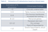
Describe TBW distribution

A

~60% TBW water.

  • age, gender, species and body condition.
  • 2 compartments.

ECF/ICF

• ICF fluid within cell

largest fluid compartment

40% of total body weight or ~ 66% of total body water. Cell membrane separatesICF from ECF

ECF(33% TBW) - comprised of

• INSTL fluid between the cells, includes lymph.

~75% of the extracellular fluid compartment volume and approximately 15% of total body weight.

• IV: plasma (the fluid in the blood vessels) ~25% of the ECF volume and ~5% of TB weight

Transcellular fluid: joint, csf, gastro = ~1%

2
Q

Describe plasma volume in relation to TBW

Total blood volume dog vs cat?

A

Plasma volume~50 mL/kg.

TBV - PCV

TBV (i.e. plasma and cells)

Dog:80- 90 mL/kg

Cat: ~ 50-60 mL/kg

3
Q

What is the 60:40:40 rule?

A

60% of body weight is water

40% of body weight is intracellular fluid

20% of body weight is extracellular fluid.

4
Q

Difference between hypovolaemia and dehydration?

A

Hypovolaemia - loss of water from the IVC

Dehydration-loss of total body water across ALL compartments.

  • does not always lead to hypovolaemia as fluid shifts from the extravascular to the intravascular compartment to maintain normovolaemia in the face of dehydration.

With severe dehydration hypovolaemia will also occur.

5
Q

Describe fluid dynamics

A

Water moves freely between body compartments, however body fluids contain not only water but various concentrations of solutes and proteins.

membranes separating compartments and the concentration of these solutes and proteins in the different compartments that govern the dynamics of fluid movement.

6
Q

Describe fluid movement from the IV to Intersitital space

A

traditionally explained by the hydrostatic and oncotic forces that govern the movement of fluid across a capillary membrane.

According to the Starling equation –> Fluid movement between the intravascular and interstitial compartments (transcapillary fluid dynamics), is governed by hydrostatic pressure and oncotic pressure.

Hydrostatic pressure: fluid pressure that pushes against a membrane

Oncotic pressure: pressure created by the colloid osmotic gradient.

Increased COP or decreased capillary HP favors resorption of fluid into the intravascular space whereas increased capillary HP and decreased capillary COP favors fluid filtration out of the vascular space.

High interstitial HP, due to the relatively non-distensible nature of the interstitial compartment, and low interstitial OP, due to the relative impermeability of the capillary membrane to proteins, favour fluid retention in the intravascular compartment.

Large differences in membrane permeability and tissue compliance occur throughout the body. This leads to great variation in fluid flux between vascular beds thus adding to the complexity and intricacy of body fluid movement.

7
Q

Define starlings equation (write it down and explain)

A

Jv ≈Kf ([Pc – Pi] – 𝝈 [πc – π i])

where

  • Jv is the net fluid movement between compartments
  • Kf([Pc–Pi]–𝝈[πc–πi]isthenetdrivingforce
  • Pc is the capillary hydrostatic pressure
  • Pi is the interstitial hydrostatic pressure
  • πc is the capillary oncotic pressure
  • π i is the interstitial oncotic pressure
  • Kf is the filtration coefficient – a proportionality constant • 𝝈𝝈 is the reflection coefficient
8
Q

Describe vascular endothelial permeability

A

Vascular endothelium is freely permeable to water and solutes, –> concentrations are almost the same in the intravascular and interstitial spaces.

However, endothelium is relatively IMpermeable to blood cells and plasma proteins (maintained within the intravascular space) –> creates a difference in protein concentration between the vasculature and the interstitial compartment and consequently a colloid osmotic gradient between the two compartments.

Oncotic pressure moves fluid in the opposite direction to hydrostatic pressure. I.e. fluid moves towards the highest value of oncotic pressure. For this reason it can be useful to think of oncotic pressure as oncotic “pull”.

9
Q

Describe EC to IC fluid movement

A

Cell membrane governs fluid distribution between the ICF & ECF space

Cell membrane is permeable to water but impermeable to most solutes.

Concentrations of various solutes (osmolality) in each space determine the volume of fluid within the space.

Ion channels and solute pumps govern solute movement into and out of the cell.

Most important pump sodium-potassium ATPase pump.

extrudes 3 sodium ions from the cell in exchange for bringing in 2 potassium ions.

Responsible for maintaining high intracellular potassium and high extracellular sodium concentrations and thus generating a concentration gradient across the cell membrane.

10
Q

Discuss transfluid flux

A

The traditional Starling equation may not hold true for transcapillary fluid flux.

The importance of the membrane glycocalyx in the regulation of fluid movement increasingly recognised.

Glycocalyx is a gel matrix layer that is secreted by the endothelial cells and lines blood vessels. Beneath the glycocalyx is the subglacial space.

Suspected that it is the relative concentrations of proteins and fluid in the intravascular space, the membrane glycocalyx, the subglyceal layer and the interstitium which govern the movement of fluids.

The colloid osmotic pressure (COP) in the intravascular space is thought to only oppose outward movement of fluid.

Thus movement of fluid from the capillaries is likely to be predominantly dependent on the capillary hydrostatic pressure. Increasing the COP by administering synthetic colloids is likely to draw water out of the glycocalyx, dehydrating and damaging it.

Disease states that alter vascular permeability cause abnormal fluid and protein movement out of the vascular space.

LEADS to interstitial oedema which interrupts normal tissue and organ homeostasis and can prove challenging to regulate.

Lymphatic vessels play a pivotal role in the prevention of oedema by returning interstitial protein and fluid to the vascular compartment.

11
Q

Define osmosis, osmolarity and osmolality

A

ne mole of any substance contains the same number of particles (Avogradro’s number = 6.023 x 10²³) regardless of their weight, size or valence. The osmotic effect exerted by solutes is dependent on the number of particles they dissociate into. One osmole is equivalent to the amount of solute that dissociates in solution to form one mole of particles. Osmolality is the number of osmoles per kilogram of solvent and is expressed as mOsm/ kg. Osmolarity is the number of osmoles per litre of solution and is expressed as mOsm/L. In the body there is very little difference between the two measurements and so ongoing I will use the term osmolality. The number of osmotically active molecules in each space and their relative permeability across membranes determine the volumes of fluid in the intracellular and extracellular spaces.

12
Q

What is calculated osmolality?

A

The calculated osmolality (in Osm) is based on the formula: 2(Na+ + K+)+ urea + glucose The reason for multiplying the electrolytes by two is to take into account the contribution of chloride and bicarbonate in the serum. This assumes the serum is electrically neutral (the concentrations of all major anions and cations are equal).

13
Q

What is effective osmolality?

A

Urea is a small molecule which readily diffuses across most membranes. As a result, it exerts very little effective osmolality. As the potassium concentration in extracellular fluid is relatively low, its effect on osmolality is negligible. This allows us to simplify the equation above to the ‘effective’ osmolality which is: 2(Na+) + glucose

14
Q

What is a normal osmolality in dog vs cat?

A

he normal osmolality in dogs ranges from 290 to 310 mOsm/kg and in cats from 290 to 330 mOsm/kg. Isotonic fluids generally have an osmolarity in the range of 270–310 mOsm/L.

15
Q

Why do we measure osmolality?

A

Measurement of osmolality with an osmometer allows you to calculate the osmolar gap, the difference between the measured and calculated osmolality. This can be useful to determine the presence of osmotically active agents not accounted for by the equation. For example, in ethylene glycol toxicity an increased osmolar gap alerts you to the presence of measured osmoles that are not included in your calculation of osmolality.

16
Q

Describe control of osmolality?

A

Osmolality is controlled by hypothalamic osmoreceptors. These receptors primarily detect changes in serum sodium concentration. Increased plasma osmolality stimulates osmoreceptors leading to the release of ADH (antidiuretic hormone, also known as vasopressin) from the posterior pituitary and increased thirst. Antidiuretic hormone acts on the V2 receptors in the renal collecting ducts of the kidney, leading to insertion of aquaporins in the collecting duct and increased water resorption by the kidney. This resorption of water occurs without concurrent sodium resorption thus the result is a decrease in serum sodium concentration (decreased osmolality).

17
Q

Describe fluid loss and their effect on osmolality?

A

In many disease states water and solutes are lost from the body. The nature of fluid loss will determine the effect on osmolality. Tonicity is a measure of the effective osmolality of two solutions that are separated by a semipermeable membrane. In the case of the body tonicity refers to the relative osmolality when compared to the intra and extra cellular fluid compartments.

18
Q

Desc isotonic fluid loss

A

Isotonic fluid losses: There is proportional loss of fluid and solutes. If we use Starling’s equation to explain inter-compartmental fluid flux: a decreased capillary hydrostatic pressure results in movement of fluid from the interstitial compartment into the vascular compartment without any change in the intracellular fluid volume. In this way the body preferentially maintains intravascular volume over interstitial fluid volume. Hypovolaemia should not result unless losses are severe or not replaced i.e. initially dehydration will occur without hypovolaemia. If volume depletion does occur along with isotonic fluid loss, the body will activate systems that act to correct hypovolaemia despite normal osmolality. Hypovolaemia leads to activation of the renin-angiotensin-aldosterone system (RAAS). The resulting production of angiotensin II leads to increased thirst and release of ADH. This means that there will be increased water intake orally and free water retention by the kidneys. Whilst these processes will correct hypovolaemia, they will result in hyponatraemia (and a hypo-osmolar state). Isotonic fluid losses are the most commonly encountered losses. Examples of isotonic fluid loss include vomiting and diarrhoea. Isotonic crystalloids should be used to replace isotonic fluid losses.

19
Q

Desc hypotonic fluid loss

A

Hypotonic fluid losses: Hypotonic losses occur with diabetes insipidus and excessive panting. This results in increased osmolarity of the extracellular fluid. Water moves along this concentration gradient with a net movement occurring from the cellular compartment into the extracellular compartment. Cell shrinkage occurs as a result. Changes in osmolality are further discussed in the tutorial ‘Electrolyte disturbances’ under the section on sodium as this electrolyte is primarily responsible for extracellular osmolality.

20
Q

Desc hypertonic fluid loss

A

Hypertonic fluid losses: These occur infrequently in small animals as a result of excessive loss of solutes in the urine or from the gastrointestinal tract. Examples include use of diuretics, hypoadrenocorticism and haemorrhagic gastroenteritis in dogs. Hypertonic fluid losses cause hypo-osmolality of the extracellular compartment. A rapid decrease in extracellular osmolality will favour net movement of water into the cells (cell swelling). Hypovolaemia will occur as fluid moves out of the hypotonic vascular compartment into the interstitium. For this reason, hypertonic fluid loss that is not replaced is likely to lead to a rapid onset of shock.

21
Q

Describe normal regulation of ECF volume

A

Fluid balance depends on the daily intake and losses of water, nutrients and minerals. Animals in homeostasis are said to be in zero balance when there is no net gain or loss of fluid, i.e., the volume of water consumed in food and water plus the water produced by metabolism equals that lost in urine, faeces, saliva, respiratory and cutaneous secretions. The intravascular volume is prioritised above interstitial and intracellular volumes to maintain tissue perfusion. This fine balance of intravascular volume is intricately associated with total body sodium which is regulated by the renin-angiotensin-aldosterone system (RAAS). This system along with baroreceptors and volume receptors of the atria are responsible for this balance. Baroreceptors are located in the walls of the carotid sinus and aortic arch (arterial side of vasculature). The arterial baroreceptors respond rapidly to acute changes in blood pressure. They play a key role in rapid, short-term regulation of blood pressure. Baroreceptors respond to changes in blood pressure by increasing or decreasing their rate of firing. In this way they send input to the cardiac and vasopressor centres of the medulla through the afferent vagal and glossopharyngeal nerves. The result of baroreceptor input is altered parasympathetic (vagal) and therefore increased sympathetic outflow. Baroreceptor input also alters release of ADH (antidiuretic hormone, vasopressin) secretion from the pituitary. Arterial baroreceptors are very sensitive to a fall in arterial blood pressure. A fall in blood pressure will lead to decreased firing of the baroreceptors and thus a decreased vagal response (and increased sympathetic response) from the medulla. At the same time there will be increased release of ADH/vasopressin from the pituitary resulting in vasoconstriction and water retention. Volume receptors (volureceptors) are located in the cardiac atria, right ventricle and the large pulmonary vessels. These receptors detect stretch and are capable of modulating sympathetic outflow and ADH release. Volume receptors are particularly sensitive to increased stretch (due to increased blood volume). Activation from increased stretch leads to decreased sympathetic outflow with ensuing vasodilation and increased renal blood flow (thus increased glomerular filtration). Increased stretch will also result in decreased ADH release from the pituitary and the release of A-type (ANP) and B-type (BNP) natriuretic peptides. ANP and BNP result in increased sodium and water excretion. All of these processes lead to increased urine output and thus result in reduced blood volume. Example A dog has been anorexic and vomiting for 12 hours. His fluid losses have caused a negative fluid balance and a decreased intravascular volume. This decreased intravascular volume leads to a reduction in blood pressure. This activates his baroreceptors and results in increased sympathetic output and vasoconstriction. In the short term this will lead to a normalisation of blood pressure for the dog despite ongoing losses and inadequate water intake.

22
Q

Desc regulation of total body sodium

A

Regulation of total body sodium (and thus total body water) is under the control of the renin-angiotensin-aldosterone system. Renin is released by the juxtaglomerular apparatus in the kidney and converts angiotensinogen to angiotensin I. This is further converted to angiotensin II and aldosterone. Aldosterone leads to Na+ resorption and thus water resorption in the kidney.

23
Q

How is RAAS activated?

A

The RAAS is activated via the following mechanisms: • reduced blood flow to the kidneys • increased sympathetic nerve activity • decreased delivery of sodium chloride to the juxtaglomerular apparatus. Renin is released by the juxtaglomerular apparatus in the kidney and converts angiotensinogen to angiotensin I. Angiotensin I is further converted to angiotensin II by the endothelium. Angiotensin II causes increased synthesis and release of aldosterone by the adrenal glands.The end result of RAAS activation is increased intravascular volume and total body water.

24
Q

What does Angiotensin II result in?

A

Angiotensin II results in: • systemic vasoconstriction • aldosterone release • vasopressin (ADH) release • increased thirst • activation of the sympathetic nervous system.

25
Q

What does Aldosterone result in?

A

Aldosterone release results in • sodium and water retention • decreased urine production • down regulation of baroreceptors.

26
Q

Describe the effects of changes in total body serum sodium vs serum sodium concentration

A
27
Q

3 main categories of fluid types

A

Crystalloids, synthetic colloids and blood products are the three main categories of fluids used, with crystalloids forming the mainstay of fluid therapy. Various types of crystalloids are available for replacement and/or maintenance fluid therapy.

28
Q

What are replacement fluids?

A

Replacement fluids have a composition similar to that of extracellular fluid or plasma, high in sodium and low in potassium. These are the most commonly used fluids in veterinary medicine with a small number of fluid types being suitable for most cases.

29
Q

Desc maintenance fluids

A

Maintenance fluids contain lower sodium and higher potassium concentrations relative to extracellular fluid. This is because there is obligate daily renal potassium loss while the kidney largely preserves sodium. Dextrose is generally added to maintenance fluids to balance the osmolality with that of extracellular fluid and thus prevent red cell lysis on administration. Once within the body this dextrose is rapidly metabolised meaning the end result is provision of a hypotonic fluid. Maintenance fluids are generally used to account for daily losses in patients that are fluid replete. The use of synthetic colloids has been the subject of much debate over recent years resulting in them falling out of favour. Indications for blood products are quite specific with haemorrhage and coagulopathies representing a majority of the veterinary patients requiring such treatments.

30
Q

Describe potassium supplementation

A

Potassium Supplementation

Prolonged use of replacement or maintenance fluids will lead to hypokalaemia unless potassium (usually in the form of potassium chloride or potassium phosphate) is added. Potassium must not be replaced at more than 0.5 mEq/hr. The following table can be used as a guide to potassium replacement.

31
Q

What are crystalloids?

A

Crystalloids can be categorised as isotonic, hypotonic and hypertonic fluids depending on their tonicity. Isotonic crystalloids, having a tonicity similar to extracellular fluid, are the most commonly used in practice. The principal osmotic component of crystalloid fluids is sodium chloride; however they also contain other physiologically active solutes (K+, Ca2+ or Mg2+, dextrose and buffers). As sodium is equally distributed between the intravascular and interstitial spaces, these fluids will also equilibrate throughout the extracellular fluid space as water freely follows sodium through a semipermeable membrane such as the endothelium. Rapid boluses of isotonic crystalloids can effectively increase plasma volume; however, this effect is short-lived with only 20–25% of the infused fluids remaining in

the intravascular space one hour after administration. Hypertonic boluses are used less commonly; again, their effect is short-lived as they will equilibrate across the endothelium. Hypotonic fluids should never be bolused as they will cause rapid fluid shifts into the cell leading to cell lysis.

32
Q

What is LRS

A

This is a buffered isotonic fluid. The lactate in this solution is metabolised in the liver
to bicarbonate and thus acts as a buffer. As LRS contains calcium it should not be administered with blood products. The citrate anticoagulant in blood products works by chelating calcium to prevent clot formation. Therefore, adding calcium to a blood product negates the effect of citrate and may lead to thrombosis.

33
Q

What is normal saline?

A

(also called NS, 0.9% saline or isotonic saline)

This solution contains a higher amount of chloride than plasma and has no buffer added. Its use can lead to acidosis. [The relationship between chloride concentrations and acidosis will be discussed in Tutorial 2.]

34
Q

What is plasmalyte and normasol R?

A

These solutions contain acetate and gluconate as buffers. Acetate and gluconate are metabolised by the muscle rather than the liver so may be preferable in cases of liver failure. Care should be exercised with rapid administration of an acetate-containing solution as it can cause vasodilation with resultant hypotension. Plasmalyte 148 is available in Australia however Normosol-R is not readily available here.

35
Q

What is 2.5% dextrose in half-strength (0.45%) saline?

A

2.5% dextrose in half-strength (0.45%) saline

This solution is generally used as a maintenance fluid in patients intolerant of a high salt load (e.g., patients with cardiac or renal failure).

36
Q

What is D5W

A

5% dextrose

This is an example of a hypotonic crystalloid. It contains no electrolytes. The dextrose is rapidly metabolised and so using it is equivalent to administering free water. It is generally used in cases of hypernatraemia. It is also commonly used as a diluent and as a solution for continuous drug infusions.

37
Q

What is HTS?

Uses?

Considerations?

Contraindications?

A

20% or 7% saline

This is a hypertonic fluid. In Australia, seven per cent saline is available in 1 litre bags and is generally used for large animals. Twenty per cent saline is available in 10 mL ampoules and can be diluted to 6.66% to provide rapid low volume resuscitation in cases of severe hypovolaemia. As this fluid is hypertonic it will initially draw fluid from the interstitial compartment into the vascular space. The saline however will eventually distribute into the interstitial space with water following it. This means that the intravascular effects of HTS are short-lived (about 40 minutes). It is always important to use isotonic crystalloids following HTS in order to avoid hypernatraemia. Always monitor serum sodium concentrations after saline administration and never use HTS in cases that you suspect to be dehydrated. The typical dose of 6.66%–7% hypertonic saline for resuscitation is 4 mL/kg. Administer this dose over 10–15 minutes. Rapid infusion can cause bronchoconstriction and vasodilation. Hypertonic saline is used for rapid correction of hypotension during anaesthesia. It is also very useful for resuscitation in cases of suspected increased intracranial pressure as it can assist with reduction of cerebral oedema.

38
Q

What are colloids?

A

While commonly used in the past, synthetic colloids have fallen out of favour with concerns over their potentially deleterious effects. In Australia, Voluven is the principal synthetic colloid available. It is a hydroxyethyl starch suspended in an isotonic crystalloid solution. The general principle behind the use of colloids is to increase colloid osmotic pressure within the vascular space and thus ultimately draw water into the vessels. Synthetic colloids contain large molecular weight particles which are unable to pass through the small fenestrations of the vascular endothelium and are so retained within the vessels. Although it is their size which governs their retention within the vascular space, it is the number of particles which determine their osmolality and thus the oncotic pressure exerted by these solutions.

39
Q

Controversies with colloid use?

A

Concer

ns over potential for coagulopathy and renal injury have brought the use of colloids into question. Coagulopathy is thought to occur as a result of platelet dysfunction. Decreased von Willebrand factor and factor VIII and decreased platelet activation are thought to be the major mechanisms responsible. Mechanical obstruction of renal tubules and direct tubular cell injury via osmotic nephrosis have been implicated in acute kidney injury after colloid use. While healthy patients may tolerate colloids, those who are coagulopathic or at risk of acute kidney injury (e.g., septic patients), may be at a much higher risk of adverse reactions.

There is also a risk of extravasation of these large synthetic molecules into the interstitium in patients with increased vascular permeability. This may lead to interstitial oedema that is unresponsive to diuretic therapy. The maximum recommended dose for synthetic colloids
is 20 mL/kg/day. Synthetic colloid boluses (5 mL/kg given over 10–15 minutes) may be used during anaesthesia to treat hypotension. Synthetic colloids will artificially alter refractometer readings. This must be taken into account when determining urine specific gravity and serum total solids.

40
Q

Blood products, what are they, how are they used?

A

Natural colloids, blood products are particularly important in emergency and critical care medicine.

Haemorrhagic shock due to trauma, haemoabdomen, gastrointestinal bleeding, vitamin K antagonist intoxication and other coagulopathies are among some of the most unstable cases we see. While crystalloids and colloids can be used in immediate resuscitation to correct hypotension, they can worsen anaemia and coagulopathies via their dilutional effect. The choice of blood product for resuscitation depends on the primary disease process and the severity of anaemia. Information on the use of these products will be covered in Subject 3 of this course.

41
Q

Indications for fluid therapy?

A

In disease, decreased food and water intake due to anorexia generally occurs along with increased water losses (for example, polyuria, vomiting, diarrhoea). Pyrexia and the increased metabolism of disease states will increase fluid requirements; animals in disease states tend to be in a negative water balance. Assessing a patient’s fluid balance is vital to enable the clinician to provide the appropriate volume of fluids over the appropriate time frame.

There are two main indications for volume replacement therapy: hypovolaemic shock requiring acute resuscitative therapy and dehydration requiring slower replacement.

42
Q

Hypovolaemic shock and fluid therapy?

A

Patients in hypovolaemic shock require rapid administration of IV fluids to restore their circulating volume and minimise tissue hypoperfusion. Shock and the treatment of shock will be addressed in tutorial “Shock; assessment and resuscitation”.

43
Q

Dehydration and fluid therapy?

A

Dehydrated patients have reduced total body water but are not necessarily hypovolaemic. Their clinical signs reflect decreased interstitial volume as the body sacrifices this fluid compartment in order to preserve intravascular volume and thus perfusion of the tissues. If severe, dehydration can lead to hypovolaemia.

44
Q

Csx of dehydration?

A

Clinical signs of dehydration:

Tacky to dry mucous membranes

Decreased skin turgor or increased skin tent as a result of decreased subcutaneous fluidity and decreased skin elasticity. Care should be exercised assessing this parameter in older, younger, cachectic or obese patients as these conditions may alter subcutaneous fluidity or skin elasticity. The head can often be the most reliable place to perform skin tent testing.

Dry corneas

Retraction of globes within the orbit

Decreasing body weight

Severe dehydration (> 12% of body weight reduction) can result in signs of hypovolaemic shock.

45
Q

Describe clinical exam findings based on % dehydration

A
46
Q

Disc laboratory findings in dehydration patients

A

When assessing hydration status, laboratory testing can be used to complement the physical exam.

Packed cell volume (PCV, haematocrit) and total solids (TS). Increased PCV and TS together indicate likely dehydration. PCV can also be increased due to polycythaemia or may be within normal reference range if a patient is both dehydrated and anaemic.

Increased urine specific gravity (USG) and urine output. Elevated USG is indicative of dehydration. However, a normal or low USG cannot rule out dehydration as conditions that lead to the kidney being unable to concentrate urine may also lead to dehydration. Reduced urine output can indicate volume deficits. It may also be due to acute kidney injury urine outflow obstruction and various other conditions that reduce perfusion to the kidneys.

Azotaemia. Dehydration that is associated with reduced renal perfusion will cause a pre-renal azotaemia. An increased USG with concurrent azotaemia is diagnostic of dehydration.

Serial assessments of volume and hydration must be carried out to assess response to any fluid therapy plan.

47
Q

An 18 year-old cat with a body condition score of 2/9 has been diagnosed with chronic renal failure. The cat is anorexic and off its water. You are treating this cat in hospital with IV fluid therapy.

How should you assess this patient’s fluid balance?

A

Four hourly assessment of cardiovascular parameters assessing carefully for signs of hypovolaemia.

Four hourly assessment for excessive wetness or dryness of mucous membranes.

Four hourly assessment of respiratory rate and lung sounds.

Twice daily monitoring of body weight – if fluid therapy is adequate, and assuming this patient is dehydrated initially, then body weight should increase with adequate volume of fluid therapy.

Once daily monitoring of PCV/TS: bear in mind that the cat is likely anaemic so look for trends from the baseline numbers.

Daily monitoring of urine output in order to note that it is regular. Whilst urine output is worth noting, if the patient is polyuric then urine output becomes a poor indicator of adequate hydration.

What parameters would not be so helpful in this case? USG is not especially helpful with a case of renal failure

The significance of skin tenting will be difficult to assess in an elderly thin cat.

Thirst may be helpful depending on how sick the cat is, if it is anorexic and depressed then it may not drink despite dehydration.

48
Q

What is a fluid therapy plan?

A

A good fluid therapy plan will take into account volume status, perfusion status, PCV, osmolality, electrolyte levels and acid base status of the patient. The first consideration
is always to maintain oxygen delivery to the tissues. When hypovolaemia leads to decreased oxygen delivery (i.e. shock) fluid therapy becomes an emergency treatment
that is administered within minutes. For animals that are dehydrated but have stable cardiovascular parameters fluid deficits should be replaced over 6–24 hours. Replacement fluids should be administered according to the patient’s estimated dehydration, maintenance needs and anticipated ongoing losses. The route of administration depends on the severity of dehydration and patient size. Oral, subcutaneous, intraosseous and IV routes are all acceptable with mild to moderate dehydration. IV fluid therapy or intraosseous routes should be used in severe dehydration as these are the most efficient methods of fluid delivery.

49
Q

Describe fluid therapy plan in a dehydrated patient without perfusion deficits?

A

A fluid plan for a dehydrated patient without perfusion deficits has the following three components:

Dehydration

The deficit of volume in millilitres is calculated using the following formula.

Body weight x 1000 x % dehydration

Generally, aim to replace half the deficit over the first 6 to 8 hours and the remainder over the following 16 - 18 hours.

50
Q

What is maintenance fluid therapy?

A

Maintenance fluid requirements refers to the volume of fluid and amount of electrolytes that must be consumed daily to maintain total body water and electrolyte content within normal limits. Approximately two-thirds of maintenance fluid requirements are to account for sensible losses (easy to measure losses such as urine output) and the remaining one third accounts for insensible losses (fluid losses which are difficult to measure such as respiratory losses or faecal losses). Although the term ’maintenance fluid therapy‘ is used in veterinary medicine, we tend to use replacement fluids for maintenance therapy more often than using fluids that are formulated for the maintenance purposes. The term ‘maintenance fluid rates’ refers to administration of fluids to a volume-replete hydrated patient who is as yet unable to meet homeostatic fluid requirements orally.

equations for daily maintenance fluid rates are extrapolated from maintenance
energy requirements as the bodies fluid requirements are thought to parallel daily energy requirements. Actual requirements of fluids for individual patients will vary greatly. Extremes of patient size with very different body surface area to lean mass ratios and with very differing metabolic rates mean that one equation will not fit all patients. Maintenance fluid requirements are often overestimated in overweight or obese patients as maintenance fluid requirements are based on lean body weight. Regardless of the equation used, we should remember that fluid therapy is not an exact science and requires much clinical judgment, hence the need for ongoing fluid balance assessments while a patient receives fluid therapy. The following equations are commonly used to calculate maintenance fluid rates.

51
Q

4 equations for working out fluid maintenance in small animals

A

(70 x body weight 0.75 )/day

(body weight × 30) +70/day

(40–60 mL/kg/day)

(2–2.5 mL/kg/hr)

52
Q

How do we estimate ongoing losses in our patients?

A

This can be hard to determine and generally requires observation over four-hour periods of hospitalisation to allow quantification of ongoing losses in the form of vomiting, diarrhoea, polyuria, excessive panting, etc.

53
Q

Moggy’s clinical signs are consistent with approximately 7% dehydration. Based on his heart rate, CRT and pulse quality, he does not appear to be hypovolaemic.

You will need to provide Moggy with fluids for maintenance and to replace his deficit. Work out a fluid plan.

A

Maintenance fluid rates for a cat ~ 2 mL/kg/hr = 10 mL/hr

Fluid deficit ~ Body weight x 1000 x % dehydration = 5 x 1000 x 7% = 350 mL

You wish to replace half of Moggy’s deficit over the next 8 hours.

i.e. 175 /8 = 22 mL/hr.

Therefore Moggy will start off with a fluid therapy rate of
32 mL hour. He will be frequently reassessed and his fluid rates adjusted as his clinical signs improve. The fluid should be an isotonic crystalloid. A balanced crystalloid such as LRS would be the best empirical choice.

54
Q

Define Oncotic pressure

A

also called colloid osmotic pressure: the osmotic pressure within the plasma that is due to plasma colloids

55
Q

Define Capillary hydrostatic pressure

A

Capillary hydrostatic pressure: the force exerted by the fluid component of blood against the internal capillary wall

56
Q

Define Glycocalyx: a gel matrix layer that is secreted by the endothelial cells and lines blood vessels

A

Glycocalyx: a gel matrix layer that is secreted by the endothelial cells and lines blood vessels

57
Q

Define mole:

A

Mole: an amount of any substance contains the same number of particles (Avogradro’s number = 6.023 x 10²³) regardless of their weight, size or valence

58
Q

Define:

Osmole

Osmolarity

Osmolality.

Effective osmolality

A

Osmole: the amount of solute that dissociates in solution to form one mole of particles

Osmolarity: the number of osmoles per litre of solution.

Osmolality: the number of osmoles per kilogram of solution.

Effective osmolality: the osmolality of a solution that will remain after equilibration with another compartment

59
Q

Define

Tonicity

Osmolar gap

Osmoreceptors

A

Tonicity: a measure of the effective osmolality of two solutions that are separated by a semipermeable membrane.

Osmolar gap: the difference between the measured and calculated osmolality

Osmoreceptors: receptors that detect changes in osmolality/osmolarity

60
Q

Define

RAAS

Baroreceptors

Volume receptors

A

RAAS: renin-angiotensin-aldosterone system

Baroreceptors: receptors that detect acute changes in pressure of the carotid and aortic arteries, especially reduced blood pressure

Volume receptors: receptors that detect changes in myocardial and pulmonary vascular stretch

61
Q

Define

Crystalloid fluid

Colloid

A

Crystalloid fluid: a fluid that is a true solution; its substances are dissolved and able to pass through a semipermeable membrane

Colloid: a homogenous substance that contains molecules that are in suspension that do not separate from the solution

62
Q

Define

Maintenance fluid requirements

A

Maintenance fluid requirements: the volume of fluid and amount of electrolytes that must be consumed daily to maintain total body water and electrolyte content within normal limits.