Foot and Ankle Flashcards

1
Q

Where is the watershed area of the PTT?

A

2-6 cm proximal to the navicular

2
Q

Obese patient comes in with feet that look like this? How can you stage this?

A
  • Stage I - Tenosynovitis
    • No deformity
    • (+) single-leg toe raise
  • Stage IIA - Flatfoot deformity
    • Exam
      • Flexible hindfoot
      • (-) single-leg heel raise
      • Mild sinus tarsi pain
    • Imaging
      • Arch collapse deformity on imaging
  • Stage IIB - Flatfoot deformity
    • Exam
      • Flexible hindfoot
      • Forefoot abduction (“too many toes”)
    • Imaging
      • >40% talonavicular uncoverage
  • Stage III
    • Exam
      • Flatfoot deformity
      • Rigid forefoot abduction
      • Rigid hindfoot valgus
    • ​Imaging
      • Arch collapse deformity
      • Subtalar arthritis
  • Stage IV
    • ​Exam
      • Flatfoot deformity
      • Rigid forefoot abduction
      • Rigid hindfoot valgus
      • Deltoid ligament compromise
      • Ankle pain
    • Imaging
      • Arch collapse deformity
      • Subtalar arthritis
      • Talar tilt in ankle mortise
3
Q

Difference between adult and juvinile hallux valgus

A
  • often bilateral and familial
  • pain usually not primary complaint
  • varus of first MT with widened IMA usually present
  • DMAA usually increased
  • often associated with flexible flatfoot
  • complications
    • recurrence is most common complication (>50%)
    • overcorrection
    • hallux varus
4
Q

Risk factors for hallux valgus

A
  • intrinsic
    • genetic predisposition
    • ligamentous laxity
    • convex metatarsal head
    • pes planus
    • rheumatoid arthritis
    • cerebral palsy
  • extrinsic
    • shoes with high heel and narrow toe box
5
Q

Pathoanatomy of hallux valgus

A
  • valgus deviation promotes varus position of metatarsal
  • sesamoid complex becomes lateral to the metatarsal head, which moves medially
  • medial MTP joint capsule becomes stretched and attenuated while the lateral capsule becomes contracted
  • adductor tendon becomes deforming force
  • inserts on fibular sesamoid
  • lateral deviation of EHL
  • plantar and lateral migration of the abductor hallucis causes muscle to plantar flex and pronate phalanx
  • windlass mechanism becomes less effective
  • leads to transfer metatarsalgia
6
Q

Radiographs for hallux valgus

A
  • views
    • weight bearing AP and Lat
    • sesamoid view can be useful
      • displacement of sesamoids
      • often displaced laterally
    • joint congruency and degenerative changes can be evaluated
  • radiographic parameters (see below) guide treatment
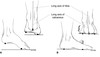
    • Hallux valgus (HVA)
      • Long axis of 1st MT and prox. phal.
      • Identifies MTP deformity
      • Normal = < 15°
    • Intermetatarsal angle (IMA)
      • Between long axis of 1st and 2nd MT
      • Normal = < 9°
    • Distal metatarsal articular (DMAA)
      • Between 1st MT long. axis and line through base of of distal articular cap
      • Identifies MTP joint incongruity
      • Normal = < 15°
    • Hallux valgus interphalangeus (HVI)
      • Between long. axis of distal phalanx and proximal phalanx
      • Normal = < 10 °
7
Q

Approach to adolescent bunions

A
  • best to wait until skeletal maturity to operate
  • can not perform metatarsal osteotomies if physis is open (cuneiform osteotomy OK)
  • surgery indicated in symptomatic patients with an IMA > 10° and HVA of > 20°
  • severe deformity with a DMAA > 20 perform a double MT osteotomy
  • technique
    • soft tissue procedure alone not successful
    • similar to adults if physis is closed (except in severe deformity)
8
Q

Options for treatment of hallux valgus

A
  • Nonoperative
    • shoe modification/ pads/ orthoses
    • silastic spacer
      • orthoses more helpful in patients with pes planus or metatarsalgia
  • Soft Tissue Procedure - modified McBride
    • never appropriate in isolation
    • in conjunction with medial eminence resection osteotomy
    • release medial capsule of 2nd MTP; can be sutured to lateral capsule of 1st
    • adductor hallucis is released and interposed
      • Can be left to scar down
    • Transverse intertarsal ligament is releases which attaches to the fibular sesamoid
    • Don’t resect the fibular sesamoid as was done with the original McBride or you will get hallux varus
  • distal metatarsal osteotomy
    • mild disease (HVA 20-40, IMA 10-13)
    • distal metatarsal osteotomies include
      • Chevron
      • biplanar Chevron
      • Mitchell
    • may be combined with proximal phalanx osteotomy
  • proximal metatarsal osteotomy
    • moderate disease (HVA >40°, IMA >13°)
    • proximal metatarsal osteotomies include
      • crescentric osteotomy
      • Broomstick osteotomy
      • Ludloff
      • Scarf
  • double (proximal and distal) osteotomy
    • severe disease (HVA 41-50°, IMA 16-20°)
    • DMAA > 15
    • Scarf + chevron
    • Lapidus + Akin
  • first cuneiform osteotomy
    • severe deformity in young patient with open physis
  • Lapidus procedure (1st metatarsocuneiform arthrodesis)
    • severe deformity
    • Metatarsus primus varus
    • hypermobile 1st tarsometatarsal joint
  • MTP Arthrodesis
    • Gout
    • Rheumatoid arthritis
    • Down’s syndrome
    • cerebral palsy
    • Severe DJD
    • Ehler-Danlos
    • Resection arthroplasty
  • proximal phalanx (Keller) resection arthroplasty
    • largely abandoned
    • still indicated in some elderly patient with reduced function demands
9
Q

Complications associated with HV

A
  • Recurrence
    • most common cause of failure is insufficient preoperative assessment and failure to follow indications
      • e.g., failure to recognize DMAA > 15°
      • e.g., failure to do adequate distal soft tissue realignment
    • more common in juvenile/adolescent population
    • noncompliant patient that bears weight
  • Avascular necrosis
    • medial capsulotomy is primary insult to blood flow to metatarsal head
    • distal metatarsal oseotomy and lateral soft tissue release inconjuction do not increase risk for AVN
  • Dorsal malunion with transfer metatarsalgia
    • due to overload of lesser metatarsal heads
    • risk associated with
      • Lapidus
      • proximal crescentric osteotomies
  • Hallux Varus
    • caused by
      • overcorrection of 1st IMA
      • excessive lateral capsular release with overtightening of medial capsule
      • overresection of medial first metatarsal head
      • lateral sesamoidectomy
  • Cock up toe deformity
    • due to injury of FHL
    • most severe complication with Keller resection
  • 2nd MT transfer metatarsalgia
    • often seen concomitant with hallux valgus
    • shortening metatarsal osteotomy (Weil) indicated with extensor tendon and capsular release
  • Neuropraxia
    • Painful incisional neuromas after bunion surgery frequently involve the dorsomedial cutaneous branch of the superficial peroneal nerve.
    • This is the medial branch of the superficial peroneal nerve that terminates as the dorsomedial cutaneous nerve to the hallux. Branches of the deep peroneal nerve to this area are rare, and no branches to this area exist from the sural nerve. The saphenous nerve branches are generally more proximal, and the medial plantar nerve lies plantarly.
10
Q

Iatrogenic causes of this deformity

A
  • overcorrection of 1st IMA
  • excessive lateral capsular release with overtightening of medial capsule
  • overresection of medial first metatarsal head
  • lateral sesamoidectomy
11
Q

Treatment

A
  • Nonoperative
    • shoe modifications to accommodate the deformity
    • extra depth and wider more flexible toe box
    • placing pads over prominent areas
    • taping or splinting the deformity
  • Abductor hallucis release and transfer of EHL or EHB to proximal phalanx
    • indications
      • flexible deformities
    • technique
      • transfer portion of EHL or EHB under the transverse intermetatarsal ligament to the distal metatarsal neck (from lateral to medial) distal portion of tendon left intact, creating static stabilizer
  • First MTP arthrodesis
    • indications
      • fixed deformity
      • significant arthrosis
12
Q

Classification of Hallux Rigidus

A

Cougling and Shurnas

  • Grade 0
    • Stiffness with normal XR
  • Grade 1
    • mild pain at extremes of motion
    • mild dorsal osteophyte, normal joint space
  • Grade 2
    • moderate pain with range of motion increasingly more constant
    • moderate dorsal osteophyte,
  • Grade 3
    • significant stiffness, pain at extreme ROM, no pain at mid-range
    • severe dorsal osteophyte, >50% joint space narrowing
  • Grade 4
    • significant stiffness, pain at extreme ROM, pain at mid-range of motion
    • same as grade III
13
Q

Options for treatment

A

Hallus Rigidus

  • *Steroid injections are often ineffective and can adversely affect the cartilage and is not recommended
  • NSAIDS, activity modification & orthotics
    • grade 0 and 1 disease
    • activity modifications
      • avoid activities that lead to excessive great toe dorsiflexion
    • types of orthotics
      • Morton’s extension with stiff foot plate is the mainstay of treatment
    • stiff sole shoe and shoe box stretching may also be used
  • Joint debridement and synovectomy
    • patients with acute osteochondral or chondral defects
  • Dorsal cheilectomy - common first approach, high success rates
    • grade 1 and 2 disease (with reports of treating even up to grade 4)
    • pain with dorsiflexion is an indicator of good results with dorsal cheilectomy
    • shoe wear irritation from dorsal prominence and pain (ideal candidate)
    • contraindication
      • pain mid-range of the joint during passive motion (grade IV)
    • technique
      • remove 25-30% of the dorsal aspect of the metatarsal head along with dorsal osteophyte resection
      • the goal of surgery is to obtain 70% to 90% dorsiflexion intraoperatively
  • Moberg procedure (dorsal closing wedge osteotomy of the proximal phalanx)
    • Sometimes done in conjunction with cheilectomy
    • runners with reduced dorsiflexion (60° is needed to run)
      • Alternatively a proximal MT osteotomy can be used to improve dorsiflexion as a joint salvage procedure, however is not recommended and is associated with metatarsalgia
    • failure of cheilectomy to provide at least 30 to 40 degrees of motion
    • technique
      • increases dorsiflexion by decreasing the plantar flexion arc of motion
  • Keller Procedure (resection arthroplasty)
    • elderly, low demand patients with significant joint degeneration and loss of motion (>70)
    • contraindicated in patients with pre-existing rigid hyperextension deformity of 1st MTP joint
    • technique
      • involves removing the base of the first proximal phalanx
    • Complications
      • requires appropirate patient selection and minimal resection
      • risk of hyperextension (cock-up deformity)
      • weakness with push-off
      • transfer metatarsalgia (decreased with capsular interposition)
      • IP arthroplasty has poor results and currently not recommended until further explored
        • Seems to do better with intact plantar plate and FDB transfer
  • MTP arthroplasty
    • indications controversial
    • technique
      • capsular interpositonal arthroplasty gaining popularity
      • silicone implants are not recommended due to poor long-term results
      • Cobalt chrome with titanium plasma spray total joint and hemi arthroplasty are available but often loosen and are unreliable
    • outcomes
      • silicone implants may have a good short term satisfaction rate but are currently not recommended
      • osteolysis and synovitis cause mid to long term pain and joint destruction
      • Total joint components have increased loosening and also are currently controversial and not often used
  • MTP joint arthrodesis - best definative option for failed chilectomy, in some patients with more severe disease can go straight to this
    • grade 3 and 4 disease (significant joint arthritis)
    • most common procedure for hallux rigidus
    • outcomes
      • 70% to 100% fusion rate
      • 15% of patients experience degeneration of IP joint after surgery (mostly asymptomatic)
14
Q

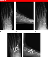
Strongest construct for first MTP arthrodesis

A

dorsal plate with compression screw is biomechanically strongest construct

15
Q

Risk factors for achilles rupture

A

Age
Steroids
Cipro
Eccentric muscle contraction

16
Q

Treatment options for acute achilles rupture

A
  • Epidemiology
    • men
    • 30-40
    • 4-6 cm above calcaneal insertion
  • Nonoperative
    • functional bracing/casting in resting equinus (20 deg plantar flexion) with protected early functional rehab
    • indications
      • sedentary patients
      • elderly patients
      • medically frail patients
      • patient desires to avoid surgery
      • increased risk of rerupture
    • outcomes
      • patient will have decreased plantar flexion strength
      • Increased risk of re-rupture
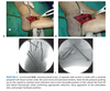
  • End-to-end Achilles tendon repair
    • acute rupture (< 3 months)
    • technique
      • Posteromedial incision
      • Avoid or beware sural nerve
      • Carefully incise periotenon
      • Find each end, debride and perform and end to end repair using krochow technique and nonabsorbable suture
      • Oversew the peritenon with 4-0 absorbable sutures
    • outcomes
      • decreased re-rupture rate
      • increase plantar flexion strength
    • disadvantages
      • skin complications including infection, sloughing (5-10%)
      • risk factors for wound complications included
        • tobacco abuse
        • steroid use
        • diabetes mellitus
        • female sex
        • sural nerve injury
  • rehab
    • initially immobilize in 20° of plantar flexion to decrease tension on skin and protect tendon repair
    • functional rehabilitation during treatment improves range of motion and outcome
  • percutaneous repair
    • weaker and not recommended
    • sural nerve at highest risk for injury

17
Q

Indications for non-operative treatment of achilles rupture

A

sedentary patients
elderly patients
medically frail patients
patient desires to avoid surgery
increased risk of rerupture

18
Q

Increased risk of wound complications of achilles repair

A

tobacco abuse
steroid use
diabetes mellitus
female sex
sural nerve injury

obesity

19
Q

Options for treatment of chronic achilles tear or re-rupture

A
  • Nonoperative
    • physical therapy - toe strengthening, gait training
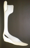
    • AFO
  • Primary repair < 3 cm
  • Reconstruction with VY advancement
    • defects 3-5 cm
    • Posteomedial incision extended proxiamlly to MC junction
    • Debride tendon edges
    • Fashion two flaps 1cm wide and 7-8cm long
    • Advance the tendon to allow end-end repair and close the proximal incision of the tendon
  • FHL transfer
    • defects > 5 cm
    • Prone position with tourniquette
    • Posterio-medial incision protecting NV bundle
    • Separate medial incision to relesae the tendon at the knot of henry
      • Some will harvest from the level of the joint
    • excise degenerative tendon edges
    • transfer FHL through osseous tunnels in the calcaneus, weave FHL to native achilles tendon
  • >10cm = Allograft
  • Gastroc turndown
    • another option but a large procedure
20
Q

Complications associated with treatment of achilles

A
  • Dehisence/Infection
    • ESR, CRP, WBC
    • Consult ID and Plastics
    • OR - I&D, deep cultures, VAC or free flap
    • Directed IV abx until normal ESR/CRP
  • Re-rupture
  • Sural nerve palsy
21
Q

Differential for Chronic Achilles inflammation

A
  • Paratenonitis
    • Inflammation of the peritendinous structures, including the paratenon and septum
  • Tendinosis
    • Asymptomatic degeneration of tendon without inflammation, with regional focal loss of tendon structure
  • Paratenonitis with tendinosis
    • Inflammation of the peritendinous structures along with intratendinous degeneration
  • Retrocalcaneal bursitis
    • Mechanical irritation of the retrocalcaneal bursa
    • Younger patient, shoe wear
    • haglund
  • Insertional tendinosis
    • Inflammatory process within the tendinous insertion of the Achilles tendon
    • middle age, boney enlargement
    • tendon calcification
22
Q

Middle aged woman with chronic heel pain. Differential? Likely Diagnosis? Helpful imaging?

A

Insertional Tendonitis

  • Differential
    • insertional tendonitis
    • tendonosis
    • retrocalcaneal bursitis
  • Pain and tendon thickening at insertion of Achilles tendon
    • repetitive trauma leads to inflammation followed by cartilaginous then bony metaplasia
  • History
    • Take a complete and AMPLE history
    • occurs in middle-aged and elderly patients with a tight heel cord
    • symptoms
      • posterior heel pain, swelling, burning, and stiffness
      • shoe wear pain due to direct pressure - pump bump
    • progressive bony enlargement of calcaneus at insertion site
  • Physical exam
    • Examine - boney enlargement at insertion
    • Palpate - midline tenderness at insertion site of Achilles tendon
    • Move - reduced passive and active ROM
  • AP, lateral and oblique views of the foot
    • lateral foot shows bone spur and intratendinous calcification
  • MRI and ultrasound
    • can demonstrate amount of degeneration
    • Early - fluid around the tendon
    • Late - intratendonous calcification, degeneration of the tendon
23
Q

Treatment of Insertional Achilles Tendonitis

A
  • activity modification, shoe wear modification, therapy
    • first line of treatment
    • techniques
      • physical therapy with eccentric training
        • Challenges muscle, to strengthen, promote repair and increase metabolic activity
      • gastrocnemius-soleus stretching
      • shoe wear
        • heel sleeves and pads (mainstay of nonoperative treatment)
      • small heel lift
      • locked ankle AFO for 6-9 months (if other nonoperative modalities fail)
  • retrocalcaneal bursa excision, debridement of diseased tendon, calcaneal bony prominence resection
    • indications
      • failure of nonoperative management
        • technique
      • midline, lateral, or medial J-shaped incisions
  • tendon augmentation or transfer (FDL, FHL, or PB) vs. suture anchor repair
    • indications
      • when > 50% of Achilles tendon insertion must be removed
24
Q

Young patient with chronic heel pain. Differential? Likely diagnosis? Helpful imaging?

A

Retrocalcaneal bursitis

  • Differential
    • insertional tendonitis
    • retrocalcaneal bursitis
    • tendosis
  • History
    • Take a complete and ample history (social, sports, smoking, PMhx, meds, allergies)
    • more common in young patients
    • Due to rubbing on shoes
  • Physical exam
    • Examine
    • Palpate
      • Two finger squeeze = pain localized to anterior and 2 to 3 cm proximal to the Achilles tendon insertion
      • fullness and tenderness medial and lateral to tendon
      • Pump bump = bony prominence at Achilles insertion
    • Move
      • pain with dorsiflexion (compresses the space)
    • Silverskiold
  • AP, lateral, oblique of the foot
    • lateral of foot demonstrates Haglund deformity
    • Loss of kager triangle due to bursitis
    • Swelling of tendon >9mm
    • _2cm above the joint lin_e
  • MRI
    • rarely needed
25
Q

Treatment of retrocalcaneal bursitis

A
  • activity modification, shoe wear modification, therapy, NSAIDs
    • first line of treatment
    • techniques
      • ice
      • shoewear - external padding of Achilles tendon
      • PT
      • no injections
  • Retrocalcaneal bursa excision and resection of Haglund deformity
    • disease refractory to nonoperative management
    • technique
      • midline, lateral, or medial J-shaped incision
26
Q

Mechanism of Achilles Tendonosis

A
  • overuse - common in soccer players
  • imbalance of dorsiflexors and plantar flexors
    • poor technique
  • poor tendon blood supply - watershed area
    • 2-6cm proximal to insertion
  • genetic predisposition
  • fluoroquinolone antibiotics
  • inflammatory arthropathy
27
Q

Classification of achilles TENDONOSIS

A
  • Achilles tendinosis
    • tendon thickening
    • thought to be caused by anaerobic degeneration in portion of tendon with poor blood supply
    • Get a mucoid degeneration of the tendon due to age or reduced vascularity
    • Inability to heal with repetative microtrauma, often seen after rupture and can be painless
    • Especially middle age men who increase their activity
  • Achilles paratenonitis
    • involves inflammation of tendon sheath
    • Younger, due to repetative injury with inflammation around the tendon
    • Runners, shoe wear issues, young athletes who overtrain
  • Tendinosis with paratenonitis
    • Progression of disease = paratenonosis < paratenonosis with tendonitis < tendonitis alone
    • Usually stranding within the tendon shows starting to develop tendonitis
    • The further along the progression, the less likely to respond to therapy
28
Q

Younger patient with chronic heel pain. Differential? Likely diagnosis? Helpful imaging?

A

Achilles Tendonosis

  • Differnetial
    • insertional tendonitis
    • retrocalcaneal bursitis
    • achilles tendonosis
      • peritenonitis
      • tendonosis
      • combined
  • History
    • Take a complete and AMPLE history
    • Age - Young (paratenonosis), old (tendinosis)
    • symptoms - gradual onset
      • Pain with activity, pain with ADL (will gradually become more painful as process continues)
    • Pain more proximal with increasing severity as time goes on
    • pain, swelling, warmth
    • worse symptoms with activity
      • difficulty running
    • Minimal pain - think tendonitis
    • PMHx - obesity, HTN, diabetes associated with tendionsis
  • Physical exam
    • Examine, Palpate - tendon thickening and tenderness 2 to 6 cm proximal to Achilles insertion
    • Move - pain throughout entire range of motion
      • Paratenonitis
        • Limited ROM
        • Pain is fixed through all ROM
    • Positive silverskiold test
  • US - fluid around the tendon with hypoechoic regions
  • MRI
    • disorganized tissue will show up as intrasubstance intermediate signal intensity
    • thickened tendon
    • paratendinosis
      • Fusiform swelling and high intesity with-in tendon (reflective of mucoid degeneration of tendinosis)
    • chronic rupture will show a hypoechoic region between tendon ends
29
Q

Options for treatment of achilles tendonosis

A
  • activity modification, shoe wear modification, physiotherapy, NSAIDs
    • first line of treatment
    • techniques
      • Retraining
      • physical therapy with eccentric training
        • Gastroc stretching
      • shoewear
      • heel lifts or shock absorber
      • cast or removable boot (severe disease)
      • Orthoses to correct pronation (tendonitis)
    • outcomes
      • nonoperative management is 65% to 90% successful
  • glyceryl trinitrate patches, prolotherapy, and aprotinin injections
    • Theory is injections to break down fibrous tissue - NOT appropriate for tenodonitis, only paratenonitis
    • indications
      • evolving indications due to lack of evidence at this time
  • **Risks for failing non-op
    • Age
    • Duration of symptoms
    • Degree of degeneration
  • Percutaneous tenotomies
    • longitudinal tenotomy made in the degenerative area
    • strip the anterior Achilles tendon with a large suture to free any adhesions
  • open excision of degenerative tendon with tubularization
    • moderate to severe disease that is refractory to non-op treatment
    • Technique
      • Posterior incision with development of full thickness flaps
      • Incise paratenon and debride posterior, medial and lateral portion of the tendon
    • outcomes
      • 70% to 100% successful
  • tendon transfer (FHL, FDL, or PB)
    • This is not used for paratenonosis, only for tendonitis
    • indications
      • degeneration of >50% of the Achilles tendon
      • >55 years of age
      • MRI evidence of diffuse tendon thickening without a focal area of disease
30
Q

Risk of failing non-operative treatment for achilles tendonosis

A

Age
Duration of symptoms
Degree of degeneration

31
Q

Indications for FDL transfer in achilles tendonosis

A
  • degeneration of >50% of the Achilles tendon
  • >55 years of age
  • MRI evidence of diffuse tendon thickening without a focal area of disease
32
Q

Name as many ossicles of the feet as you can

A
  • Up to 40 accessory ossicles and multiple sesamoids have been described in the foot and ankle
  • Definition
    • accessory ossicles - are secondary ossification centers that remain separated from the normal bone
    • sesamoids - are bones that are incorporated into tendons and move with normal and abnormal tendon motion
  • Most common ossicles
    • os trigonum
    • accessory navicular (os tibiale externum)
    • os intermetatarseum
  • Most common sesamoids
    • os peroneum
      • located in the peroneus longus tendon
    • hallux sesamoids
      • located in the flexor hallucis brevis tendon at the base of the 1st metatarsal head
  • Os trigonum
    • 10-25%
    • Posterior ankle impingement, FHL entrapment
    • DDx - Shepherd’s fracture
  • Type II accessory navicular
    • 2-12%
    • Posterior tibial tendon dysfunction
    • DDx - Navicular tuberosity avulsion frx, type I accessory navicular
  • Os subfibulare
    • 2%
    • Painful os subfibulare
    • DDx - Lateral malleolus avulsion frx
  • Os peroneum
    • 9-20%
    • Painful os peroneum, fracture, diastasis
    • DDx - Painful os vesalianum, bipartite os peroneum
  • Os vesalianum
    • 2%
    • Painful os vesalianum
    • Ddx - Avulsion frx of the 5th metatarsal base
  • Hallux sesamoids
    • ~100%
    • Fracture, stress fracture
    • DDx - Bipartite tibial sesamoid
33
Q

Dancer, pain with “en pointe” plantarflexion. Diagnosis? Treatment?

A

Os Trigonum

  • Definition
    • sesamoid bone representing the seperated posterolateral tubercle of the talus
    • usually asymptomatic, but can become symptomatic and cause os trigonum syndrome
  • Epidemiology
    • 10-25% of the population have os trigonum
    • commonly symptomatic in ballet dancers due to extreme plantar flexion (“en pointe” toe position)
  • Pathophysiology of os trigonum syndrome
    • repetitive microtrauma (ankle plantarflexion)
      • may present as a stress fracture
    • acute forced plantarflexion
      • may present as an acute fracture
  • Associated conditions
    • FHL tenosynovitis or entrapment
  • Osteology
    • the secondary ossification center forms posterior to the talus between 8-13yrs
    • normally fuses with talus within 1yr
    • if the ossicle fails to fuse, it articulates with the talus through a synchondrosis
    • the os lies lateral to FHL, tibial nerve, PTT, and posterior tibial artery
  • Presentation
    • pain in “en pointe” position
  • physical exam
    • posterior lateral ankle pain with passive ankle plantar flexion
    • may have swelling and tenderness over FHL if associated with FHL tendinitis
  • radiographs
    • lateral radiograph with foot in plantar flexion
    • shows os trigonum impinged between posterior tibial malleolus and calcaneal tuberosity
    • os trigonum can be round, oval or triangular and of variable size
  • MRI
    • shows os trigonum and associated inflammation and edema in FHL tendon
  • Differential diagnosis
    • fracture of the posterior process of the talus (Shepherd’s fracture)
    • FHL and posterior tibialis tendinitis
      • produce posterior medial ankle pain and tenderness
  • NSAIDS, rest, immobilization, restricted weightbearing
  • surgical excision
    • if nonoperative management fails
    • techniques
      • through open lateral approach or posterior ankle arthroscopy
34
Q

Risk factors for diabetic ulcers

A
  • factors associated with decreased healing potential
    • uncontrolled hyperglycemia
    • inability to offload the affected area
    • poor circulation
    • infection
    • poor nutrition
  • factors associated with increased healing potential
    • serum albumin > 3.0 g/dL
    • total lymphocyte count > 1,000/mm3
  • associated conditions
    • angiopathy
      • lesser effect than neuropathy
      • >60% of diabetic ulcers have decreased blow flow due to peripheral vascular disease
    • infection / osteomyelitis
      • high rates of associated osteomyelitis if bone is able to be probed, or is exposed at the base of the ulcer
      • 67% of ulcers that probe to bone have osteomyelitis
      • Organisms (polymicrobial)
        • S. aureus most common
        • gram negative in setting of abx
35
Q

Wagner classification diabetic foot ulcer

A
  • Grade 0
    • Skin intact but bony deformities lead to “foot at risk”
    • Shoe modifications with serial exams
  • Grade 1
    • Superficial ulcer
    • Office debridement and contact casting
  • Grade 2
    • Deeper, full thickness extension
    • Operative formal debridement and contact casting
  • Grade 3
    • Deep abscess formation or osteomyelitis
    • Operative formal debridement and contact casting
  • Grade 4
    • Partial Gangrene of forefoot
    • Local vs. larger amputation
  • Grade 5
    • Extensive Gangrene
    • Amputation
36
Q

Brodsky classification diabetic foot ulcer

A
  • Depth
    • 0
      • At risk foot, no ulceration
      • Patient education, accommodative footwear, regular clinical examination
    • 1
      • Superficial ulceration, not infected
      • Off-loading with total contact cast, walking brace or special footwear
    • 2
      • Deep ulceration, exposing tendons or joints
      • Surgical debridement, wound care, off-loading, culture-specific antibiotics
    • 3
      • Extensive ulceration or abscess
      • Debridement or partial amputation, off-loading, culture-specific antibiotics
  • Ischemia
    • A
      • Not ischemic
    • B
      • Ischemia without gangrene
      • Non-invasive vascular testing and vascular reconstruction with angioplasty/bypass
    • C
      • Partial forefoot gangrene
      • Vascular reconstruction and partial foot amputation
37
Q

Work-up for this patient

A

Diabetic Ulcer

  • History
    • often painless
    • time, first noticed, treatment
    • hx of neuropathy or associated diabetic symptoms
    • sugar control history
    • PMHx
  • Physical exam
    • depth of ulcer
      • probe from bone
      • presence of infection
        • look for cellulities, pus
        • check for gangrene
    • Silfverskiöld test
    • Semmes-Winstein
      • _​_go from smaller to larger
      • 5.07 = diabetes
    • Circulation
      • assess dorsalis pedis and posterior tibialis pulses
      • ABI’s > 0.45
      • toe pressure >40mm Hg
    • Transcutaneous oxygen pressures (TcpO2)
      • considered gold standard to assess wound healing potential
      • > 30 mm Hg is a good sign of healing potential
  • Labs
    • ​WBC > 1000
    • ESR, CRP
    • albumin > 3.0
    • HgA1C
  • Radiographs
    • AP, lateral, and oblique of foot and ankle
    • evidence of flatfoot, boney prominence, charcot joint
    • OM
  • MRI
    • useful to determine presence of osteomyelitis
  • Bone scan
    • obtain with technetium Tc99m, gallium (Ga)67, or indium (In) 111
    • useful to differentiate between
      • soft tissue infection
      • osteomyelitis
      • Charcot arthropathy
  • Bone biopsy
    • If grade 3 or you suspect infection
38
Q

What are important factors for wound healing

A
  • albumin > 3.0 g/dl
  • lymphocyte > 1000 mm3
  • transcutaneous oxygen > 30mmHg
    • _​_gold standard
  • ABI > 0.45
  • toe pressure > 40mmHg
39
Q

Treatment plan for diabetic foot

A
  • General - factors important in deciding a treatment plan include
    • angiopathic vs. neuropathic
    • deep vs. superficial
    • +/- osteomyelitis, antibiotics based on bone biopsy culture sensitivities
    • +/- pyarthrosis
  • Nonoperative
    • wound care
      • goals of wound care and dressings
        • provide moist environment
        • absorb exudate
        • act as a barrier
        • off-load pressure at ulcer
    • shoe modifications
      • includes deep or wide shoes, custom insoles, rocker bottom soles, etc.
      • of the available shoe only modifications, rocker sole shoes best reduce the plantar pressure on the forefoot
      • medicare will cover modifications and custom shoes/insoles yearly
  • total contact casting (TCC)
    • gold standard for mechanical relief plantar ulcerations
    • technique
      • weekly change with wound care
      • often necessary for up to 4 months
      • TCC followed by Charcot restraint walker then custom shoe
    • pneumatic walking brace
      • alternative to TCC, same principal
      • allows better wound surveillance
      • compliance is an issue with significant foot deformity
    • outcomes
      • if ulcer recurs, it is typically 3-4 weeks after cast removal
  • surgical debridement, antibiotics, local wound care, contact casting
    • grade 3 or greater ulcers should undergo I&D with antibiotic treatment before casting
    • outcomes
      • high rates of associated osteomyelitis if bone is able to be probed, or is exposed at the base of the ulcer
  • ostectomy +/- TAL (tendon-achilles lengthening)
    • bony prominence causing internal pressure
    • technique
      • TAL indicated if tight achilles
      • several studies have shown TAL to be effective to help heal and prevent recurrence of plantar forefoot ulcers
  • partial calcanectomy +/- TAL
    • large heel ulcers with associated calcaneal osteomyelitis
    • outcomes
      • preserves limb length and decreases morbidity compared to higher level amputations
  • syme amputation
    • forefoot gangrene and a palpable posterior tibial artery pulse
40
Q

Causes of charcot joint

A
  • Diabetes
  • syrnix/syringomelia (UE)
  • spinal tumor (LE)
  • EtOH
  • syphilis (knee)
  • leprosy
  • dialysis
  • congential insensitivity to pain
41
Q

Pathophysiology

A

Charcot Foot

  • Mechanism
    • Charcot flare may occur as a response to local trauma in a patent with neuropathy
    • Charcot neurpathy of midfoot may occur following ORIF of ankle fracture
  • Epidemiology
    • 50% of diabetics have neuropathy within 25 years of diagnosis
      • sensory neuropathy is most common (70%)
      • motor neuropathy presents as claw toes
    • intrinsic weakness and Achilles tendon contracture
    • Type I diabetes
      • Charcot neuropathy presents in 5th decade (duration of diabetes of 20-24years)
    • Type II diabetes
      • Charcot neuropathy presents in 6th decade (duration of diabetes of 5-9 years)
  • incidence
    • 0.1-0.4% of patients with diabetes
    • 7.5% of patients with diabetes and neuropathy
    • 9% to 35% have bilateral disease
  • Pathophysiology
    • neuropathy
      • severe sequelae result from presence of both sensory and autonomic involvment
      • sensory dysfunction leads to lack of protective sensation.
      • autonomic dysfunction leads to drying of skin due to lack of normal glandular function.
    • angiopathy
      • < 33% of patients with diabetic ulcers have decreased arterial flow
  • Theories
    • neuroanatomic
      • insensate joints subjected to repetitive microtrauma
    • neurovascular
      • autonomic dysfunction increases blood flow through AV shunting, resulting in bone resorption and weakening
  • Ulcer location
    • forefoot
      • skin breakdown secondary to increased pressure, exacerbated by tight Achilles tendon or gastrocnemius
      • treatment is total contact casting
    • midfoot
      • increased pressure from architectural collapse
      • treatment is resection of offending bone (cuboid or cuneiform) through a separate incision from the ulcer
    • hindfoot
      • vascular etiology
  • Molecular biology
    • RANK/RANKL/OPG triad pathway thought to be involved
    • involves IL-1 and TNFa
    • markers
      • bone turnover markers increased (increased osteoclastic activity)
      • bone formation markers unchanged
42
Q

Brodsky classification charcot foot

A
  • Type 1: involves tarsometatarsal and naviculocuneiform joints
    • most common location (60% of cases)
    • collapse leads to fixed rocker-bottom foot with valgus angulation
  • Type 2: involves subtalar, talonavicular or calcaneocuboid joints
    • 10% of cases
    • unstable, requires long periods of immobilization (up to 2 years)
  • Type 3A: involves tibiotalar joint
    • 20% of cases
    • late varus or valgus deformity produces ulceration and osteomyelitis of malleoli
  • Type 3B: follows fracture of calcaneal tuberosity
    • late deformity results in distal foot changes or proximal migration of the tuberosity
  • Type 4: involves a combination of areas
  • Type 5: occurs solely within forefoot
43
Q

Eichenholtz classification charcot foot

A
  • Stage 0
    • Joint edema
    • Radiographs are negative
    • Bone scan may be positive in all stages
  • Stage 1 - fragmentation
    • Joint edema
    • Radiographs show osseous fragmentation with joint dislocation
  • Stage 2 - coalescence
    • Decreased local edema
    • Radiographs show coalescence of fragments and absorption of fine bone debris
  • Stage 3 - reconstruction
    • No local edema
    • Radiographs show consolidation and remodeling of fracture fragments
44
Q

Diabetic patient comes in with painful foot. Work-up?

A

Charcot Foot

  • History
    • Take a complete and AMPLE history
    • Characterize the event - trauma?
      • swollen foot and ankle
      • pain in 50%, painless in 50%
    • loss of function
    • PMHx - diabetes (1 vs 2), length of time, PVD
  • Physical exam
    • Look
      • swollen, warm, erythematous joint
      • mimics infection
        • skin temperature 3.3degC higher than unaffected side
      • Put foot up on chair, if erythema resolves it’s charcot
      • Look for any evidence of prominences or ulcerations
    • Feel
      • joint may be mechanically unstable
      • Silfverskiöld test
    • Neuro
      • Semmes-Winstein
        • ​go from smaller to larger
        • 5.07 = diabetes
    • Circulation
      • assess dorsalis pedis and posterior tibialis pulses
      • ABI’s > 0.45
      • toe pressure >40mm Hg
    • Transcutaneous oxygen pressures (TcpO2)
      • considered gold standard to assess wound healing potential
      • > 30 mm Hg is a good sign of healing potential
  • Laboratory
    • HgA1C
    • ESR and WBC can be elevated making it difficult to differentiate from osteomyelitis
    • Wound healing levels
      • absolute lymphocyte count >1500/mm3
      • serum albumin >3.0g/dL
      • ABI >0.45
      • toe pressure >40mmHg
    • Wound culture
      • obtain biopsy, ulcer curettage, or aspiration (not wound swab) to rule out osteomyelitis before starting antibiotics
    • Histology
      • synovial hypertrophy
      • detritic synovitis (cartilage and bone distributed in synovium)
  • Radiographs
    • obtain standard AP and lateral of foot, complete ankle series
    • findings
      • early changes
        • degenerative changes may mimic osteoarthritis
      • late changes
        • obliteration of joint space
        • fragmentation of both articular surfaces of a joint leading to subluxation or dislocation
        • scattered “chunks” of bone in fibrous tissue
        • surrounding soft tissue edema
        • joint distension by fluid
        • heterotopic ossification
  • Bone scan
    • technetium bone scan
      • may be misleading
      • positive for neuropathic joint and osteomyelitis
    • indium WBC scan
      • will be negative (cold) for neuropathic joints and positive (hot) for osteomyelitis
      • this is a valuable study to differentiate from osteomyelitis
  • MRI
    • best for differentiating abscess from soft-tissue swelling
    • difficult to differentiate infection from Charcot arthropathy on MRI
  • Angiography may be considered if there is though of PVD
45
Q

Treatment approach?

A

Charcot Joint

  • diabetic team consult for blood glucose management
  • contact casting, medications
    • technique
      • use for 2-4 months, change every 2-4 weeks
      • Hindfoot and ankle (Type 3) will require a longer immobilization
      • Charcot restraint orthotic walker (CROW) boot can be used after contact casting
      • later, can be fitted with custom shoe with orthotics
    • medications
      • bisphosphonates - promising
      • calcitonin
      • neuropathic pain medications
        • gabapentin
        • antidepressants
        • topical anesthetics
    • outcomes
      • 75% healing rate
  • resection of bony prominences (exostectomy) and TAL
    • indications
      • suitable for patient with viable healing potential and stable deformity creating skin at risk of continued ulceration
    • technique
      • goal is to acheive plantigrade foot that allows ambulation without skin compromise
  • arthrodesis
    • indications
      • for severe deformity
    • outcomes
      • average time to fusion is 3 to 5 months
  • amputations
    • indications
      • severe deformity not ammenable to arthrodesis
    • goal is for a partial or limited amputation if vascularity allows it
46
Q

Options for fixation for arthrodesis of charcot joint

A
  • internal fixation
    • screw, pin, plate, tibiocalcaneal nail
    • high complication rate
  • external fixation
    • indications
      • morbid obesity
      • poor bone quality
      • poor soft-tissue envelope
      • ulcers with underlying osteomyelitis
    • high complication rate (up to 70%)
      • infection (superficial and deep)
      • hardware malposition
      • recurrent ulceration
      • fracture
      • commonest complication is pin track infection
47
Q

Acceptable amputation for charcot joint

A

acceptable limited amputation that lead to a functionable and braceable foot include (forefoot stability is preserved if not more than 2 rays are resected)

lateral three ray amputation
medial two ray amputation
two central ray amputation
Lisfranc (with TAL) amputation
Chopart amputation
Syme amputation

only perform in younger patient with good pulses at ankle)

48
Q

Options for achilles tendon lengthening

A
49
Q

What are the phases of gait?

A
  • Overview
    • one gait cycle is measured from heel-strike to heel-strike
    • consists of
      • stance phase
        • period of time that the foot is on the ground
        • ~60% of one gait cycle is spent in stance
      • swing phase
        • period of time that the foot is off the ground moving forward
        • ~40% of one gait cycle is spent in swing
  • Stance phase (“I Like My Tea Presweetened”)
    • initial contact
      • occurs when foot contacts the ground
      • FIRST ROCKER METATARSALGIA- heel - occurs only in the context of congenital deformity or tight heelcord
      • muscular contractions
        • hip extensors contract
    • loading response (initial double limb support)
      • occurs after initial contact until elevation of opposite limb
      • SECOND ROCKER - stiff ankle, plantarflexed MT will cause increase pressure threw the foot
      • muscular contractions
        • ankle dorsiflexors (tibialis anterior) contract eccentrically to control plantar flexion moment
        • quads contracts to stabilize knee and counteract the flexion moment (about the knee)
    • mid-stance (single limb support)
      • from elevation of opposite limb until both ankles are aligned in coronal plane
      • muscular contractions
        • hip extensors and quads undergo concentric contraction
    • terminal stance (single limb support)
      • from when ankles are aligned to until the opposite to when heel strikes in contralateral limb
      • muscular contractions
        • toe flexors contract
        • The post tib will invert the subtalar joint to lock the hindfoot and provide a solid lever arm for toe-off
      • THIRD ROCKER - forefoot on ground - progressive MTP deformities
    • Pre-swing phase
      • from initial contact of opposite limb to just prior to elevation of ipsilateral limb
      • muscular contractions
        • hip flexors contract to propel advancing limb
  • swing phase (“In My Teapot”)
    • initial swing (toe off)
      • from elevation of limb to point of maximal knee flexion
      • muscular contractions
        • hip flexors contract
    • mid-swing (foot clearance)
      • following knee flexion to point where tibia is vertical
      • muscular contractions
        • ankle dorsiflexors contract to ensure foot clearance - mostly tib ant
        • Anything comprimising tib ant will cause the extensors to fire to help recruit
        • Tight gastrocs will increase subtalar extension with the knee in extension, plantar fascia and achilles are overloaded
    • terminal swing (tibia vertical)
      • from point where tibia is vertical to just prior to initial contact
      • muscular contractions
        • hamstring muscles decelerate forward motion of thigh
50
Q

Where is the center of gravity in the body

A
  • 5cm anterior to S2 vertebral body
  • displaced 5cm horizontally and 5cm vertically during adult male step
51
Q

What are the determinants of gait?

A
  • pelvic rotation
    • pelvis rotates 4 degrees medially (anteriorly) on swing side
    • lengthens the limb as it prepares to accept weight
  • pelvic tilt
    • pelvis drops 4 degrees on swing side
    • lowers COG at midstance
  • knee flexion in stance
    • early knee flexion (15 degrees) at heel strike
      • lowers COG, decreasing energy expenditure
      • also absorbs shock of heel strike
  • foot mechanisms
    • ankle plantar flexion at heel strike and first part of stance
  • knee mechanisms
    • at midstance, the knee extends as the ankle plantar flexes and foot supinates
    • restores leg to original length
    • reduces fall of pelvis at opposite heel strike
  • lateral displacement of pelvis
    • pelvis shifts over stance limb
    • COG must lie over base of support (stance limb)
52
Q

Etiology and pathoanatomy

A
  • Etiology
    • Neuromuscular
      • _​_CMT/HMSN
      • CP
      • stroke
      • anterior horn cell
    • Traumatic
      • _​_compartment syndrome
      • crush
      • talus nonunion
      • peroneal nerve
    • Idiopathic
    • Residual clubfoot
  • Deformity caused by
    • spasticity - contracted plantar fascia
    • Peroneus longous is stronger than tibialis anterior - plantarflexes first ray
    • Strong tibialis posterior, weak peroneus brevis
    • Weak flexor intrinsics overpowered by strong extensor extrinsics
  • Pain
    • Overloading of metatarsal heads - metatarsaliga
    • Prone to ankle sprains, peroneal injury, IT syndrome, medial knee arthrosis
    • Lateral stress fractures, claw toes, ankle impingement
53
Q

Work-up for cavus foot

A
  • History
    • Pain on the lateral side of the foot
      • Stress fracture of the base of the fifth MT
      • Lateral ankle instability (from varus)
      • Metatarsaligia (distal migration of the fat pads)
      • Early degeneration of the medial side of the ankle
    • history for trauma, stroke, family history for HNMD
    • Goals of the patient
  • Physical exam
    • Assess for Calluses along metatarsal heads and lateral foot
    • Gait
      • High stepping gait due to equinus contracture or foot drop
    • Feel
      • Assess for pain along any tendons
      • Assess for pain along the ATFL or base 5th
      • Assess the tripod of the foot
    • Move
      • Map the strength of each tendon, look for deficiency
      • Feel stability of the ankle
    • Neuro exam
      • Assess for bilateral pathology suggestive of CMT or polio
      • Assess for UMN or LMN pathology
      • Look at peroneal distribution
    • Peak-a-Boo
      • Can see the heel from the front due to varus
    • Coleman block test
      • flexible hindfoot will correct to neutral when block placed under lateral aspect of foot
    • Silverskiold
      • Test for tight achilles/gastroc
    • always remove shirt and look for spinal dysraphism
  • Consider referal to neurologist
  • EMGs
    • If you suspect a neuromuscular condition
  • Plain Radiographs
    • AP
      • Talo-1st metatarsal angle (0-3 deg)
      • Measures the forefoot position
    • Lateral
      • Meary’s angle
      • Calcaneal pitch
      • Subtalar arthritis
    • AP ankle
      • Varus of talus under mortise - hindfoot
    • Canale
      • Can look for any varus malunion or evidence of previous fracture
  • Bone scan or sequence of lidocaine injections might help to find the pathology
  • CT
    • Can be done if there is presence of boney deformity
  • MRI
    • To assess vascularity of talus or other bones
54
Q

Approach to conservative treatment

A
  • Conservative - non-rigid deformity
    • Accomodative orthotics
      • custom, full length
      • _​_high shoe box for claw toes
      • Recessed first ray
      • Low arch support
      • Heel cushion
      • MT pad if metatarsalgia present
    • Ankle brace for instability
    • AFO for any foot drop or substantial weakness
    • Baclofen, diazepam, Botulinum toxin
    • PT - Gastrocnemius stretching, proprioceptive retraining
    • Address any associated injuries
      • Stress fractures, ankle instability, peroneal tendon
  • general surgical indications
    • Surgery should be delayed until progression of the deformity begins to cause symptoms and/or weakness of the muscular units resulting in contractures of the antagonistic muscle units
    • Some will say you should do it right away because it is often a progressive deformity
55
Q

Surgical treatment options for cavus foot

A
  • plantar fascia release, posterior tibial tendon transfer, tendoachilles lengthening (TAL), and +/- 1st metatarsal dorsiflexion osteotomy
    • indications
      • flexible hindfoot cavus deformities (normal Coleman block test)
    • Plantar fascia release through a medial incision
    • +/- transfer posterior tibialis to dorsum of foot to improve foot drop (augment weak tibialis anterior)
    • +/- transfer of peronues longus to brevis (to help offset pull threw arch and improve eversion)
      • In stroke patients you can use an ‘out of phase’ muscle - FDL or FHL
    • Hallux Clawing
      • 1st metatarsal osteotomy and transfer of EHL to neck of 1st MT when hallux clawing combined with cavus foot
    • Other options
      • TAL - depending on results of silverskiold (gastroc recession vs achilles lengthening)
      • Split anterior tibialis transfer
        • For a neurologic deformity
      • FDL/FHL release if the toes are clawed in a neutral position
        • Often done for stroke patients
      • Posteromedial release
        • Free the NV bundle
        • Subtalar and ankle capsule, deltoid ligament
        • Medial TN release
        • Following trauma or compartment syndrome may need to release or lengthen tendons
      • TSF
        • For a very severe deformity can use a frame for correction to prevent stretch of the NV structures
  • calcaneal valgus producing osteotomy
    • indications
      • rigid hindfoot cavus deformities (abnormal Coleman block test)
    • technique
      • combine with soft tissue procedure discussed above, and dorsiflexion 1st metatarsal osteotomy
  • Supramalleolar osteotomy +/- derotation
    • For a more severe deformity about the hindfoot
  • Midfoot osteotomy
    • For a more severely deformed foot
  • Lateral ray lengthening
    • Out of favour but an option for a severe deformity
    • Cuboid or calcaneous
  • Dorsiflexion osteotomy first ray
    • If the forefoot is pronated following hindfoot correction
    • Closing wedge dorsal osteotomy
    • Dorsal wedge 5mm = 12 deg change
      • Secure with dorsal plate and screws
  • concomitant weil for second ray
    • shortening > 5mm
  • triple arthrodesis
    • severe rigid deformities
    • may be helpful in select cases but is falling out of favor
    • A calcaneal osteotomy may still be required if you can’t get enough varus correction via the subtalar joint
    • Should be done in conjunction with tendon rebalancing or will see stress threw other joints
56
Q

Common reasons to have pain following ankle instability

A
  • up to 50% continue to experience symptoms following an acute ankle sprain
  • most common cause of chronic pain is a missed injury, including
    • injury to the anterior process of calcaneus
    • injury to the lateral or posterior process of the talus
    • injury to the base of the 5th metatarsal
    • osteochondral lesion
    • injuries to the peroneal tendons
    • injury to the syndosmosis
    • tarsal coalition
    • impingement syndromes
57
Q

Factors that contribute to chronic ankle instability

A
  • Mechanical
    • Pathologic laxity
    • Arthrokinetic restriction
    • Synovial changes
    • Degenerative changes
  • Functional
    • Impaired proprioception
    • Impaired neuromuscular control
    • Impaired postural control
    • Strength deficits
58
Q

Classification of ankle sprains and associated ligamentous injuries

A
  • Injured ligaments
    • ATFL - 100%
    • CFL - 75%
    • PTFL - 10%
  • Grade I
    • no ligament disruption
    • minimal swelling
    • WBAT
  • Grade II
    • stretch without tear
    • moderate eccymosis
    • mild difficulty with WB
  • Grade III
    • complete tear
    • severe eccymosis
    • severe pain with WB
59
Q

Patient comes in with recurrent ankle sprains? Work-up?

A
  • History
    • pain with weight bearing
    • recurrent instability
    • catching or popping sensation may occur following recurrent sprains
    • Level of function, level of play
      • Disability with chronic injury
    • PMHx
  • Physical exam
    • focal tenderness and swelling over involved ligament(s)
    • Can use ottawa ankle rules
    • anterior drawer test
      • possible laxity with anterior drawer and eversion/inversion stress testing
      • Plantar flexion - ATFL
      • Dorsiflexion - CFL
    • full NV exam, test all liagments
    • examine for alternative pathology
  • Radiographs
    • indications for radiographs with an ankle injury include (Ottawa ankle rules)
      • inability to bear weight
      • medial or lateral malleolus point tenderness
      • 5MT bases tenderness
      • navicular tenderness
    • ER rotation stress view
      • useful to diagnosis syndesmosis injury in high ankle sprain
      • look for asymmetric mortise widening
      • medial clear space widening > 4mm
      • tibiofibular clear space widening of 6 mm
    • varus stress view
      • used to diagnose injury to ATFL or CFL
      • measures ankle instability by looking at talar tilt and anterior talar translation
      • Not useful in acute injury as doesn’t change treatment
  • MRI
    • indications
      • consider MRI if pain persists for 8 weeks following sprain
      • useful to evaluate chronic injury
        • peroneal tendon pathology
        • osteochondral injury
        • Bone bruising, occult fracture, sinus tarsi, degeneration, impingement
        • Ligamentous injury
60
Q

Surgical techniques for chronic ankle instability (JAAOS)

A
  • Anatomic repair
    • imbrication of the lateral ankle liagments
    • Augmentation of the Broström repair with extensor retinaculum
  • Tenodesis stabilization
    • Peroneous brevis graft tenodesis to fibula and talus
    • Peroneus brevis graft tenodesis to fibula
    • Split peroneus brevis graft tenodesis to fibula and calcaneus
61
Q

Management of acute ankle sprain

A
  • RICE, elastic wrap to minimize swelling, followed by therapy
    • Should always be used in acute injury
    • Should always be the first line of treatment in chronic injury
    • technique
      • may require short period of weight-bearing immobilization in a walking boot or cast
      • but early mobilization facilitates a better recovery
    • therapy
      • once swelling and pain have subsided and patient has full range of motion begin neurmuscular training with a focus on peroneal muscles strength and propioception training
      • a functional brace that controls inversion and eversion is typically used during the strengthening period and used as prophylactic treatment during high risk activities thereafter
  • early functional rehabilitation allows for quickest return to physical activity
62
Q

Approach to chronic ankle instability

A
  • Suggested approach for chronic instability
    • Ankle arthroscopy to assess concurrent pathology
    • Anatomic bronstum repair of the ligaments
    • Subsidize with reconstruction with ispilateral free gracilis if ligaments are elongated
      • Other options include hamstring or allograft
  • Long term data lacking supporting anatomic repair vs reconstruction
  • arthroscopy
    • indications
      • recurrent ankle sprains and chronic pain caused by impingement lesions
      • anteriorinferior tibiofibular ligament impingement
      • posteromedial impingement lesion of ankle
    • procedure
      • debride impinging tissue
  • Gould modification of Brostrom anatomic reconstruction
    • procedure
      • an anatomic shortening and reinsertion of the ATFL and CFL
      • reinforced with inferior extensor retinaculum (gould modification) and distal fibular periosteum
    • results
      • good to excellent results in 90%
  • Tendon transfer and tenodesis (Watson-Jones, Chrisman-Snook, Colville, Evans)
    • procedure
      • a nonanatomic reconstruction using a tendon transfer
    • technique
      • Uses peroneal tendon to reconstruct the ligament
      • any malalignment must be corrected to acheive success during a lateral ligament reconstruction
      • Coleman block testing used to distinguish between fixed and flexible hindfoot varus
    • results
      • subtalar stiffness is a common complication
      • Not recommended due to loss of function of peroneals
63
Q

Work-up for midfoot OA

A
  • History
    • Take a complete and AMPLE history
    • Characterize the pain
    • midfoot pain (and in arch) with push off (second rocker)
    • History of trauma
    • PMHx - gout, inflammatory, DM2
  • Physical exam
    • Look (examine)
      • longitudinal arch collapse with weight bearing
    • midfoot collapse (look like PTTI)
      • forefoot abduction
      • hindfoot valgus
      • equinuus contracture of achilles tendon
      • halux valgus
    • Feel - palpation of arch/midfoot leads to pain
    • Ankle ROM
    • Silverskiold
  • AP
    • arthritic signs in midfoot
    • abduction of forefoot
  • Lateral
    • loss of co-linearity between talus-1st MT (Meary’s line)
    • apex of deformity is at the level of the midfoot
    • may show collapse of longitudinal arch
64
Q

Management

A

Midfoot arthritis

  • NSAIDS, activity modification, orthotic/bracing
    • modalities
      • steroid injections under xray guidance; can be diagnostic/therapeutic
      • should include _longitudinal arch support_s, a stiff sole & possibly a rocker bottom
      • AFO can also be used with a rocker bottom
  • Midfoot arthrodesis with bonegraft and internal fixation
    • Bone graft is not commonly used and there is really no evidence to support it (unless the foot is flat and you want to plantarflex it)
      • then use a tricortical iliac crest graft
    • Medial Column
      • Considerations
        • failure of non operative management
        • Which joints to fuse
          • Selective cortisone injection
          • Usually the first 2 rays, then the 3rd is often not necessary, but this varies
        • If there is sag at nav-cun joint, might need to extend fusion proximally
      • outcomes
        • midfoot joints are non essential joints with fusion typically resulting in near normal foot function
        • **Anatomic realignment is the most important predictor of good outcomes in post-traumatic OA
    • Lateral Column
      • More mobile, at risk of nonunion and stress fractures
      • Options are limited and difficult
        • Peroneus brevis or tertius interposition arthroplasty
        • Spherical arthroplasty implants
    • Post-op
      • 3 months NWB
      • Start walker at 12 weeks, with PT
      • Full recovery in one year
  • Achilles tendon lengthening/hindfoot realignment
    • may need to be done concomitantly
65
Q

Approach to midfoot arthrodesis

A
  • Approach
    • Incision between 1/2 and over 4th, make sure you have an adequate skin bridge
      • Be careful, the NV bundle is over the base of the second
        • The perforator of dorsalis pedis is in between the first and second
    • Dissect with scissors, once down to bone can make flaps
  • Clean the joint
    • Laminar spreader
    • Clean the joint with curette
    • K-wires to trephanate the bone to promote healing
  • First fuse 1/2, then once they are stable to can fuse the talocuneiform joint if you need to
  • realign & fuse the 1st ray through the TMT & 2nd/3rd ray via the naviculocuneiform and intercuneiform joints
    • There are some small studies to suggest plates are better than lag screws alone
  • tarsometatarsal joints are 2-3 cm deep and warrant appropriate preparation prior to fusion
  • may use screws, staples, plates designed for midfoot fusions
  • Close with Nylon
  • Postoperative management
    • Bulky dressing and cast
    • Should remain NWB for 3 months
  • Complications
    • Fusion Nonunion
    • Neuroma
    • Stress fracture
    • Adjacent joint OA
66
Q

Complications of midfoot fusion. How can you improve outcomes?

A
  • Complications
    • Fusion Nonunion
    • Neuroma
    • Stress fracture
    • Adjacent joint OA
  • Anatomic alignement of the joint is most predictive of good outcomes
  • Plates are stronger than screws (only small studies)
67
Q

Options for treatment

A
  • activity modification, bracing to immobilize the ankle, and NSAIDS
    • indicated as first line of treatment in mild disease
    • single rocker sole shoe modification can improve gait and pain symptoms
    • AFO
    • medications to manage arthropathy
  • Operative
    • indicated upon failure of conservative treatment in a patient with radiographic evidence of ankle arthritis
    • Address proximal deformity and foot deformity
      • Supramalleolar osteotomy
      • otherwise fusion will fail
    • Address any soft tissue deficit if there is only early OA
      • IE - cavus with bronstrum
  • Ankle debridement with anterior tibial/dorsal talar exostectomy
    • mild disease with pain during push off
  • Distraction arthroplasty
    • controversial but may be beneficial for mild disease
    • Releives the pressure off the joint
  • Supramalleolar osteotomy
    • mild to moderate disease with malalignment of ankle, with near-normal ROM
      • Dome osteotomy with plate fixation
      • Wedge ostetomy
  • Arthrodesis
    • Options
      • External fixation - good for septic or osteopenic bone
      • Arthroscopic
        • older, less active
        • less deformity (<15deg)
        • quicker recovery/DC from hosptial
      • Open
        • better debridement for more severe deformity
    • Important - adequate debridement, alignment, compression, ? Need for augmentation (graft)
  • Arthroplasty
    • indications
      • posttraumatic or inflammatory arthritis
      • elderly patient
    • contraindications
      • uncorrectable deformity
      • severe osteoporosis
      • talus osteonecrosis
      • charcot joint
      • ankle instability
      • obesity, and young laborers increase the risk of failure and revision
  • Some studies show arthrodesis is equivalent to arthoplasty, but there are no good long term data
  • Arthroscopic arthrodesis has shorter time to DC from hospital, but similar outcomes
  • Arthoplasty may be better with more severe adjacent joint disease - unclear in the lit, but more complications and revision surgery in the long term
68
Q

You are planning an ankle arthroplasty for this patient. Indications? Contraindications? Outcomes?

A
  • patient selection is crucial
  • indications
    • posttraumatic or inflammatory arthritis
    • elderly patient
  • contraindications
    • uncorrectable deformity
    • severe osteoporosis
    • talus osteonecrosis
    • charcot joint
    • ankle instability
    • obesity, and young laborers increase the risk of failure and revision
  • techniques
    • new generation arthroplasty minimizes bony resection, retains soft tissue stabilizers, and relies on anatomic balancing
    • must balance the ankle at the time of arthroplasty
  • outcomes
    • recent 5-10 year outcome studies demonstrate up to 90% good to excellent clinical results
    • long-term studies are still pending on the newest generation of ankle arthroplasty
  • complications
    • syndesmosis nonunion
      • include wound infection, deep infection, and osteolysis
69
Q

Indications for ankle arthrodesis

A
  • Indications - painful arthritis following
    • infection
    • trauma (most common cause)
    • chronic instability
    • AVN of the talus
    • inflammatory arthropathy
    • primary OA
    • neuropathic arthropathy
    • tumor resection
    • salvage for failed ORIF
    • salvage for failed TAA
  • assess hindfoot joint and consider TTC or arthoplasty if there is invovlement
70
Q

Complications of ankle arthrodesis

A
  • Nonunion
    • incidence
    • 10% non union rate
    • tobacco users have 2.7x risk
  • Lateral plantar nerve injury
  • Superficial peroneal nerve
    • injury to superficial peroneal nerve during transfibular approach
  • Hindfoot arthritis
    • 50% at 10 years
    • adjacent hindfoot arthritis commonly occurs following fusion
    • isolated hindfoot arthritis due to chronic pes planus is treated with subtalar joint arthrodesis
71
Q

Technique for ankle arthrodesis

A
  • Optimal Position
    • neutral dorsiflexion
    • 5-10° of external rotation
    • 5° of hindfoot valgus
  • Talocalcaneal, calcanealcuboid and cubonavicular should be in good condition
    • Otherwise consider replacement or multiple fusion
  • If ankle malalignment present may need to stage the fusion
    • Assess all soft tissues, previous scars, NV status
      • Neuropathy may require TTC
    • Alignment of the foot
    • Hindfoot ROM
    • All tendons
      • Still need function of plantarflexion and dorsiflexion
  • Imaging
    • Full foot and ankle views
    • Get full length standing views
    • Consider CT or MRI for AVN talus
  • Arthroscopic arthrodesis
    • only indicated if minimal deformity present(<15 degrees)
    • Outcomes shown to be similar to open
    • poor soft tissue envelope, multiple medical co-morbidities
    • Establish Portals
      • Burr down subchondral bone
      • Percutaneous 6.5mm or 7mm screws using flouro - both medial
      • Can also use acutwist screws
      • 3 screws is ok - use the rule of thirds
  • Mini-open - minimal soft tissue stripping, but also doesn’t allow for deformity correction
  • Open arthrodesis
    • Anterior approach is easiest
      • Between Tib Ant/EHL or EHL/EDL
      • Preserves ability to do conversion to TAA if there is degerative joint disease later
      • 10 cm anterior to the joint
      • Carefully identify EHL and incise the sheath, take it laterally to protect the NV bundle
      • Incisie the capsule
      • Try to debride the bone without flat cuts, this will preserve length; want to also be able to make slight adjustments for positioning
      • Bone graft deficiet, otherwise unecessary
    • Lateral approach with fibular osteotomy - with greater deformity and you need to correct the valgus, need to ostetomize the fibula to correct this
      • Incision between sural and superficial peroneal 10-12 cm
      • Osteotomize the fibula 6cm above the joint
      • Can make flat cuts or decorticate the joint
    • Medial approach - can ostetomize the med mal
      • Two incisions
  • Fixation
    • screw fixation - 2-4 screw - better than plates due to less soft tissue stripping and can be difficult to get compression threw a plate
      • From lateral process to medial cortex
      • Lateral tibia to medial talus
      • posterior tibia to talar neck
    • plate and screw construct
    • external fixation - lower fusion rates
72
Q

Indications for TTC

A
  • diabetic neuropathy
  • osteoarthritis
  • posttraumatic injury
  • talar AVN
  • RA involving the ankle and subtalar joints
  • failed TAR or TAA with insufficient talus for ankle fusion

You will maintain the same dorsi/plant with TTC or ankle fusion, but will loose 40% subtalar motion with TTC

73
Q

Approach to work-up and treatment

A

TTC

  • History
    • Pain, function, baseline function
    • Previous trauma, surgery
    • PMHx - diabetc control, PVD
  • Physical
    • Previous scars, obvious deformity
    • Ankle ROM, subtalar ROM
    • Function of tendons
    • NV status
      • Complete insensate foot is contraindication
  • Imaging
    • WB AP/lateral, oblique of both foot and ankle
    • consider full length standing
    • CT to assess boney antomy
    • MRI as needed
  • Indications for TTC
    • diabetic neuropathy
    • osteoarthritis
    • posttraumatic injury
    • talar AVN
    • RA involving the ankle and subtalar joints
    • failed TAR or TAA with insufficient talus for ankle fusion
  • Contraindictions
    • Insensate foot
    • Active infection
  • Technique
    • Approach
      • Medial Mall, anterior, posterior
      • Lateral fibular osteotomy
        • Useful if there is severe deformity to get access to both the ankle and subtalar joints
        • Don’t use if there is any consideration that you might convert to TAR later
        • Can be used for bone graft for significant bone loss
    • Decorticate your cartilage to bleeding bone, try to presever bone
    • Bone graft as necessary
    • Alignment
      • Valgus if possible
      • 10 deg ER
      • Neutral plantarflexion, no dorsiflexion
    • Heel incision- between malleoli and 1 cm behind the chopart joint
    • Guide wire into the medullary canal of the tibia with flouroscopic guidance
    • Assess position
    • Drill 1mm over your nail to prevent fracture
    • Insert nail
  • Post-op
    • NWB 6 weeks
    • WBAT in air cast 6 weeks
    • fit for brace at 10-12 weeks with rocker bottom sole for transition
74
Q

subtalar/triple arthrodesis

A
  • Assess and realease achilles PRN
  • Approach - 7-8 cm
    • ​lateral
      • tip of the fibula to base 4th
      • between peroneals and EDC
      • elevate EDB
      • release calcaneocubiod ligament and capsule
    • medial
      • tip of medial mall to base of 1st
      • protect tib ant, cauterize the saphenous
      • release the spring ligament, incise TN capsule
  • ​Prepare the joints
    • chisel or osteotome to remove the cartilage, preserve the subchondral architecture
    • bone graft deficit only as necessary
  • Stabilize with k-wires
  • Cannulated 6.3mm screws
    • Calcanal-talar - compression
    • 2 in TN joint - retrograde
      • first compression
      • second postitional
    • Then antegrade anterioprocess into cuboid
  • Layered closure
  • Post-op
    • Assess wounds at 2 weeks
    • NWB 10 weeks
    • If healing then can start WB in an air cast for several weeks, then progress out of cast
  • Complications
    • ​nonunion
    • maluion
    • persistent pain
    • hardware irritaiton
    • infection
    • sural neuralgia
75
Q

Compare medial and lateral OCD lesions of the talus

A
  • medial talar dome
    • usually no history of trauma
    • more common
    • more posterior
    • larger and deeper than lateral lesions
  • lateral talar dome
    • usually have a traumatic history
    • more superficial and smaller
    • lower incidence of spontaneous healing
76
Q

Berdt and Hardy staging system for OCD of the talus

A

Plain Radiographs

Stage 1: Small area of subchondral compression
Stage 2: Partial fragment detachment.
Stage 3: Complete fragment detachment but not displaced.
Stage 4: Displaced fragment.

77
Q

Ferkel grading system for talus OCD

A

CT

Stage 1: Cystic lesion within dome of talus with an intact roof on all view
Stage 2a: Cystic lesion communication to talar dome surface
Stage 2b: Open articualr surface lesion with overlying nondisplaced fragement
Stage 3: Nondisplaced lesion with lucency
Stege 4: Displaced fragment

used for grading and determining treatment

78
Q

Prognostic factors for outcomes following OCD talus drilling/microfracture

A
  • Good
    • younger patient
    • traumatic lesion
    • short duration
  • Bad
    • arthritis
    • subchondral cysts
    • large lesion
79
Q

Diagnosis? Work-up?

A

Lesion of the Talus

  • Take a complete and AMPLE history
    • Characterize the pain, previous trauma, surgeries, treatment
    • instability
    • pain, swelling, and mechanical symptoms such as catching or locking
    • Previous treatment, injections
    • PMHx
  • Physical exam
    • Effusion, alignment, gait
      • Cavus, planus can predisposed to trauma
    • Swelling, tender points
    • Rule out instability - anterior drawer
    • Assess tendon strenth, NV status
  • Radiographs
    • may be normal
    • may see subtle luceny or bone fragmentation
    • Make sure to rule out evidence of arthritis
    • Assess alignment
  • CT
    • The best way to observe and address bone loss
    • Used for staging and treatment planning
  • MRI
    • indicated in ankle sprains that do not heal with time
    • Can identify other pathology and qualify any cartilage damage
    • Often overestimates the boney loss and should not be used for staging
  • Diagnostic Injection
    • No relief should seek alternative treatment
80
Q

Options for treament

A

OCD Talus

  • Short leg cast and non weight bearing for 6 weeks
    • nondisplaced fragment with incomplete fracture
  • Operative
    • Indicated for patients that have failed non-operative treatment
    • Identify and address any malalignment of the foot
    • CT to address bone loss and guide treatment
      • subchondarl cysts = bad outcome with microdrilling
      • Consider fusion if sclerosis or large talar deficiet
  • arthroscopy with removal of the loose fragment and drilling of the base
    • Should always try arthroscopic treatment first
    • indications
      • size < 1 cm
      • displaced smaller fragment with minimal bone on the osteochondral fragment (poor healing potential)
    • Technique
      • Standard approach anteriorly
      • Assess for other pathology and treat​
      • Can use a posteolateral portal to help get a more posterior
      • Assess and debride the defect; try to maintain containment (don’t debride lateral or medial wall)
      • If posterior can use a transmalleolar drilling technique
    • Post-op
      • NWB 2 weeks, the PT to progress activity
  • retrograde drilling and or bone grafting
    • indications
      • size > 1 cm with intact cartilage cap
  • ORIF
    • used for an acute dislpaced fragment with adequate ossoeus fragment
  • osteochondral grafting
    • indications
      • size > 1 cm and displaced
      • Failed arthroscopic treatment
      • May want to consider this as primary treatment for patients with subchondral cysts
    • Contraindications
      • Kissing lesions
      • Arthritis
      • Diffuse osteonecrosis
    • Approach - requires perpendicular visulaization
      • Medial malleolar osteotomy
      • Modified bronstrum
        • Lateral lesions often associated with ATFL attenuation
        • Approach/procedure can be combined to address both
        • Suture anchors to repair ATFL and CFL following the procedure
    • Options
      • OATS
        • From ipsilateral knee, allograft talus
      • ACI
        • Two stage procedure to harvest chondrocytes
        • Second stage lesion is prepared; periosteal flap is secured to lesion; chodrocytes are injected under the flap
      • Structural allograft
        • Measured from CT
          • Last resort
          • failed ACI/OATS
          • large lesion/axillary lesion
    • rehabilitation
      • emphasize peroneal strengthening, range of motion, and proprioceptive training
81
Q

Sequelae of missed foot compartment syndrome

A

Claw toe - due to extrinsics overpowering intrinsics (**MOST COMMON)
Cavus foot - instrinsic contractures
Hammer toe
Ischemic neuropathy associated with chronic pain
Insensate foot
Neuropathic pain, neuropathic ulceration

82
Q

What value of compartment pressure do you worry about compartment syndrome

A
  • indicated in patients with altered mental status
  • absolute value of 30 to 45 mm Hg or
  • within 30 mm Hg of diastolic BP (delta p)
    • if delta p is less than 30 mmHg intraoperatively, check preoperative diastolic pressure and follow postoperatively as intraoperative pressures may be low and misleading
83
Q

Approach to foot compartment syndrome

A

Emergent Fasciotomy

  • 9 major compartments of foot
    • medial
    • lateral
    • interosseous (x4)
    • central (x3: superfical, middle, and deep)
    • posterior tibial neurovascular bundle and quadratus plantae are in the deep central compartment
  • Can be done through 1-3 incisions, a dual incision approach is the gold standar
  • dorsal medial incision
    • in between the 1/2 ray
    • allows decompression of 1st and 2nd interosseous (lateral), medial, and deep central compartments
  • dorsal lateral incision
    • in line with 4th ray
    • allows decompression of 3rd and 4th interosseous (lateral), superfical central, middle and central)
  • some add an additional medial incision
  • Treatment of sequale
    • try to treat non-opearatively
    • can try posterior tibial nerve neurolysis
    • treat claw toes with IP fusion and MTP capsulectomy
    • treat cavus as deformity requires
84
Q

Indications for surgical treatment of infections around the foot

A

cellulitis with abcess

felon

septic arthritis

OM with abcess

OM refractory to antibiotics

nec fasc

85
Q

Approach to punture wound of the foot

A
  • tetanus booster and I&D
    • indications
      • recent (within hours) puncture wound with no evidence of infection
      • if open wound, bedside irrigation and debridement
    • no standard prophylactic abx for acute (within hours) injury, but should cover for Pseudomonas if evidence of infection
  • surgical debridement
    • indications
      • indicated in late/delayed presentation with deep infection with or without osteomyelitis
      • foreign body removal
      • no improvement with PO antibiotics
    • technique
      • tract and soft tissue debridement
      • deep culture
      • bony curretage (if osteo)
      • packing with wick to allow for healing by secondary intention
    • postoperative
      • follow with IV antibiotics (coverage for pseudomonas)
      • convert to PO antibiotics once clinical picture improves
    • antibiotic choice
      • preferred antibiotics
        • ciprofloxacin or levofloxacin (except in children)
      • alternative antibiotics: ceftazidime or cefepime
86
Q

Anatomy associated with peroneal subluxation

A
  • Longitudinal tears of the peroneus brevis and or longus
    • usually brevis at the level of the groove
  • Presence of peroneus quartus
  • Long brevis muscle belly
  • Large peroneal tubercle
87
Q

Grading of peroneal tendon tears

A

Grade 1 - both strained
Grade 2 - both torn, repairable
Grade 3 - both torn, irrepairable - FHL or gracilis graft

88
Q

Where do peroneus longus tears usually occur

A
  • can have an ossicle (os peroneum) located within the tendon body
    • Usually located just under the cuboid
  • Less common, associated with DM, RA, HTN
    • Usually tear at the cuboid

brevius tears are more common and usually occur at the level of the fibula

89
Q

Describe the anatomy of the peroneal tendons behind the fibula

A
  • peroneal longus is posterior in the sulcus (longus takes the long way around)
  • deepened by a fibrocartilaginous rim (still only about 5 millimeters deep)
  • covered by superior peroneal retinaculum (SPR)
    • originates from the posterolateral ridge of the fibula and inserts onto the lateral calcaneus (peroneal tubercle)
    • the inferior aspect of the SPR blends with the inferior peroneal retinaculum
    • is the primary restraint the peroneal tendons within the retromalleolar sulcus
      • Cruciate, crural ligament
  • SPR is re-enforced by ligaments
    • Tibotalar ligament
    • Calcaneofibular ligament
    • PITFL
90
Q

Ogden classification of peroneal subluxation

A

Grade 1

The SPR is partially elevated off of the fibula allowing for subluxation of both tendons

Grade 2

The SPR is separated from the cartilofibrous ridge of the lateral malleolus, allowing the tendons to sublux between the SPR and the cartilofibrous ridge

Grade 3

There is a cortical avulsion of the SPR off of the fibula, allowing the subluxed tendons to move underneath the cortical fragment

Grade 4

The SPR is torn from the calcaneous, not the fibula

91
Q

Work-up for peroneal tendon subluxation

A
  • History
    • Take a complete and AMPLE history
    • Usually young patient,
    • patients often report they felt a pop with a dorsiflexion ankle injury
      • clicking, popping and feelings of instability or pain on the lateral aspect of the ankle
  • Physical exam
    • Examine (Look)
      • swelling posterior to the lateral malleolus - for grade1 and 2 when there is only a partial tear
      • Look for hindfoot varus with associated cavus foot - block test
    • Palpate (feel)
      • tenderness over the tendons
      • ‘pseudotumor’ over the peroneal tendons
      • Anterior drawer for CFL and ATFL
    • ROM (move)
      • voluntary subluxation of the tendons +/- a popping sound
    • apprehension tests
      • the sensation of apprehension or subluxation with active dorsiflexion and eversion against resistance cause subluxation/dislocation and apprehension
    • compression test
      • pain with passive dorsiflexion and eversion of the ankle
  • Radiographs - AP, lateral, mortise weight bearing views
    • recommended views - best recognized on an internal rotation view
    • Get foot XR - needed to evaluate for varus hindfoot or evidence of cavus, other boney injuries
    • may see a cortical avulsion off the distal tip of the lateral malleolus (rim fracture)
    • Hypertrophy of peroneal tubercle
    • Rule out os peroneum fracture
  • US
    • Can assess for fluid around the tendons
    • Can do a dynamic test to assess for subluxation of the tendons
  • MRI
    • best evaluated with axial views of a slightly flexed ankle
    • Look for fluid in the tendon sheath, abnormalities to tendons
    • can demonstrate anatomic anomalies leading to pathology
  • peroneus quartus muscle
  • low-lying peroneus brevis muscle belly
  • Hypertrophied peroneal tubcerle
92
Q

Treatment of peroneal subluxation

A
  • Rest, ice, NSAIDS, orthoes, PT
    • PT for stretching and eccentric training
    • short leg cast immobilization and protected weight bearing for 6 weeks
      • all acute injuries in nonprofessional athletes
      • technique
        • tendons must be reduced at the time of casting
    • outcomes
      • success rates for nonsurgical management are only marginally better than 50%.
  • Approach to operative treatment
    • Consider damage to tendons
    • Consider predisposition to subluxation
      • repair of superior peroneal retinaculum and deepening of the fibular groove
      • Debride peroneus quartus
      • Debride peroneal tubercle
    • Address any foot deformities
      • Make sure if they have hindfoot varus that you do a corrective osteotomy to take the tension off your repair
  • Excise longitudinal tears indications
    • acute tendon dislocations in serious athletes who desire a quick return to a sport or active lifestyle
    • presence of a longitudinal tears
  • groove-deepening with soft tissue transfer and/or osteotomy
    • indications
      • chronic/recurrent dislocation refractor to non-operative treatment
    • technique
      • Supine, lateral approach
      • Address tendons
        • Debride and tubularize brevis
        • Debride brevis and tenodes brevis to longus
      • less able to reconstruct SPR so treatment focuses on other aspects of peroneal stability
        • typically involves groove-deepening in addition to soft tissue transfers or bone block techniques (osteotomies to further contain the tendons within the sulcus)
      • plantaris grafts can act to reinforce the SPR, can reconstruct with CFL
      • hindfoot varus must be corrected prior to any SPR reconstructive procedure
  • Post-op care
    • SLC, NWB for 6 weeks
    • WBAT in air cast with start ROM at 8 weeks
    • Resume strength and return to activity at 3 months
93
Q

Approach and managment for painful os peroneum

A
  • Usually associated with less healthy - DM, RA, HTN
  • Chronic pain over the lateral foot
  • Pain distal to the fibula with weakened eversion and platarflexion of the first ray
  • Painful os peroneum will have pain over the latearl side of the foot
  • Imaging
    • Harris views to assess enlargement of the peroneal tubercle
    • Foot views to look at where and if there is an os peroneum, assess for any fracture
  • Treatment
    • Conservative
    • Operative
      • Debridement with tubularization
      • Debridement of os with tubularization
      • End-to-end repair
      • Debridement with tenodesis to brevis
94
Q

Differential diagnosis of heel pain

A
  • Plantar Fascitis
  • Heel Pad atrophy
    • Difficult to diagnose and treat
  • Baxter’s nerve entrapments
    • Distal to heel pad but can often occur in conjunction with plantar fascitis
    • Will see atrophy of abductor digiti quinti
    • Always try conservative measures first
    • Then if > 3 months with refractory cases
      • Can release abductor hallucis
  • Calcaneal Stress Fracture
    • Gradual increasing pain in calcaneus
    • XR - interuption in trabecular lines with sclerosis at cal tub
    • MRI and bone scan can be diagnostic
    • Tx
      • Period of immobilization with gradual return to activities
  • Tarsal Tunnel Syndrome
    • Overdiagnosed
    • Pain in posteromedial ankle
    • MRI for space occupying lesion
    • Sx for very refractory cases or space occupying lesion
      • Release behind posterior mall and abductor hallucis sheath
95
Q

What are the layers of the foot

A
  • DORSAL
    • Extensor digitorum brevis
  • FIRST
    • Abductor hallucis
    • Flexor digitorum brevis (FDB)
    • Abductor digiti minimi
  • SECOND
    • Quadratus plantae
    • Lumbrical muscles
    • Flexor digitorum longus (FDL)
    • Flexor hallucis longus (FHL)
  • THIRD
    • Flexor hallucis brevis
    • Adductor hallucis
    • Flexor digiti minimi brevis
  • FOURTH
    • Dorsal interosseous
    • Plantar interosseus
    • Peroneus longus
    • Tibialis Posterior
96
Q

Muscles that share insertion onto the calcaneous with the plantar fascia? What nerve is usually affected?

A
  • abductor hallucis
  • flexor digitorum brevis
  • quadratus plantae
  • Irritation of the first branch of the lateral planter nerve (baxter’s nerve) found between first and second layers
97
Q

Work-up and treament for plantar fascitis

A
  • History
    • Take a complete and ample history - PQRST of pain
    • sharp heel pain
    • insidious onset of heel pain when first getting out of bed
      • worse at the end of the day after prolonged standing
      • relieved by ambulation, warming up
    • No weakness or numbness
    • PMHx - often history of obesity
  • Physical exam
    • tender to palpation at medial tuberosity of calcaneus
    • Silverskiold - tight Achilles tendon (limited ankle dorsiflexion)
  • Radiographs
    • often normal
    • may show plantar heel spur
  • Bone Scan
    • useful to rule out stress fracture of the calcaneus
  • EMG
    • useful to rule out entrapment of baxter’s nerve
  • night splinting & stretching programs
    • indications
      • first line of treatment
    • technique
      • plantar fascia-specific stretching
      • achilles stretching
      • pre-fabricated shoe inserts
    • outcomes
      • stretching programs have equally successful satisfaction outcomes at 2 years
      • pre-fabricated shoe inserts shown to be more effective than custom orthotics in relieving symptoms when used in conjunction with achilles and plantar fascia stretching
  • shock wave treatment
    • indications
      • second line of treatment
    • technique
      • painful for patients
    • outcomes
      • efficacious at 6 month f/u
  • surgical release with plantar fasciotomy
    • indications
      • refractory disease
    • technique
      • can be done open or arthroscopically
      • resection of heel spurs does not improve outcomes
      • Can consider achilles release if positive silverskiold
    • outcomes
      • complications common and recovery can be protracted
98
Q

Diagnosis? Epidemiology?

A

Freiberg’s Infraction

  • Osteonecrosis most often seen in 2nd second metatarsal (MT) head
    • term is a combination of infarction and fracture
  • demographics
    • most commonly seen in patients 13-18 years
    • more common in female adolescent athletes
  • Risk factors
    • more common in patients with long 2nd metatarsals
  • Pathophysiology
    • thought to be related to a disruption in the blood supply due to microtrauma and stress overloading
    • leads to eventual collapse of 2nd MT head
99
Q

Smille Classification of Freiberg infraction

A
  • Stage 1
    • Subchondral fracture visible only on MRI
  • Stage 2
    • Dorsal collapse of articular surface on plain radiographs (best seen on oblique)
  • Stage 3
    • Collapse of dorsal MT head, with plantar articular portion intact
  • Stage 4
    • Collapse of entire MT head, joint space narrowing
  • Stage 5
    • Severe arthritic changes and joint space obliteration
100
Q

Treatment of Freiberg Disease

A
  • activity limitations, NSAIDS, immobilization
    • indications
      • early stage of disease
    • technique
      • short leg walking cast for 3-4 weeks
        • can be used if symptoms are severe and do not improve with orthotics
      • stiff-soled shoe with MT bars or pads
        • typically used after period of casting
  • metatarsophalangeal arthrotomy with removal of loose bodies
    • indications
      • very rarely indicated
    • only if extensive nonoperative management fails
  • dorsiflexion closing and shortening osteotomy
    • indications
      • dorsal disease involvement of bone and cartilage
  • DuVries arthroplasty (partial MT head resection)
    • indications
      • severe stage 4 or 5 disease
    • plantar cartilage is not sufficient to reconstruct joint
101
Q

Classficiation of bunionette deformity

A
  • Type I
    • Enlarged 5th MT head or lateral exostosis
    • Treatment - Condylectomy (excision of lateral bony eminence)
  • Type II
    • Congenital bow of 5th MT, normal 4-5 IMA
    • Chevron (distal) osteotomy +/- lateral eminence resection
  • Type III
    • Increased 4-5 IMA (most common)
    • Oblique mid-diaphyseal metatarsal osteotomy
      • shave plantal aspect 5th MT head if plantar callosity present (never excise 5th MT head)
102
Q

Differential diagnosis for pain under the great toe

A
  • Stress fracture (40%)
    • Repetitive loading
  • Chondromalacia/sesamoiditis (30%)
    • Pain on palpation and PROM with no XR changes
    • May have crepitus
    • Predisposed by malalignment (cavus)
  • Acute fracture (10%)
    • Need to differentiate from bipartate
  • osteochondritis dissecans (10%)
    • Female, usually adolescent but all ages
    • Bone scan may be cold until revascularization
  • OA (5%)
    • Osteophytes on sesamoids can lead to intractable platar keratosis
  • Bursitis (5%)
  • Symptomatic bipartate
  • Dislocation
  • sprain “turf toe”
  • FHB tendonitis, infection, nerve impingement must be considered
103
Q

Mechanism of injury and conditions associated with sesamoid injuries

A
  • Epidemiology
    • tibial sesamoid more commonly injured
      • has greater weight bearing status
      • larger than lateral sesamoid
  • Mechanism
    • forced dorsiflexion of first MTP
    • potential avulsion of plantar plate off base of phalanx
    • proximal migration of sesamoids
  • Associated conditions
    • bilateral sesamoiditis should raise alarm and concern for reiter’s disease (urethritis, conjuctivitis / iritis, inflammatory bowel disease)
    • psoriatic arthritis
    • seronegative RA
104
Q

Anatomy of the sesamoids

A
  • Inserts into the plantar plate, the fibular sesamoid inserts also onto the base of the MT head
    • tibial is larger and takes more weight
  • Blood supply
    • Planter arch (25%), medial arch (25%), both (50%)
    • Medial and proximal
  • Function
    • absorbing weight-bearing pressure (50%)
    • reducing friction at MT head
    • protect FHL tendon
      • glides between sesamoids
    • provide fulcrum for flexor hallucis brevis that increases MTP flexion power
  • bipartite sesamoid present in 10-25%, apparent at age 8-10yo
    • 97% are in the tibial sesamoid
    • 25% bilateral
  • Biomechanics
    • sesamoid function is analogous to the patella as they increase the mechanical advantage of the FHB
105
Q

Name 4 ways to differentiate sesamoid fracture from bipartate patella

A

Irregular cortices
Fragmentation
Callus

Bone scan
Evidence of fracture in OR

106
Q

Work-up and Treatment of sesamoid injury

A
  • Radiographs
    • Views
      • AP and lateral of foot
      • medial oblique (sesamoid view)
      • axial sesamoid view
    • findings
      • proximal migration of sesamoids - be suspicious of intrinsic minus hallux
  • Bone scan
    • helps distinguish a bipartite sesamoid from a fracture
    • use caution with interpretation as 25%-30% of asymptomatic patients can have increased uptake
      • increased uptake compared to uninjured side helps diagnosis
  • NSAIDs, reduced weightbearing, activity modification, orthoses
    • indicated as first line of treatment
  • short leg cast with toe extension
    • acute fracture (controversial)
  • Shaving keratotic lesion
    • keratotic lesion present increasing pressure on sesamoids
  • *Acute fractures can be fixed with 2.0mm screws
  • partial or complete sesamoidectomy
    • nonoperative management fails after 3-12 months
    • Historically was the most common form of treatment
    • Bilateral excision leads to cock-up deformity and unilateral can lead to valgus/varus
    • Theoretically will put FHL at risk of rupture
    • Should be kept available only as a salvage procedure
    • technique (see below)
  • autologous bone grafting
    • nonunion or fracture
    • Good outcomes reported
  • dorsiflexion osteotomy
    • plantar-flexed first ray with sesamoid injury
107
Q

Complications associated with complete or partial sesamoidectomy

A
  • Cock-up deformity
    • removal of both sesamoids is associated with a high incidence of cock-up deformity of the great toe
    • caused by weakening of the flexor hallucis brevis tendon, which should be meticulously repaired after sesamoid excision
    • Can also be caused by iatrogenic injury to FHL
    • excision of both sesamoids should be avoided
  • Hallux valgus
    • may be caused from tibial sesamoid excision
  • Hallux varus
    • may be caused by fibular sesamoid excision
    • Meticulous repair of adductor and FHB is required
  • Fracture
108
Q

Technique of sesamoidectomy or fixation

A
  • 4 approaches total, 2 for each sesamoid
  • approach to tibial sesamoid
    • Intra or extra medial artricular
    • Medial approach
      • Protect plantar nerve
      • Go intra or extra-articular
      • Protect FHL
      • high risk of injuring proper branch of medial plantar nerve
  • approach to fibular sesamoid
    • Dorsolateral
      • protect the common digital nerve and the transverse metatarsal ligament
      • Incise the adductor off the MT and sesamoid, incision the sesamoid ligament
      • Protect FHL
      • Need to repair the adductor/ligament after
    • plantar approach
      • Medial to base 1st MT, protect lateral plantar nerve branch under sesamoid
      • Incisie adductor/FHB conjoint tendon to expose sesamoid - repair this after procedure
      • beware for proper branch to lateral side of hallux first common branch to first web space
  • technique
    • may be partial or complete sesamoidectomy
    • sesamoid shaving (contraindicated in a patient with a plantar flexed 1st MT)
    • Grafting for OCD or stress fracture
    • Fixation of acute fracture with 2.0mm screws
  • Post-operative
    • Cast, NWB, gradual return to activity
    • Can help prevent complications
109
Q

Mechanism and injuries associated with turf toe

A
  • Mechanism & pathophysiology
    • hyperextension of the great toe MTP joint
    • hyperextension compounded by axial loading of the posterior hindfoot
    • occurs in collision and contact sports when athlete pushes off to accelerate
  • Associated injuries
    • sesamoid fracture
    • stress fracture of proximal phalanx
    • hallux rigidus (late sequelae)
  • Prognosis
    • can be a devastating injury to the professional athlete
110
Q

Treatment of turf toe

A
  • XR - will show proximal migration of the sesamoids
  • MRI - will show rupture of the volar plate
  • rest, stiff-sole shoe or walking boot
    • nonoperative modalities indicated in most injury patterns
      • early icing
      • stiff-sole shoe or rocker bottom sole to limit motion
      • taping in plantar flexion
      • more severe injuries may require walker boot or short leg cast
    • therapy
      • progressive motion once the injury is stable
  • surgical repair
    • indications (rarely necessary)
      • retraction of sesamoids
      • fracture of sesamoids with diastasis
      • traumatic bunions
      • loose fragments in the joint
    • technique
      • abductor hallucis transfer may be required if plantar plate or flexor tendons cannot be restored
111
Q

What is the effect of a complete plantar fascial release

A
  • decrease tarsal arch height
  • increased strain of the cuboid
  • intensify stress in the midfoot and metatarsal bones
  • Post- operatively
    • acute plantar fasciitis
    • forefoot stress fractures
    • calcaneal and cuboid fractures
    • medial or lateral column foot pain

Release of >40% of the plantar fascia can have biomechanical consequences on the foot

112
Q

What structures are released during plantar fascial release

A
  • divide supericial fascia of the abductor hallucis
  • fascia between abductor hallucis and plantar quadratus is released
    • releases the nerve between the two
  • a portion of the plantar fascia is incised
  • medial heel spur is resected if present
113
Q

Name 7 radiographic findings of PTTI

A

AP foot

  • Increased talonavicular uncoverage
  • Increased talo-1st MT angle (Simmon angle)

Lateral

  • Increased talo-1st MT angle (Meary angle)
  • Decreased calcaneal pitch
  • Decreased medial cuneiform-floor height (loss of arch)
  • Subtalar arthritis (Stage 3)

Ankle mortise

  • Talar tilt (stage IV)
114
Q

Type of orthosis for PTTI?

A

In-shoe orthosis: Stage I, II

Custom molded in-shoe orthosis with medial forefoot post

UCBL with medial posting

AFO

Stage > II patients who are not operative candidates

115
Q

What is an Evans osteotomy?

A

calcaneus lateral opening wedge osteotomy

For lateral column lengthening

116
Q

What is a Dwyer osteotomy?

A

Medial calcaneal closing wedge

for correction of varus hindfoot

(Not lengthening)

117
Q

Classic exam findings in PTTI

A

Collapse of medial longitudinal arch

Hindfoot valgus

Forefoot abduction and varus (too many toes sign)

Achilles contracture

118
Q

What is a Cotton osteotomy?

A

Plantarflexion medial cuneiform osteotomy via dorsal opening wedge osteotomy

Done for residual forefoot varus/supination after hindfoot correction in PTTI

119
Q

Where is the PTT watershed zone?

A

2-6cm proximal to navicular insertion

between medial malleolus and navicular

120
Q

This is best for what disease?

A

AFO

best for posterior tibial tendon insufficiecy

Classic indication for AFO

121
Q

3 signs of tib ant rupture

A

pseudotumour at ankle (palpable mass)

Weakness in dorsiflexion (will recruit toes)

Loss of contour of tendon

122
Q

4 surgical options for tib ant tendon rupture

A

Direct repair

sliding tendon graft

EHL transfer or tenodisis

Free tendon graft (autograft vs. allograft)

123
Q

5 risks for tib ant rupture

A

Older age

Diabetes

Fluoroquinolone use

Local steroid injection

Inflammatory arthritis

124
Q

Risks for postop infection after Achilles tendon repair

A

tobacco abuse

steroid use

female gender

diabetes mellitus.

125
Q

What is the primary restraint to the peroneal tendons?

A

Superior peroneal retinaculum

126
Q

Mechanism peroneal tears or subluxation

A

rapid dorsiflexion of an inverted foot

reflexive contraction of PL and PB tendons leads to tears in the tendon or tear of the superior retinacular ligament

127
Q

Which peroneal tendon most commonly tears

A

Brevis

128
Q

Provocative test for peroneal subluxation?

What’s the best x-ray for peroneal subluxation?

A

Apprehension or subluxation with resisted active dorsiflexion and eversion

Internal rotation view is best x-ray

129
Q

1st line of treatment in plantar fascitis

Name 2 specific things

A

Plantar fascia stretching program (better than Achilles stretching program)

Pre-fabricated shoe inserts (better than custom orthotics)

130
Q

What bug to cover in puncture wounds of the foot

A

Pseudomonas aeruginosa

131
Q

Risk factors for plantar fascitis

A

Obesity

Decreased ankle dorsiflexion in non-athletic population (Tightness of the foot and calf)

Weight bearing endurance activity (dancing, running)

132
Q

Anterior drawer laxity of ankle in plantarflexion. Diagnosis?

A

ATFL injury

133
Q

+ Anterior drawer of ankle in dorsiflexion. Diagnosis?

A

CFL injury

134
Q

In addition to standard views, what 2 additional x-rays may be helpful in diagnosing ankle instability

A

ER stress test

>4mm medial clear space

>6mm tibiofibular clear space

135
Q

Most common cause of continued ankel pain following a “sprain”

A

injury to the anterior process of calcaneus

injury to the lateral or posterior process of the talus

injury to the base of the 5th metatarsal

osteochondral lesion

injuries to the peroneal tendons

injury to the syndesmosis

tarsal coalition

impingement syndromes

136
Q

What is a modified Brostrom procedure?

A

anatomic lateral ligament (ATFL±CFL) reconstruction with augmentation from inferior extensor retinaculum

137
Q

Name the foot deformity associated the myelomeningocoele at:

L1

L2

L3

L4

L5

S1

A

Think about what is knocked out by level:

L1: equniovarus

L2: equinovarus

L3: equinovarus

L4: cavovarus

L5: calcaneovalgus

S1: Foot deformity (no ankle)