Friday [08/10/2021] Flashcards

(502 cards)

1
Q

How do Lacunar strokes typically present? [2]

A

Lacunar stroke is a type of stroke that results from occlusion of one of the penetrating arteries that provides blood to the brain’s deep structures. Lacunar strokes most commonly present as a pure motor hemiparesis, pure sensory stroke, sensorimotor stroke, ataxic hemiparesis or dysarthria/clumsy hand syndrome.

2
Q

What is the classification system for strokes? [1]

A

Oxford Stroke classification

3
Q

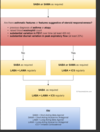
Criteria assessed by the Oxford Stroke Classification [3]

A
  1. unilateral hemiparesis and/or hemisensory loss of the face, arm & leg
  2. homonymous hemianopia
  3. higher cognitive dysfunction e.g. dysphasia
4
Q

How do TACI [c.15%] typically present? Which arteries do they involve? [2]

A

involves middle and anterior cerebral arteries
all 3 of the above criteria are present

5
Q

Which arteries do PACI [c.25%] typically effect and how do they present? [2]

A

involves smaller arteries of anterior circulation e.g. upper or lower division of middle cerebral artery
2 of the above criteria are present

6
Q

Which arteries do lacunar infarcts typically effects [c.25%] and how they present? [4]

A

involves perforating arteries around the internal capsule, thalamus and basal ganglia
presents with 1 of the following:
1. unilateral weakness (and/or sensory deficit) of face and arm, arm and leg or all three.
2. pure sensory stroke.
3. ataxic hemiparesis

7
Q

Which arteries do POCI [c.25%] typically effect and what is the presentation? [4]

A

involves vertebrobasilar arteries
presents with 1 of the following:
1. cerebellar or brainstem syndromes
2. loss of consciousness
3. isolated homonymous hemianopia

8
Q

What is a lateral medullary stroke? [3]

A

Lateral medullary syndrome (posterior inferior cerebellar artery)
aka Wallenberg’s syndrome
ipsilateral: ataxia, nystagmus, dysphagia, facial numbness, cranial nerve palsy e.g. Horner’s
contralateral: limb sensory loss

9
Q

What is Weber’s syndrome and how does it present? [2]

A

ipsilateral III palsy
contralateral weakness

10
Q

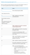
COPD still breathless despite using SABA/SAMA and a LABA+ICS, what should you add? [1]

A

A LAMA like inhaled triotropium

11
Q

General Mx of COPD [4]

A

>smoking cessation advice: including offering nicotine replacement therapy, varenicline or bupropion
annual influenza vaccination
one-off pneumococcal vaccination
pulmonary rehabilitation to all people who view themselves as functionally disabled by COPD (usually Medical Research Council [MRC] grade 3 and above)

12
Q

bronchodilator therapy initially in COPD [2]

A

a short-acting beta2-agonist (SABA) or short-acting muscarinic antagonist (SAMA) is first-line treatment
for patients who remain breathless or have exacerbations despite using short-acting bronchodilators the next step is determined by whether the patient has ‘asthmatic features/features suggesting steroid responsiveness’

13
Q

Criteria to determine if patient has asthmatic/steroid responsiveness features []4

A

any previous, secure diagnosis of asthma or of atopy
a higher blood eosinophil count - note that NICE recommend a full blood count for all patients as part of the work-up
substantial variation in FEV1 over time (at least 400 ml)
substantial diurnal variation in peak expiratory flow (at least 20%)

14
Q

How to Tx patient if no asthmatic features/features suggesting steroid responsiveness? [2]

A

add a long-acting beta2-agonist (LABA) + long-acting muscarinic antagonist (LAMA)
if already taking a SAMA, discontinue and switch to a SABA

15
Q

How to Tx a patient if they have asthmatic features/features suggesting steroid repsiveness? [3]

A

LABA + inhaled corticosteroid (ICS)
if patients remain breathless or have exacerbations offer triple therapy i.e. LAMA + LABA + ICS
if already taking a SAMA, discontinue and switch to a SABA
NICE recommend the use of combined inhalers where possible

16
Q

When to include oral theophylline in Tx for COPD? []2

A

NICE only recommends theophylline after trials of short and long-acting bronchodilators or to people who cannot used inhaled therapy
the dose should be reduced if macrolide or fluoroquinolone antibiotics are co-prescribed

17
Q

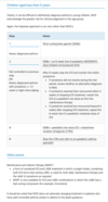
When to include prophylactic antibiotic therapy for COPD Tx [4]

A

azithromycin prophylaxis is recommended in select patients
patients should not smoke, have optimised standard treatments and continue to have exacerbations
other prerequisites include a CT thorax (to exclude bronchiectasis) and sputum culture (to exclude atypical infections and tuberculosis)
LFTs and an ECG to exclude QT prolongation should also be done as azithromycin can prolong the QT interval

18
Q

What should be done before giving azithromycin prophylaxis for COPD patients? [2]

A

LFTs and an ECG to exclude QT prolongation should also be done as azithromycin can prolong the QT interval

19
Q

When should mucolytics be considered in COPD patients? [1]

A

should be ‘considered’ in patients with a chronic productive cough and continued if symptoms improve

20
Q

Features of Cor pulmonale and how to Tx [3]

A

features include peripheral oedema, raised jugular venous pressure, systolic parasternal heave, loud P2
use a loop diuretic for oedema, consider long-term oxygen therapy
ACE-inhibitors, calcium channel blockers and alpha blockers are not recommended by NICE

21
Q

Factors which may improve survival in patients with stable COPD [3]

A

smoking cessation - the single most important intervention in patients who are still smoking
long term oxygen therapy in patients who fit criteria
lung volume reduction surgery in selected patients

22
Q

WHen is electroconvulsive therapy considered? [2]

A

Electroconvulsive therapy is a useful treatment option for patients with severe depression refractory to medication (e.g. catatonia) those with psychotic symptoms. For catatonic schizophrenai and severe mania too.

23
Q

What is the only absolute CI to ECT? [1]

A

The only absolute contraindications is raised intracranial pressure.

24
Q

Short-term SE of ECT? [5]

A

headache
nausea
short term memory impairment
memory loss of events prior to ECT
cardiac arrhythmia

25
Long-term SE of ECT [1]
some patients report impaired memory
26
What is done first-line during endoscopy to stop bleeding? [1]
Band ligation
27
Acute Tx of variceal haemorrhage [7]
ABC: patients should ideally be resuscitated prior to endoscopy correct clotting: FFP, vitamin K vasoactive agents: terlipressin is currently the only licensed vasoactive agent and is supported by NICE guidelines. It has been shown to be of benefit in initial haemostasis and preventing rebleeding octreotide may also be used although there is some evidence that terlipressin has a greater effect on reducing mortality prophylactic IV antibiotics have been shown to reduce mortality in patients with liver cirrhosis. Quinolones are typically used. NICE support this in their 2016 guidelines: 'Offer prophylactic intravenous antibiotics for people with cirrhosis who have upper gastrointestinal bleeding.' both terlipressin and antibiotics should be given before endoscopy in patients with suspected variceal haemorrhage endoscopy: endoscopic variceal band ligation is superior to endoscopic sclerotherapy. NICE recommend band ligation Sengstaken-Blakemore tube if uncontrolled haemorrhage Transjugular Intrahepatic Portosystemic Shunt (TIPSS) if above measures fail connects the hepatic vein to the portal vein exacerbation of hepatic encephalopathy is a common complication
28
What should be given before endoscopy if being given? [2]
both terlipressin and antibiotics should be given before endoscopy in patients with suspected variceal haemorrhage [Abx given those with liver cirrhosis]
29
If endoscopic measures fail to control variceal bleed,what is done next? [2]
Sengstaken-Blakemore tube if uncontrolled haemorrhage Transjugular Intrahepatic Portosystemic Shunt (TIPSS) if above measures fail connects the hepatic vein to the portal vein exacerbation of hepatic encephalopathy is a common complication
30
What are prophlyactic measures for variceal haemorrhages? [2]
propranolol: reduced rebleeding and mortality compared to placebo endoscopic variceal band ligation (EVL) is superior to endoscopic sclerotherapy. It should be performed at two-weekly intervals until all varices have been eradicated. Proton pump inhibitor cover is given to prevent EVL-induced ulceration. This is supported by NICE who recommend: 'Offer endoscopic variceal band ligation for the primary prevention of bleeding for people with cirrhosis who have medium to large oesophageal varices.'
31
third step for asthma Mx in 5-16 y/o
1 - SABA 2 - SABA + paediatric low-dose inhaled ICS 3 - SABA + paediatric low-dose inhaled ICS + LTRA
32
Prophylaxis for migraines [2]
NICE advise either topiramate or propranolol 'according to the person's preference, comorbidities and risk of adverse events'. Propranolol should be used in preference to topiramate in women of child bearing age as it may be teratogenic and it can reduce the effectiveness of hormonal contraceptives
33
Acute Tx for migraines [2]
first-line: offer combination therapy with an oral triptan and an NSAID, or an oral triptan and paracetamol
34
Which cancer are transplant patient at paritcular risk from? [1]
Patients who have received an organ transplant are at risk of skin cancer (particularly squamous cell carcinoma) due to long-term use of immunosuppressants
35
Which renal transplant drugs can cause CVD? [2]
Cardiovascular disease - tacrolimus and ciclosporin can cause hypertension and hyperglycaemia. Tacrolimus can also cause hyperlipidaemia. Patients must be monitored for accelerated cardiovascular disease.
36
Which renal transplant drugs can cause renal failure? [2]
Renal failure - due to nephrotoxic effects of tacrolimus and ciclosporin/graft rejection/recurrence of original disease in transplanted kidney
37
What must be monitored with eryhroderma? [3]
SoB: - Inpatient treatment for erythroderma must be monitored for complications like dehydration, infection and high-output heart failure
38
What is erythroderma? [1]
When more than 95% of the skin is involved in a rash of any kind
39
Causes of erythroderma [5]
eczema psoriasis drugs e.g. gold lymphomas, leukaemias idiopathic
40
What is erythrodermic psoriasis and how should it be Mx? [2]
may result from progression of chronic disease to an exfoliative phase with plaques covering most of the body. Associated with mild systemic upset more serious form is an acute deterioration. This may be triggered by a variety of factors such as withdrawal of systemic steroids. Patients need to be admitted to hospital for management
41
Sx of liver abscesses [2]
Raised inflammatory markers, RUQ pain and fever
42
How are liver abscesses Mx? [2]
Combination of antibiotics and drainage
43
Most common orgnaisms found in pyogenic liver abscesses [2]
The most common organisms found in pyogenic liver abscesses are Staphylococcus aureus in children and Escherichia coli in adults.
44
Which Abx are used in Mx of liver abscesses? [3]
drainage (typically percutaneous) and antibiotics amoxicillin + ciprofloxacin + metronidazole if penicillin allergic: ciprofloxacin + clindamycin
45
Target levels for blood glucose in pregnant women [3]
Pregnant women with GDM should be advised to maintain their CBGs below the following target levels: fasting: 5.3mmol/L AND 1 hour postprandial: 7.8 mmol/L or 2 hours postprandial: 6.4 mmol/L
46
If blood glucose targets are not met by metformin/lifestyle, what should be done? [2]
If these targets are not met with diet, exercise and metformin, then insulin should be offered as add-on therapy.
47
When do Sx start of alcohol withdrawal? [1]
Between 6-12 hours
48
when do seizures, adn when does delirium tremens begin with alcohol? [2]
Alcohol withdrawal symptoms: 6-12 hours seizures: 36 hours delirium tremens: 72 hours
49
Frist-line Mx of delirium tremens [2]
first-line: long-acting benzodiazepines e.g. chlordiazepoxide or diazepam. Lorazepam may be preferable in patients with hepatic failure. Typically given as part of a reducing dose protocol
50
Sx of alcohol withdrawal [1]
tremor, sweating, tachycardia, anxiety
51
Which drugs can cause cataracts? [1]
Steroids
52
Which drugs can cause corneal opacities? [2]
amiodarone indomethacin
53
Which drugs can cause optic neuritis? [3]
ethambutol amiodarone metronidazole
54
Which drugs can cause retinopathy? [2]
chloroquine, quinine
55
What type of herpes are genital ulcers caused by? [1]
Genital herpes is most often caused by the herpes simplex virus (HSV) type 2 (cold sores are usually due to HSV type 1)
56
Sx of primary attacks from Herpes [2]
Primary attacks are often severe and associated with fever whilst subsequent attacks are generally less severe and localised to one site. There is typically multiple painful ulcers.
57
What is syphilis caused by and what is the presentation of it? [2]
Syphilis is a sexually transmitted infection caused by the spirochaete Treponema pallidum. Infection is characterised by primary, secondary and tertiary stages. A painless ulcer (chancre) is seen in the primary stage. The incubation period= 9-90 days.
58
What is Chancroid caused by and how does it present? [2]
Chancroid is a tropical disease caused by Haemophilus ducreyi. It causes painful genital ulcers associated with unilateral, painful inguinal lymph node enlargement. The ulcers typically have a sharply defined, ragged, undermined border.
59
What is LGV caused by and how does it present? [2]
Lymphogranuloma venereum (LGV) is caused by Chlamydia trachomatis. Typically infection comprises of three stages stage 1: small painless pustule which later forms an ulcer stage 2: painful inguinal lymphadenopathy stage 3: proctocolitis
60
How is LGV Tx? [1]
Doxycycline
61
Immediate frist aid for burns [4]
airway, breathing, circulation burns caused by heat: remove the person from the source. Within 20 minutes of the injury irrigate the burn with cool (not iced) water for between 10 and 30 minutes. Cover the burn using cling film, layered, rather than wrapped around a limb electrical burns: switch off power supply, remove the person from the source chemical burns: brush any powder off then irrigate with water. Attempts to neutralise the chemical are not recommended
62
How to assess the extent of the burn [3]
Wallace's Rule of Nines: head + neck = 9%, each arm = 9%, each anterior part of leg = 9%, each posterior part of leg = 9%, anterior chest = 9%, posterior chest = 9%, anterior abdomen = 9%, posterior abdomen = 9% Lund and Browder chart: the most accurate method the palmar surface is roughly equivalent to 1% of total body surface area (TBSA). Not accurate for burns \> 15% TBSA
63
When to refer to secondary care? [3]
all deep dermal and full-thickness burns. superficial dermal burns of more than 3% TBSA in adults, or more than 2% TBSA in children superficial dermal burns involving the face, hands, feet, perineum, genitalia, or any flexure, or circumferential burns of the limbs, torso, or neck any inhalation injury any electrical or chemical burn injury suspicion of non-accidental injury
64
What is the PP of severe burns? [3]
Following the burn, there is a local response with progressive tissue loss and release of inflammatory cytokines. Systemically, there are cardiovascular effects resulting from fluid loss and sequestration of fluid into the third space. There is a marked catabolic response. Immunosupression is common with large burns and bacterial translocation from the gut lumen is a recognised event. Sepsis is a common cause of death following major burns.
65
How to Mx more severe burns [3]
The initial aim is to stop the burning process and resuscitate the patient. Intravenous fluids will be required for children with burns greater than 10% of total body surface area. Adults with burns greater than 15% of total body surface area will also require IV fluids. The fluids are calculated using the Parkland formula which is; volume of fluid= total body surface area of the burn % x weight (Kg) x4. Half of the fluid is administered in the first 8 hours. A urinary catheter should be inserted. Analgesia should be given. Complex burns, burns involving the hand perineum and face and burns \>10% in adults and \>5% in children should be transferred to a burns unit.
66
When to conservatively manage burns? [2]
Conservative management is appropriate for superficial burns and mixed superficial burns that will heal in 2 weeks. More complex burns may require excision and skin grafting. Excision and primary closure is not generally practised as there is a high risk of infection.
67
What are escharotomies and when are they used? [2]
Indicated in circumferential full thickness burns to the torso or limbs. Careful division of the encasing band of burn tissue will potentially improve ventilation (if the burn involves the torso), or relieve compartment syndrome and oedema (where a limb is involved)
68
Mx of scabies [4]
Treating the affected person and all household members, close contacts, and sexual contacts with a topical insecticide (e.g. permethrin 5% cream or malathion 0.5% liquid), even in the absence of symptoms.
69
Guidance of Tx for households with scabies [4]
avoid close physical contact with others until treatment is complete all household and close physical contacts should be treated at the same time, even if asymptomatic launder, iron or tumble dry clothing, bedding, towels, etc., on the first day of treatment to kill off mites.
70
Features of scabies
widespread pruritus linear burrows on the side of fingers, interdigital webs and flexor aspects of the wrist in infants, the face and scalp may also be affected secondary features are seen due to scratching: excoriation, infection
71
What is obese class 1? [1]
Overweight is 25-30, obese class 1 is 30-35
72
Step-wise approach to managing obesity [3]
conservative: diet, exercise medical surgical
73
MoA of Orlistat and adverse effects of the drug [2]
Orlistat is a pancreatic lipase inhibitor used in the management of obesity. Adverse effects include faecal urgency/incontinence and flatulence
74
What is the lower dose version now available [2]
A lower dose version is now available without prescription ('Alli').
75
Which adults can be given orlistat? [4]
BMI of 28 kg/m^2 or more with associated risk factors, or BMI of 30 kg/m^2 or more continued weight loss e.g. 5% at 3 months orlistat is normally used for \< 1 year
76
When are women covered by the lactational amenorrhoea method? [LAM] [1]
She is having unprotected intercourse and will not be covered by the lactational amenorrhea method (LAM) as the baby is getting less than 85% of its feeds as breast milk (NICE CKS: Contraception - natural family planning)
77
If not covered by LAM, when are women art risk of prognancy from unprototected sex? [2]
If not covered by the LAM, women are at risk of pregnancy from unprotected intercourse from day 21 postpartum
78
When is COCOP CI? [1]
The COCP is absolutely contraindicated (UKMEC 4) for women who are breastfeeding less than 6 weeks post-partum
79
Are women allowed to have the COCP 7w post-partum? [2]
However, this lady is seven weeks postpartum meaning she falls into the 6 weeks - 6 months postpartum guidance, in which the COCP is categorised as UKMEC2 for breastfeeding women. This means that advantages of prescribing the COCP would generally outweigh the disadvantages and based on the FRSH advice regarding the COCP, NICE clinical knowledge summaries recommend
80
WHen can women start the PIP? [3]
the FSRH advise 'postpartum women (breastfeeding and non-breastfeeding) can start the POP at any time postpartum.' after day 21 additional contraception should be used for the first 2 days a small amount of progestogen enters breast milk but this is not harmful to the infant
81
When can women start the COC? [4]
absolutely contraindicated - UKMEC 4 - if breast feeding \< 6 weeks post-partum UKMEC 2 - if breast feeding 6 weeks - 6 months postpartum\* the COC may reduce breast milk production in lactating mothers may be started from day 21 - this will provide immediate contraception after day 21 additional contraception should be used for the first 7 days
82
When can an IUD or IUD be inserted owmne a women post-partum? [1]
The intrauterine device or intrauterine system can be inserted within 48 hours of childbirth or after 4 weeks.
83
How effective is LAM? [2]
is 98% effective providing the woman is fully breast-feeding (no supplementary feeds), amenorrhoeic and \< 6 months post-partum
84
Problems with Inter-pregnancy interval being shortened? [2]
An inter-pregnancy interval of less than 12 months between childbirth and conceiving again is associated with an increased risk of preterm birth, low birthweight and small for gestational age babies.
85
What is pre-eclampsia defined as? [2]
Pre-eclampsia is defined as new-onset BP ≥ 140/90 mmHg after 20 weeks AND ≥ 1 of proteinuria or organ dysfunction
86
Consequences of pre-eclampsia [4]
eclampsia other neurological complications include altered mental status, blindness, stroke, clonus, severe headaches or persistent visual scotomata fetal complications intrauterine growth retardation prematurity liver involvement (elevated transaminases) haemorrhage: placental abruption, intra-abdominal, intra-cerebral cardiac failure
87
Features of severe pre-eclampsia [4]
hypertension: typically \> 160/110 mmHg and proteinuria as above proteinuria: dipstick ++/+++ headache visual disturbance papilloedema RUQ/epigastric pain hyperreflexia platelet count \< 100 \* 106/l, abnormal liver enzymes or HELLP syndrome
88
High RF for pre-eclampsia [5]
hypertensive disease in a previous pregnancy chronic kidney disease autoimmune disease, such as systemic lupus erythematosus or antiphospholipid syndrome type 1 or type 2 diabetes chronic hypertension
89
Moderate RF for pre-eclamspia [4]
first pregnancy age 40 years or older pregnancy interval of more than 10 years body mass index (BMI) of 35 kg/m² or more at first visit family history of pre-eclampsia multiple pregnancy
90
What should be given and whom to to reduce the risk of pre-eclampsia in pregnannt women? [2]
Reducing the risk of hypertensive disorders in pregnancy women with the following should take aspirin 75-150mg daily from 12 weeks gestation until the birth ≥ 1 high risk factors ≥ 2 moderate factors
91
Which women will admitted and observed for risk of pre-eclampsia in pregnancy? [2]
Initial assessment NICE recommend arranging emergency secondary care assessment for any woman in whom pre-eclampsia is suspected women with blood pressure ≥ 160/110 mmHg are likely to be admitted and observed
92
First-line, and defintive Mx of pre-eclampsia in pregnancy [2]
oral labetalol is now first-line following the 2010 NICE guidelines. Nifedipine (e.g. if asthmatic) and hydralazine may also be used delivery of the baby is the most important and definitive management step. The timing depends on the individual clinical scenario
93
What can be used for pre-eclampsia if women if asthmatic and can;t have oral labetolol? [1]
Nifedipine
94
Which type of Herpes causes oral lesions? [1]
There are two strains of the herpes simplex virus (HSV) in humans: HSV-1 and HSV-2. Whilst it was previously thought HSV-1 accounted for oral lesions (cold sores) and HSV-2 for genital herpes it is now known there is considerable overlap
95
Features of Herpes Simplex virus [3]
primary infection: may present with a severe gingivostomatitis cold sores painful genital ulceration
96
Mx of gingivomastitis vs cold sores vs genital herpes [3]
gingivostomatitis: oral aciclovir, chlorhexidine mouthwash cold sores: topical aciclovir although the evidence base for this is modest genital herpes: oral aciclovir. Some patients with frequent exacerbations may benefit from longer term aciclovir
97
How should women with Herpes at pregnancy be Tx? [2]
elective caesarean section at term is advised if a primary attack of herpes occurs during pregnancy at greater than 28 weeks gestation women with recurrent herpes who are pregnant should be treated with suppressive therapy and be advised that the risk of transmission to their baby is low
98
What are patietns with Addison's disease given? [2]
Patients who have Addison's disease are usually given both glucocorticoid and mineralocorticoid replacement therapy.
99
Which drugs does that mean they are given [2]
hydrocortisone: usually given in 2 or 3 divided doses. Patients typically require 20-30 mg per day, with the majority given in the first half of the day fludrocortisone
100
Which part of patient educatio is important with Addison disease Tx? [3]
emphasise the importance of not missing glucocorticoid doses consider MedicAlert bracelets and steroid cards patients should be provided with hydrocortisone for injection with needles and syringes to treat an adrenal crisis discuss how to adjust the glucocorticoid dose during an intercurrent illness (see below)
101
Mx of intercurrent illness of Addison's disease [2]
in simple terms the glucocorticoid dose should be doubled, with the fludrocortisone dose staying the same the Addison's Clinical Advisory Panel have produced guidelines detailing particular scenarios - please see the CKS link for more details
102
Associations of adhesive capsulitis [1]
diabetes mellitus: up to 20% of diabetics may have an episode of frozen shoulder
103
Features of adhesive capsulitis [4]
external rotation is affected more than internal rotation or abduction both active and passive movement are affected patients typically have a painful freezing phase, an adhesive phase and a recovery phase bilateral in up to 20% of patients the episode typically lasts between 6 months and 2 years
104
Mx of adhesive capsulitis [2]
no single intervention has been shown to improve outcome in the long-term treatment options include NSAIDs, physiotherapy, oral corticosteroids and intra-articular corticosteroids
105
What is Trichomonas vaginalis? [1]
Trichomonas vaginalis is a highly motile, flagellated protozoan parasite. Trichomoniasis is a sexually transmitted infection (STI).
106
Features of Trichomonas vaginalis [5]
vaginal discharge: offensive, yellow/green, frothy vulvovaginitis strawberry cervix pH \> 4.5 in men is usually asymptomatic but may cause urethritis
107
Microscopy of TV [2]
microscopy of a wet mount shows motile trophozoites
108
Mx of TV [2]
oral metronidazole for 5-7 days, although the BNF also supports the use of a one-off dose of 2g metronidazole
109
Which receptors are attacked at the NMJ in myasthenia gravis? [2]
Myasthenia gravis is an autoimmune condition whereby the patient's immune system attacks the acetylcholine receptors at the neuromuscular junction. Symptoms include weakness, especially in the evening. Treatment is focused on increasing the concentration of acetylcholine in the synaptic cleft using acetylcholinesterase inhibitors.
110
WEhat would GBS look like on NCS? [2]
Guillan-barre syndrome is an autoimmune condition. It has no known causes but often occurs after an episode of infection or immunization. Both conditions are associated with slowed motor conduction on nerve conduction studies.
111
Does MND present as LMN or UMN? [1]
Motor neuron disease is a neurological condition of unknown cause which can present with both upper and lower motor neuron signs. It rarely presents before 40 years and various patterns of disease are recognised including amyotrophic lateral sclerosis, progressive muscular atrophy and bulbar palsy.
112
Are there sensory signs/Sx in MND? [4]
fasciculations the absence of sensory signs/symptoms\* the mixture of lower motor neuron and upper motor neuron signs wasting of the small hand muscles/tibialis anterior is common
113
Are eyes effected in MND? [1]
doesn't affect external ocular muscles no cerebellar signs abdominal reflexes are usually preserved and sphincter dysfunction if present is a late feature
114
What will NCS, electromyography and MRI in MND show? [3]
The diagnosis of motor neuron disease is clinical, but nerve conduction studies will show normal motor conduction and can help exclude a neuropathy. Electromyography shows a reduced number of action potentials with increased amplitude. MRI is usually performed to exclude the differential diagnosis of cervical cord compression and myelopathy
115
Which signs are seen in ALS? [2]
affects both upper (corticospinal tracts) and lower motor neurons results in a combination of upper and lower motor neuron signs
116
Which neuron signs are seen in poliomyelitis? [1]
affects anterior horns resulting in lower motor neuron signs
117
Which tracts are affected in Brown-Sequard syndrome [spinal cord hemisection? [3]
1. Lateral corticospinal tract 2. Dorsal columns 3. Lateral spinothalamic tract
118
How does Brown-Sequard syndrome present? [3]
1. Ipsilateral spastic paresis below lesion 2. Ipsilateral loss of proprioception and vibration sensation 3. Contralateral loss of pain and temperature sensation
119
Which tracts are affected in Freidrich's ataxia? [1]
Same as subacute combined degeneration of the spinal cord (see above)
120
How does Friedrich's ataxia present? [2]
Same as subacute combined degeneration of the spinal cord (see above) In addition cerebellar ataxia → other features e.g. intention tremor
121
Which tracts are affected in MS? [2]
Asymmetrical, varying spinal tracts involved
122
How does MS present? [2]
Combination of motor, sensory and ataxia symptoms
123
Name a condition that is just a snesory lesion? [3]
Disorder Tracts affected Clinical notes Neurosyphilis (tabes dorsalis) 1. Dorsal columns 1. Loss of proprioception and vibration sensation
124
What is conveyed by the dorsal column? [3]
The sensation of fine touch, proprioception and vibration are all conveyed in the dorsal column
125
What is conveyed by the lateral spinothalamic tract? [2]
It is primarily responsible for transmitting pain and temperature as well as coarse touch.
126
What is conveyed by the lateral corticospinal tract? [2]
The primary responsibility of the lateral corticospinal tract is to control the voluntary movement of contralateral limbs
127
What is th efunction of the spinocerebellar tract? [2]
The spinocerebellar tracts carry unconscious proprioceptive information gleaned from muscle spindles, Golgi tendon organs, and joint capsules to the cerebellum
128
Asympatomic patient w/o any Sx of gour should be treated how? [1]
No Tx as not recommedned by NICE. Not proven to be beneficial.Lifestyle changes (less red meat, alcohol and sugar) can reduce uric acid levels without drug treatment and so can be advised.
129
What is associated with hyperuricaemia? [2]
Hyperuricaemia may be associated with hyperlipidaemia and hypertension. It may also be seen in conjunction with the metabolic syndrome
130
What causes increased synthesis of uric acid? [3]
Lesch-Nyhan disease myeloproliferative disorders diet rich in purines exercise psoriasis cytotoxics
131
What causes decreased excretion of uric acid? [4]
drugs: low-dose aspirin, diuretics, pyrazinamide pre-eclampsia alcohol renal failure lead
132
What is idiopathic pulmonary fibrosis? [2]
Idiopathic pulmonary fibrosis (IPF, previously termed cryptogenic fibrosing alveolitis) is a chronic lung condition characterised by progressive fibrosis of the interstitium of the lungs. Whilst there are many causes of lung fibrosis (e.g. medications, connective tissue disease, asbestos) the term IPF is reserved when no underlying cause exists.
133
Whom is idiopathic pulmonary firbosis typically seen in? [1]
IPF is typically seen in patients aged 50-70 years and is twice as common in men.
134
Features of idiopathic pulmonary fibrosis [4]
progressive exertional dyspnoea bibasal fine end-inspiratory crepitations on auscultation dry cough clubbing
135
Dx pulmonary fibrosis [4]
spirometry: classically a restrictive picture (FEV1 normal/decreased, FVC decreased, FEV1/FVC increased) impaired gas exchange: reduced transfer factor (TLCO) imaging: bilateral interstitial shadowing (typically small, irregular, peripheral opacities - 'ground-glass' - later progressing to 'honeycombing') may be seen on a chest x-ray but high-resolution CT scanning is the investigation of choice and required to make a diagnosis of IPF ANA positive in 30%, rheumatoid factor positive in 10% but this does not necessarily mean that the fibrosis is secondary to a connective tissue disease. Titres are usually low
136
What is the investigation of choice and required to make a diagnosis for IPF? [2]
imaging: bilateral interstitial shadowing (typically small, irregular, peripheral opacities - 'ground-glass' - later progressing to 'honeycombing') may be seen on a chest x-ray but high-resolution CT scanning is the investigation of choice and required to make a diagnosis of IPF
137
Mx of IPF [3]
pulmonary rehabilitation very few medications have been shown to give any benefit in IPF. There is some evidence that pirfenidone (an antifibrotic agent) may be useful in selected patients (see NICE guidelines) many patients will require supplementary oxygen and eventually a lung transplant
138
Prognosis of IPF [1]
poor, average life expectancy is around 3-4 years
139
What is a restrictive lung picture in studies? [2]
classically a restrictive picture (FEV1 normal/decreased, FVC decreased, FEV1/FVC increased)
140
What will a CXR show in IPF [2]
A chest x-ray will show interstitial shadowing, and a high resolution CT will show 'honeycomb' lungs.
141
WHat is Lynch syndreom [HNPCC] characterised by? [1]
Lynch syndrome (HNPCC) is characterised by development of bowel cancer (among other cancers) with little formation of adenomatous polyps
142
How does familial adenomatous polyposis presetn? [2]
Typically over 100 colonic adenomas Cancer risk of 100% 20% are new mutations
143
Hoes does MYH associated polyps present? [3]
Multiple colonic polyps Later onset right sided cancers more common than in FAP 100% cancer risk by age 60
144
How does Puetz-Jeghers syndreom present? [3]
Multiple benign intestinal hamartomas Episodic obstruction and intussusception Increased risk of GI cancers (colorectal cancer 20%, gastric 5%) Increased risk of breast, ovarian, cervical pancreatic and testicular cancers
145
How does Cowden disease develop? [3]
Macrocephaly Multiple intestinal hamartomas Multiple trichilemmomas 89% risk of cancer at any site 16% risk of colorectal cancer
146
How does HNPCC disease develop? [3]
Colo rectal cancer 30-70% Endometrial cancer 30-70% Gastric cancer 5-10% Scanty colonic polyps may be present Colonic tumours likely to be right sided and mucinous
147
What is sick euthyroid syndrome? [2]
In sick euthyroid syndrome (now referred to as non-thyroidal illness) it is often said that everything (TSH, thyroxine and T3) is low. In the majority of cases however the TSH level is within the \>normal range (inappropriately normal given the low thyroxine and T3).
148
Mx of sick euthyroid syndrome [1]
In sick euthyroid syndrome (now referred to as non-thyroidal illness) it is often said that everything (TSH, thyroxine and T3) is low. In the majority of cases however the TSH level is within the \>normal range (inappropriately normal given the low thyroxine and T3). Changes are reversible upon recovery from the systemic illness and hence no treatment is usually needed.
149
A 22-year-old male falls of a ladder. He complains of neck pain and cannot feel his legs. His GCS suddenly deteriorates and a CT head confirms an extradural haematoma. What is the best imaging for his neck?
The correct answer is: CT c-spine40% This man needs a CT scan of his c-spine. A CT scan will give the best resolution of any bony injury.
150
A 25-year-old teacher falls down the stairs. She complains of a headache and has vomited 3 times. She has a GCS of 15/15.
CT head within 1h72% This lady has a head injury and vomiting \> 1, therefore an urgent CT head is indicated.
151
An 18-year-old student is shot in the back of the head.
Urgent neurosurgical review (even before CT head performed)86% A penetrating injury needs urgent neurosurgical review.
152
Which patients need a CT head immediately? [6]
GCS \< 13 on initial assessment GCS \< 15 at 2 hours post-injury suspected open or depressed skull fracture. any sign of basal skull fracture (haemotympanum, 'panda' eyes, cerebrospinal fluid leakage from the ear or nose, Battle's sign). post-traumatic seizure. focal neurological deficit. more than 1 episode of vomiting
153
Which patietns need a CT head within 8h? [4]
CT head scan within 8 hours of the head injury - for adults with any of the following risk factors who have experienced some loss of consciousness or amnesia since the injury: age 65 years or older any history of bleeding or clotting disorders dangerous mechanism of injury (a pedestrian or cyclist struck by a motor vehicle, an occupant ejected from a motor vehicle or a fall from a height of greater than 1 metre or 5 stairs) more than 30 minutes' retrograde amnesia of events immediately before the head injury
154
If a patient is on warfarin who have sustained a head injury with no other indications for a CT head scan, how should they be maanged? [1]
Perform CT head within 8h
155
How should thoracic back pain be maanged? [1]
Red flag if presneting with this, should be rederred to hospital immediately
156
Red flags for lower back pain [5]
age \< 20 years or \> 50 years history of previous malignancy night pain history of trauma systemically unwell e.g. weight loss, fever
157
Presentation of spinal stensosis [5]
Usually gradual onset Unilateral or bilateral leg pain (with or without back pain), numbness, and weakness which is worse on walking. Resolves when sits down. Pain may be described as 'aching', 'crawling'. Relieved by sitting down, leaning forwards and crouching down Clinical examination is often normal Requires MRI to confirm diagnosis
158
Presentationr AS [3]
Typically a young man who presents with lower back pain and stiffness Stiffness is usually worse in morning and improves with activity Peripheral arthritis (25%, more common if female)
159
Presentation for PAD [3]
Pain on walking, relieved by rest Absent or weak foot pulses and other signs of limb ischaemia Past history may include smoking and other vascular diseases
160
Karyotype of Klinfelter's syndrome [1]
Klinefelter's syndrome is associated with karyotype 47, XXY
161
Features of Klinefelter's syndrome [5]
often taller than average lack of secondary sexual characteristics small, firm testes infertile gynaecomastia - increased incidence of breast cancer elevated gonadotrophin levels
162
Dx of Klinefelter's syndrome [1]
Diagnosis is by chromosomal analysis
163
Inheritance and cause of Kallman's syndrome [2]
Kallman's syndrome is a recognised cause of delayed puberty secondary to hypogonadotrophic hypogonadism. It is usually inherited as an X-linked recessive trait. Kallman's syndrome is thought to be caused by failure of GnRH-secreting neurons to migrate to the hypothalamus.
164
Features of Kallman's syndrome
Features 'delayed puberty' hypogonadism, cryptorchidism anosmia sex hormone levels are low LH, FSH levels are inappropriately low/normal patients are typically of normal or above average height Cleft lip/palate and visual/hearing defects are also seen in some patients
165
Exam question often of Kallman's syndrome [1]
The clue given in many questions is lack of smell (anosmia) in a boy with delayed puberty
166
Inheritance of androgen insensitivity syndrome [1]
Androgen insensitivity syndrome is an X-linked recessive condition due to end-organ resistance to testosterone causing genotypically male children (46XY) to have a female phenotype. Complete androgen insensitivity syndrome is the new term for testicular feminisation syndrome
167
Features of androgen insensitvity syndrome [3]
'primary amenorrhoea' undescended testes causing groin swellings breast development may occur as a result of conversion of testosterone to oestradiol
168
Dx of androgen insensitivty syndrome [1]
buccal smear or chromosomal analysis to reveal 46XY genotype
169
Mx of Androgen insensitivty syndrome [3]
``` counselling - raise child as female bilateral orchidectomy (increased risk of testicular cancer due to undescended testes) oestrogen therapy ```
170
What will Lh and testerone be in primary hypogonadism? [1]
Klinefelter's syndrome - high LH - low testerone
171
What will LH and testerone be in hpyogonadotrophic hypogonadism? [1]
Kallman's - low LH - low testerone
172
What will Lh and testerone be in androgen insensitivty syndrome [1]
AIS - high LH - normal/high testerone
173
Adverse effects of depolarising NM blocking drugs [3]
Malignant hyperthermia Hyperkalaemia [normally transient] May cause fasciculations
174
Why may succinylcholine [or suxamethonium cause fasciculations? [2]]
Binds to nicotinic acetylcholine receptors resulting in persistent depolarization of the motor end plate
175
CI for suxamethonium [2]
Suxamethonium is contraindicated for patients with penetrating eye injuries or acute narrow angle glaucoma, as suxamethonium increases intra-ocular pressure
176
Non-depolarizing NM blocking drugs MoA, examples, adverse effects and reversal of them
Competitive antagonist of nicotinic acetylcholine receptors Tubcurarine, atracurium, vecuronium, pancuronium Hypotension Reversal by Acetylcholinesterase inhibitors (e.g. neostigmine)
177
In aortic dissection, pulse deficit may be seen when? [2]
In aortic dissection, a pulse deficit may be seen: weak or absent carotid, brachial, or femoral pulse variation in arm BP
178
Pan-systolic murmur seen when? [1]
MR
179
Slow rising pulse seen when? [1]
AS
180
Splinter haemorrhages seen when? [1]
infective endocarditis
181
RV heave seen when? [1]
RVH such as in cor pulmomale
182
PP of aortic dissection [1]
tear in the tunica intima of the wall of the aorta
183
Associations of aortic dissection [5]
hypertension: the most important risk factor trauma bicuspid aortic valve collagens: Marfan's syndrome, Ehlers-Danlos syndrome Turner's and Noonan's syndrome pregnancy syphilis
184
Features of aortic dissection [5]
chest pain: typically severe, radiates through to the back and 'tearing' in nature pulse deficit weak or absent carotid, brachial, or femoral pulse variation (\>20 mmHg) in systolic blood pressure between the arms aortic regurgitation hypertension other features may result from the involvement of specific arteries. For example coronary arteries → angina, spinal arteries → paraplegia, distal aorta → limb ischaemia the majority of patients have no or non-specific ECG changes. In a minority of patients, ST-segment elevation may be seen in the inferior leads
185
Stanford classication of aortic dissection [2]
type A - ascending aorta, 2/3 of cases type B - descending aorta, distal to left subclavian origin, 1/3 of cases
186
Debakey classification of aortic dissection
type I - originates in ascending aorta, propagates to at least the aortic arch and possibly beyond it distally type II - originates in and is confined to the ascending aorta type III - originates in descending aorta, rarely extends proximally but will extend distally
187
Most common trigger of anaphylaxis in children [1]
Food!
188
Define anaphylaxis [2]
Anaphylaxis may be defined as a severe, life-threatening, generalised or systemic hypersensitivity reaction.
189
Common identified triggered of anaphylaxis [3]
food (e.g. nuts) - the most common cause in children drugs venom (e.g. wasp sting)
190
How does the Resus Council IUK define anaphyaxis? [2]
The Resus Council UK define anaphylaxis as: the sudden onset and rapid progression of symptoms Airway and/or Breathing and/or Circulation problems Airway problems may include: swelling of the throat and tongue →hoarse voice and stridor Breathing problems may include: respiratory wheeze dyspnoea Circulation problems may include: hypotension tachycardia
191
What other features will around 80-90% of pts with anaphlyaxius have? [2]
generalised pruritus widespread erythematous or urticarial rash
192
Recommended adrenaline dose for \<6m old compared to adult? [2]
\<6m then 100-150mcg Adult then 500mcg
193
How often can adrenaline be repeated? [1]
Adrenaline can be repeated every 5 minutes if necessary
194
What is the best site for IM injection of adnrealine? [1]
The best site for IM injection is the anterolateral aspect of the middle third of the thigh.
195
What is refractory anaphylaxis and hwo should this be Mx? [3]
defined as respiratory and/or cardiovascular problems persist despite 2 doses of IM adrenaline IV fluids should be given for shock expert help should be sought for consideration of an IV adrenaline infusion
196
How should the patient be managed following stabilisation? [5]
non-sedating oral antihistamines, in preference to chlorphenamine, may be given following initial stabilisation especially in patients with persisting skin symptoms (urticaria and/or angioedema) sometimes it can be difficult to establish whether a patient had a true episode of anaphylaxis. Serum tryptase levels are sometimes taken in such patients as they remain elevated for up to 12 hours following an acute episode of anaphylaxis all patients with a new diagnosis of anaphylaxis should be referred to a specialist allergy clinic an adrenaline injector should be givens an interim measure before the specialist allergy assessment (unless the reaction was drug-induced) patients should be prescribed 2 adrenaline auto-injectors training should be provided on how to use it
197
What can happen in around 20% of patients with anaphlaxis? [2]
- a risk-stratified approach to discharge should be taken as biphasic reactions can occur in up to 20% of patients
198
How long should a patient be kept if they needed 2 doses of IM adnrealine? [1]
fast-track discharge (after 2 hours of symptom resolution): good response to a single dose of adrenaline complete resolution of symptoms has been given an adrenaline auto-injector and trained how to use it adequate supervision following discharge minimum 6 hours after symptom resolution: 2 doses of IM adrenaline needed, or previous biphasic reaction minimum 12 hours after symptom resolution: severe reaction requiring \> 2 doses of IM adrenaline patient has severe asthma possibility of an ongoing reaction (e.g. slow-release medication) patient presents late at night patient in areas where access to emergency access care may be difficult observation for at 12 hours following symptom resolution
199
Factors favouring pseudoseizures[5]
pelvic thrusting family member with epilepsy much more common in females crying after seizure don't occur when alone gradual onset
200
Factors favouring true epileptic seizures [2]
Ongue biting Raised serum prolactin [why is not fully understood]
201
What is a useful tool for differentiating NES from ES [1]
Video telemetry
202
What causes HFM? [1]
Hand, foot and mouth disease is a self-limiting condition affecting children. It is caused by the intestinal viruses of the Picornaviridae family (most commonly coxsackie A16 and enterovirus 71)
203
Can HFM be spread? [1]
It is very contagious and typically occurs in outbreaks at nursery
204
Clinical features of HFM disease [3]
mild systemic upset: sore throat, fever oral ulcers followed later by vesicles on the palms and soles of the feet
205
Mx of HFM disease [3]
symptomatic treatment only: general advice about hydration and analgesia reassurance no link to disease in cattle children do not need to be excluded from school the HPA recommends that children who are unwell should be kept off school until they feel better they also advise that you contact them if you suspect that there may be a large outbreak.
206
A 16-year-old patient presents to the emergency department with shortness of breath and is referred for an x-ray. The scan shows the presence of a bony growth extending from the C7 vertebrae unilaterally. While not immediately concerning, this could cause problems for the patient in future. Which condition is more likely to develop in this patient?
cervical rib is a common cause of thoracic outlet syndrome [about 10% will develop cervical rib syndrome]
207
What is a Hangman's fracture? [2]
Hangman's fracture is another incorrect answer. This is a fracture to the C2 vertebrae commonly as a result of hanging. It is unlikely that this 16-year-old boy has a hangman's fracture, especially as there is no history of neck pain or suicide attempts.
208
What is thoracic outlet syndrome? [2]
Thoracic outlet syndrome (TOS) is a disorder involving compression of brachial plexus, subclavian artery or vein at the site of the thoracic outlet. TOS can be neurogenic or vascular; the former accounts for 90% of the cases.
209
Epidemiology of thoracic outlet syndrome [3]
given the lack of widely agreed diagnostic criteria, the epidemiology of TOS is not well documented patients are typically young thin women possessing long neck and drooping shoulders peak onset occurs in the 4th decade
210
Aetiology of thoracic outlet syndrome [5]
TOS develops when neck trauma occurs to individuals with anatomical predispositions neck trauma can either be a single acute incident or repeated stresses anatomical anomalies can either be in the form of soft tissue (70%) or osseous structures (30%) a well-known osseous anomaly is the presence of cervical rib examples of soft tissue causes are scalene muscle hypertrophy and anomalous bands there is usually a history of neck trauma preceding TOS
211
CLinical presentation of neurogenic TOS [3]
painless muscle wasting of hand muscles, with patients complaining of hand weakness e.g. grasping sensory symptoms such as numbness and tingling may be present if autonomic nerves are involved, the patient may experience cold hands, blanching or swelling
212
Clinical presentation of vascular TOS [2]
subclavian vein compression leads to painful diffuse arm swelling with distended veins subclavian artery compression leads to painful arm claudication and in severe cases, ulceration and gangrene
213
Ix for TOS [4]
chest and cervical spine plain radiographs to check for any obvious osseous abnormalities e.g. cervical ribs, exclude malignant tumours or other differentials e.g. cervical spine degenerative changes other imaging modalities may be helpful e.g. CT or MRI to rule out cervical root lesions venography or angiography may be helpful in vascular TOS an anterior scalene block may be used to confirm neurogenic TOS and check the likelihood of successful surgical treatment
214
Tx for TOS [5]
there is a limited evidence base conservative management with education, rehabilitation, physiotherapy, or taping is typically the first-line management for neurogenic TOS surgical decompression is warranted where conservative management has failed especially if there is a physical anomaly. Early intervention may prevent brachial plexus degeneration in vascular TOS, surgical treatment may be preferred other therapies being investigated include botox injection
215
What should be used for urge incontinence is older frail women? [1]
``` bladder retraining (lasts for a minimum of 6 weeks, the idea is to gradually increase the intervals between voiding) bladder stabilising drugs: antimuscarinics are first-line. NICE recommend oxybutynin (immediate release), tolterodine (immediate release) or darifenacin (once daily preparation). Immediate release oxybutynin should, however, be avoided in 'frail older women' mirabegron (a beta-3 agonist) may be useful if there is concern about anticholinergic side-effects in frail elderly patients ```
216
First-line stress incontinence Mx [2]
pelvic floor muscle training: NICE recommend at least 8 contractions performed 3 times per day for a minimum of 3 months surgical procedures: e.g. retropubic mid-urethral tape procedures duloxetine may be offered to women if they decline surgical procedures a combined noradrenaline and serotonin reuptake inhibitor mechanism of action: increased synaptic concentration of noradrenaline and serotonin within the pudendal nerve → increased stimulation of urethral striated muscles within the sphincter → enhanced contraction
217
What is HL? [2]
Hodgkin's lymphoma is a malignant proliferation of lymphocytes characterised by the presence of the Reed-Sternberg cell. It has a bimodal age distributions being most common in the third and seventh decades
218
Staging tool of HL [1]
Ann-Arbor
219
Nodes on both sides of diaphragm what is the stage of HL? [4]
I: single lymph node II: 2 or more lymph nodes/regions on same side of diaphragm III: nodes on both sides of diaphragm IV: spread beyond lymph nodes
220
How can each stage of HL be subdivided? [2]
``` A = no systemic symptoms other than pruritus B = weight loss \> 10% in last 6 months, fever \> 38c, night sweats (poor prognosis ```
221
What is Takotsubo cardiomyopathy associated with?on an echo? [2]
Takotsubo cardiomyopathy is associated with apical ballooning of myocardium (resembling an octopus pot)
222
What is Takotsubo cardiomyopathy? [2]
Takotsubo cardiomyopathy is a type of non-ischaemic cardiomyopathy associated with a transient, apical ballooning of the myocardium. It may be triggered by stress.
223
PP of Takotsubo cardiomyopathy [2]
Takotsubo is a Japanese word that describes an octopus trap the apical ballooning appearance occurs due to severe hypokinesis of the mid and apical segments with preservation of activity of the basal segments. In simple terms, the bottom of the heart (the apex) does not contract and therefore appears to balloon out. However, the area closer to the top (the base) continues to contract (creating the neck of the octopus trap)
224
Features of Takotsubo cardiomyoapthy [4]
chest pain features of heart failure ECG: ST-elevation normal coronary angiogram
225
Prognosis and Tx of Takutsubo cardiomyoapthy [2]
Treatment is supportive. Prognosis the majority of patients improve with supportive treatment
226
An 80-year-old man presents to the emergency department with back pain. He has no documented past medical history. An x-ray spine shows vertebral wedge compression fractures and focal sclerotic bony lesions. Which of the following diagnoses is most likely?
The most likely diagnosis in this case is metastatic prostate cancer. This disease is the most common cancer to metastasise to bone and patients often present with pathological fractures or bone pain as the first sign of disease. Prostate cancer metastases typically have a sclerotic appearance on x-ray.
227
How does MM and Paget's disease present? [2]
Multiple myeloma and Paget's disease are typically associated with osteolytic lesions rather than sclerotic, however.
228
How does osteosarcoma typically present? [2]
Osteosarcoma is a rare cancer most commonly affecting the long bones of children/young adults. It would be highly unusual to be present in the spine of man of this age. It often presents with pathological fractures of long bones such as the femur.
229
What are the most common tumours causing bone mets? [3]
Most common tumour causing bone metastases (in descending order) prostate breast lung
230
Where are the most common sites of bone mets? [5]
Most common site (in descending order) spine pelvis ribs skull long bones
231
Features of bone mets
pathological fractures -\> sclerotic appearnace, not osteolytic lesions hypercalcaemia raised ALP
232
What biopsy results would Crohn's disease have? [2]
Colonoscopy Deep ulceration affecting terminal ileum and colon, with skip lesions Biopsy sample from terminal ileum Granulomatous inflammation extending from serosa to mucosa [transmural]
233
What is the first-line for inducing remission in Crohn's disease? [1]
GLucocorticoids [oral, topical or IV] -\> e.g. 300mg of predninsolone
234
Which part of the GI tract does Crohn's effect? [2]
Crohn's disease is a form of inflammatory bowel disease. It commonly affects the terminal ileum and colon but may be seen anywhere from the mouth to anus. NICE published guidelines on the management of Crohn's disease in 2012.
235
What should all patients with Crohn's stop doing immediately? [1]
Smoking
236
Frist and second line Tx for Crohn's [2]
``` First-line = glucocorticoids (oral, topical or intravenous) are generally used to induce remission. Budesonide is an alternative in a subgroup of patients Second-line = 5-ASA drugs (e.g. mesalazine) are used second-line to glucocorticoids but are not as effective ```
237
What can also be done apart from medication to reduce chacnes of remission? [2]
enteral feeding with an elemental diet may be used in addition to or instead of other measures to induce remission, particularly if there is concern regarding the side-effects of steroids (for example in young children)
238
What can be used as an add-on in Crohn;s? [1]
azathioprine or mercaptopurine\* may be used as an add-on medication to induce remission but is not used as monotherapy. Methotrexate is an alternative to azathioprine
239
When is infliximab and metronidazole useful in Crohn's? [2]
infliximab is useful in refractory disease and fistulating Crohn's. Patients typically continue on azathioprine or methotrexate metronidazole is often used for isolated peri-anal disease
240
What is used to maintain remission in Crohn's? [2]
as above, stopping smoking is a priority (remember: smoking makes Crohn's worse, but may help ulcerative colitis) azathioprine or mercaptopurine is used first-line to maintain remission methotrexate is used second-line 5-ASA drugs (e.g. mesalazine) should be considered if a patient has had previous surgery
241
What should be assessed before starting azathioprine for Crohn's? [1]
+TMPT activity should be assessed before starting
242
How common is surgery for Crohn's? [2]
around 80% of patients with Crohn's disease will eventually have surgery see below for further detail
243
First-line remission drug for crohns? [1]
Azathioprine or mercaptopurine
244
Commonest disease pattern and Mx for Crohn;s? [2]
The commonest disease pattern in Crohn's is stricturing terminal ileal disease and this often culminates in an ileocaecal resection
245
Other procedures seen in Crohn's [2]
Other procedures performed include segmental small bowel resections and stricturoplasty. Colonic involvement in patients with Crohn's is not common and, where found, distribution is often segmental. However, despite this distribution segmental resections of the colon in patients with Crohn's disease are generally not advocated because the recurrence rate in the remaining colon is extremely high, as a result, the standard options of colonic surgery in Crohn's patients are generally; sub total colectomy, panproctocolectomy and staged sub total colectomy and proctectomy. Restorative procedures such as ileoanal pouch have no role in therapy.
246
What is Crohn;s disease notorious for developing [2]
Crohn's disease is notorious for the developmental of intestinal fistulae; these may form between the rectum and skin (perianal) or the small bowel and skin. Fistulation between loops of bowel may also occur and result in bacterial overgrowth and malabsorption. Management of enterocutaneous fistulae involves controlling sepsis, optimising nutrition, imaging the disease and planning definitive surgical management.
247
What are Crohn;s patietns at an increased risk of? [4]
``` As well as the well-documented complications described above, patients are also at risk of: small bowel cancer (standard incidence ratio = 40) colorectal cancer (standard incidence ration = 2, i.e. less than the risk associated with ulcerative colitis) osteoporosis ```
248
How common is each type of diabetes in pregnancy? [3]
87.5% have gestational diabetes 7.5% have type 1 diabetes 5% have type 2 diabetes
249
How common is gestational diabetes? [1]
Gestational diabetes is the second most common medical disorder complicating pregnancy (after hypertension), affecting around 4% of pregnancies.
250
How often should pregnant women with T1DM be tested for their glucose? [2]
Pregnant patients with type 1 diabetes should monitor their blood glucose levels closely. They should test their levels multiple times during the day. NICE NG3 Daily fasting, pre-meal, 1-hour post meal and bedtime tests.
251
Mx of pre-existing DM? [5]
weight loss for women with BMI of \> 27 kg/m^2 stop oral hypoglycaemic agents, apart from metformin, and commence insulin folic acid 5 mg/day from pre-conception to 12 weeks gestation detailed anomaly scan at 20 weeks including four-chamber view of the heart and outflow tracts tight glycaemic control reduces complication rates treat retinopathy as can worsen during pregnancy
252
A 10-year-old boy is brought to the GP by his mother after two weeks of a productive cough and fevers. The GP who saw him last week sent him away advising to come back in a week if he was still no better. The patient is documented to be allergic to penicillin. Which antibiotic should be used to treat his respiratory infection?
Clarithromycin -This patient was likely sent away initially due to the high probability he had a self-limiting viral illness. Due to the persistence of his symptoms the GP would be justified in treating the child for a bacterial lower respiratory tract infection. Without penicillin allergy then amoxicillin is the first choice treatment, however in this case this must be avoided, clarithromycin is the antibiotic of choice instead.
253
Inheritance pattern of essential tremor? [1]
Essential tremor (previously called benign essential tremor) is an autosomal dominant condition which usually affects both upper limbs
254
Features of essential tremor? [3]
postural tremor: worse if arms outstretched [6-8Hz] improved by alcohol and rest most common cause of titubation (head tremor)
255
Mx of essential tremor
Management propranolol is first-line primidone is sometimes used
256
Important risk factors used by major tools like FRAX? [6]
history of glucocorticoid use rheumatoid arthritis alcohol excess history of parental hip fracture low body mass index current smoking
257
Other risk factors for osteoporsis [5]
sedentary lifestyle premature menopause Caucasians and Asians endocrine disorders: hyperthyroidism, hypogonadism (e.g. Turner's, testosterone deficiency), growth hormone deficiency, hyperparathyroidism, diabetes mellitus multiple myeloma, lymphoma gastrointestinal disorders: inflammatory bowel disease, malabsorption (e.g. Coeliac's), gastrectomy, liver disease chronic kidney disease osteogenesis imperfecta, homocystinuria
258
Medciations that may worsen OP [5]
SSRIs antiepileptics proton pump inhibitors glitazones long term heparin therapy aromatase inhibitors e.g. anastrozole
259
When should a patient with OP have further testing? [4]
If a patient is diagnosed with osteoporosis or has a fragility fracture further investigations may be warranted. NOGG recommend testing for the following reasons: exclude diseases that mimic osteoporosis (e.g. osteomalacia, myeloma); identify the cause of osteoporosis and contributory factors; assess the risk of subsequent fractures; select the most appropriate form of treatment
260
Ix recommended by NOGG for OP? [4]
History and physical examination Blood cell count, sedimentation rate or C-reactive protein, serum calcium, albumin, creatinine, phosphate, alkaline phosphatase and liver transaminases Thyroid function tests Bone densitometry ( DXA)
261
Other Ix recommended by NOGG if indicated in OP [3]
Lateral radiographs of lumbar and thoracic spine/DXA-based vertebral imaging Protein immunoelectrophoresis and urinary Bence-Jones proteins 25OHD PTH Serum testosterone, SHBG, FSH, LH (in men), Serum prolactin 24 hour urinary cortisol/dexamethasone suppression test Endomysial and/or tissue transglutaminase antibodies (coeliac disease) Isotope bone scan Markers of bone turnover, when available Urinary calcium excretion
262
As such, what should be ordered as a minimum for P{ further Ix? [5]
full blood count urea and electrolytes liver function tests bone profile CRP thyroid function tests
263
What malignancy is most associated with the SIADH? [1]
Small cell lung cancer
264
What is SIADH characterised by? [2]
The syndrome of inappropriate ADH secretion (SIADH) is characterised by hyponatraemia secondary to the dilutional effects of excessive water retention.
265
Mx of SIADH [4]
correction must be done slowly to avoid precipitating central pontine myelinolysis fluid restriction demeclocycline: reduces the responsiveness of the collecting tubule cells to ADH ADH (vasopressin) receptor antagonists have been developed
266
A 55-year-old man with end-stage renal failure is due to have a renal transplant. He has read that having a renal transplant will increase his risk of cancer and would like to know more about this. The risk of which one of the following cancers is he most at risk of following renal transplantation?
Squamous cell carcinoma of the skin [also cervical and lymphoma cancer risk increased] Research shows that 25% of patients who live for 20 years after a transplant develop some type of cancer. This occurs due to the immunosuppressive effects of the medication given to prevent transplant rejection. The risk of all skin cancers increases following kidney transplantation, evidence has shown that in particular the risk of squamous cell carcinoma is increased. The risks of lymphoma and cervical cancer are also increased.
267
Which monitoring do patients on long-term immunosuppression need? [3]
CVD, renal disease and malignany monitoring
268
Why should their CVD be monitored for patients with an organ transplant?
Cardiovascular disease - tacrolimus and ciclosporin can cause hypertension and hyperglycaemia. Tacrolimus can also cause hyperlipidaemia. Patients must be monitored for accelerated cardiovascular disease.
269
Why should their renal failure be monitored for patients with an organ transplant?
Renal failure - due to nephrotoxic effects of tacrolimus and ciclosporin/graft rejection/recurrence of original disease in transplanted kidney
270
How should their malignancy be monitored for patients with an organ transplant?
Malignancy - patients should be educated about minimising sun exposure to reduce the risk of squamous cell carcinomas and basal cell carcinomas
271
What is first-line for menorrhagia? [1]
IUS [Mirena]
272
Define menorrhagia [1]
Previously defined as over 80ml blood loss per menses, now has become whatever the women feels is heavy
273
Ix for menorrhagia -important [2]
a full blood count should be performed in all women NICE recommend arranging a routine transvaginal ultrasound scan if symptoms (for example, intermenstrual or postcoital bleeding, pelvic pain and/or pressure symptoms) suggest a structural or histological abnormality. Other indications include abnormal pelvic exam findings.
274
Which Rx should be given in women experiencing menorrhagia that does not require contraception? [2]
either mefenamic acid 500 mg tds (particularly if there is dysmenorrhoea as well) or tranexamic acid 1 g tds. Both are started on the first day of the period if no improvement then try other drug whilst awaiting referral
275
Which Rx should be given if women does require contraception? [3]
``` intrauterine system (Mirena) should be considered first-line combined oral contraceptive pill long-acting progestogens ```
276
What can be used in the short-term to stop HMB? [1]
Norethisterone 5 mg tds can be used as a short-term option to rapidly stop heavy menstrual bleeding.
277
An obese 14-year-old boy presents with difficulty running and mild knee and hip pain. There is no antecedent history of trauma. On examination internal rotation is restricted but the knee is normal with full range of passive movement possible and no evidence of effusions. Both the C-reactive protein and white cell count are normal.
Slipped upper femoral epiphysis is the commonest adolescent hip disorder. It occurs most commonly in obese males. It may often present as knee pain which is usually referred from the ipsilateral hip. The knee itself is normal. The hip often limits internal rotation. The diagnosis is easily missed. X-rays will show displacement of the femoral epiphysis and the degree of its displacement may be calculated using the Southwick angle. Treatment is directed at preventing further slippage which may result in avascular necrosis of the femoral head.
278
A 6-year-old boy presents with pain in the hip it is present on activity and has been worsening over the past few weeks. There is no history of trauma. He was born by normal vaginal delivery at 38 weeks gestation On examination he has an antalgic gait and limitation of active and passive movement of the hip joint in all directions. C-reactive protein is mildly elevated at 10 but the white cell count is normal.
Perthes disease78% This is a typical presentation for Perthes disease. X-ray may show flattening of the femoral head or fragmentation in more advanced cases.
279
A 30-year-old man presents with severe pain in the left hip it has been present on and off for many years. He was born at 39 weeks gestation by emergency caesarean section after a long obstructed breech delivery. He was slow to walk and as a child was noted to have an antalgic gait. He was a frequent attender at the primary care centre and the pains dismissed as growing pains. X-rays show almost complete destruction of the femoral head and a narrow acetabulum.
Developmental dysplasia of the hip63% Developmental dysplasia of the hip. Usually diagnosed by Barlow and Ortolani tests in early childhood. Most Breech deliveries are also routinely subjected to USS of the hip joint. At this young age an arthrodesis may be preferable to hip replacement.
280
Compare mild, moderately severe and severe pancreatitis [3]
Mild = no organ failure, no local complications M. severe = no or transient organ failure [\<48h], possible local Cx Severe = persistent [\>48h] organ failure, possible local Cx
281
What are the 5 key aspects to Mx pancreatitis? [5]
1. Fluid resus 2. Analgesia 3. Nutrition 4. Abx 5. Surgery
282
Fluid resus for pancreatitis [3]
aggressive early hydration with crystalloids. In severe cases 3-6 litres of third space fluid loss may occur aim for a urine output of \> 0.5mls/kg/hr may also help relieve pain by reducing lactic acidosis
283
Analgesia for pancreatitis [2]
pain may be severe so this is a key priority of care intravenous opioids are normally required to adequately control the pain
284
Nutrition for pancreatitis [3]
patients should not routinely be made 'nil-by-mouth' unless there is a clear reason e.g. the patient is vomiting enteral nutrition should be offered to anyone with moderately severe or severe acute pancreatitis within 72 hours of presentation parental nutrition should only be used if enteral nurition has failed or is contraindicated
285
Role of Abx in pancreatitis [2]
NICE state the following: 'Do not offer prophylactic antimicrobials to people with acute pancreatitis' potential indications include infected pancreatic necrosis
286
Role of surgery in pancreatitis [4]
Patients with acute pancreatitis due to gallstones should undergo early cholecystectomy Patients with obstructed biliary system due to stones should undergo early ERCP Patients who fail to settle with necrosis and have worsening organ dysfunction may require debridement, fine needle aspiration is still used by some Patients with infected necrosis should undergo either radiological drainage or surgical necrosectomy. The choice of procedure depends upon local expertise
287
Which age do febrile convulsions typically occur at? [1]
6m to 5y
288
Clinical features of febrile convulsions [3]
usually occur early in a viral infection as the temperature rises rapidly seizures are usually brief, lasting less than 5 minutes are most commonly tonic-clonic
289
Simple febrile convulsion [4]
\<15m, generalised seizure, typically no recurrence within 24h, should be complete recovery within an hour
290
Complex febriel convulsions
15-30m, focal seziure, may have repeated seizures
291
Febrile status epilepticus [1]
Time seizure lasts over 30m
292
Mx for febrile seizure [1]
children who have had a first seizure OR any features of a complex seizure should be admitted to paediatrics
293
Do regular antipyreitcs reduce the chance of febriel seizures occuring? [1]
regular antipyretics have not been shown to reduce the chance of a febrile seizure occurring
294
If recurrences of seizures, how should parents be advised to Mx? [2]
if recurrences, try teaching parents how to use rectal diazepam or buccal midazolam. Parents should be advised to phone for an ambulance if the seizure lasts \> 5 minutes
295
Prognosis of febrile seizures [3]
the overall risk of further febrile convulsion = 1 in 3. However, this varies widely depending on risk factors for further seizure. These include: age of onset \< 18 months, fever \< 39ºC, shorter duration of fever before seizure and a family history of febrile convulsions if recurrences, try teaching parents how to use rectal diazepam or buccal midazolam. Parents should be advised to phone for an ambulance if the seizure lasts \> 5 minutes regular antipyretics have not been shown to reduce the chance of a febrile seizure occurring
296
Link to epilepsy for febrile seziures [3]
risk factors for developing epilepsy include a family history of epilepsy, having complex febrile seizures and a background of neurodevelopmental disorder children with no risk factors have 2.5% risk of developing epilepsy if children have all 3 features the risk of developing epilepsy is much higher (e.g. 50%)
297
Meningitis causes 0-3m [3]
Group B Streptococcus (most common cause in neonates) E. coli Listeria monocytogenes
298
Meningitis causes 3m-6y [3]
Neisseria meningitidis Streptococcus pneumoniae Haemophilus influenzae
299
Meningitis causes 6y-60m [2]
Neisseria meningitidis Streptococcus pneumoniae
300
Meningitis causes over 60y [3]
Streptococcus pneumoniae Neisseria meningitidis Listeria monocytogenes
301
Meningitis cause for the immunosuppressed? [1]
Listera monocytogenes
302
What type of bacteria is N.meninigitis, S. pneumoniae, E.coli, H.influenzae, L.monocytognese [5]
Neisseria meningitis and Streptococcus pneumoniae would be most common in this age group but it is N.meningitis that is a gram negative diplococci. S. pneumoniae is a gram positive diplococci/chain E. coli is a gram negative bacilli H. influenzae is a gram negative coccobacilli L. monocytogenes is a gram positive rod
303
Mx for DCM [2]
Management of patients with cervical myelopathy should be by specialist spinal services (neurosurgery or orthopaedic spinal surgery). Decompressive surgery is the mainstay of treatment and has been shown to stop disease progression
304
Sx of DCM [5]
Pain (affecting the neck, upper or lower limbs) Loss of motor function (loss of digital dexterity, preventing simple tasks such as holding a fork or doing up their shirt buttons, arm or leg weakness/stiffness leading to impaired gait and imbalance Loss of sensory function causing numbness Loss of autonomic function (urinary or faecal incontinence and/or impotence) - these can occur and do not necessarily suggest cauda equina syndrome in the absence of other hallmarks of that condition Hoffman's sign: is a reflex test to assess for cervical myelopathy. It is performed by gently flicking one finger on a patient's hand. A positive test results in reflex twitching of the other fingers on the same hand in response to the flick.
305
What is DCM often confuse with? [1]
Carpal tunnel syndrome
306
What Ix is the gold standard for DCM? [1]
An MRI of the cervical spine is the gold standard test where cervical myelopathy is suspected. It may reveal disc degeneration and ligament hypertrophy, with accompanying cord signal change.
307
Why is it important for early Tx with DCM? [2]
All patients with degenerative cervical myelopathy should be urgently referred for assessment by specialist spinal services (neurosurgery or orthopaedic spinal surgery). This is due to the importance of early treatment. The timing of surgery is important, as any existing spinal cord damage can be permanent. Early treatment (within 6 months of diagnosis) offers the best chance of a full recovery but at present, most patients are presenting too late. In one study, patients averaged over 5 appointments before diagnosis, representing \>2 years.
308
What is the only Tx currently available for DCM? [2]
Currently, decompressive surgery is the only effective treatment. It has been shown to prevent disease progression. Close observation is an option for mild stable disease, but anything progressive or more severe requires surgery to prevent further deterioration. Physiotherapy should only be initiated by specialist services, as manipulation can cause more spinal cord damage.
309
A 75-year-old man presents with dysphagia and halitosis. On the left side of the neck is a small, fluctuant swelling which gurgles when palpated.
Pharyngeal puch
310
. A 54-year-old woman presents with a neck swelling. She is systemically well apart from some recent weight loss. On examination she is noted to have a midline, non-tender neck swelling which moves upwards when she swallows.
Goitre47% Patients with a goitre are often euthyroid or have minor symptoms. A thyroglossal cyst would be unusual at this age - they typically present in patients \< 20 years old.
311
A newborn baby is noted to have a large swelling on the left-side of the neck. On examination a soft, fluctuant and highly transilluminable lump is noted just beneath the skin.
Cystic hygroma
312
By far the most common cause of neck swellings. There may be a history of local infection or a generalised viral illness
Reactive lyphmadenopathy
313
Rubbery, painless lymphadenopathy The phenomenon of pain whilst drinking alcohol is very uncommon There may be associated night sweats and splenomegaly
Lymphoma
314
May be hypo-, eu- or hyperthyroid symptomatically Moves upwards on swallowing
Thyroid swelling
315
More common in patients \< 20 years old Usually midline, between the isthmus of the thyroid and the hyoid bone Moves upwards with protrusion of the tongue May be painful if infected
Thyroglossal cyst
316
More common in older men Represents a posteromedial herniation between thyropharyngeus and cricopharyngeus muscles Usually not seen but if large then a midline lump in the neck that gurgles on palpation Typical symptoms are dysphagia, regurgitation, aspiration and chronic cough
Pharyngeal pouch
317
A congenital lymphatic lesion (lymphangioma) typically found in the neck, classically on the left side Most are evident at birth, around 90% present before 2 years of age
Cystic hygroma
318
An oval, mobile cystic mass that develops between the sternocleidomastoid muscle and the pharynx Develop due to failure of obliteration of the second branchial cleft in embryonic development Usually present in early adulthood
Brnachial cyst
319
More common in adult females Around 10% develop thoracic outlet syndrome
Cervical rub
320
Pulsatile lateral neck mass which doesn't move on swallowing
Carotid aneurysm
321
What can amenorrhoea be divided into? [2]
primary: defined as the failure to establish menstruation by 15 years of age in girls with normal secondary sexual characteristics (such as breast development), or by 13 years of age in girls with no secondary sexual characteristics secondary: cessation of menstruation for 3-6 months in women with previously normal and regular menses, or 6-12 months in women with previous oligomenorrhoea
322
Causes of primary amenorrhoea [5]
gonadal dysgenesis (e.g. Turner's syndrome) - the most common causes testicular feminisation congenital malformations of the genital tract functional hypothalamic amenorrhoea (e.g. secondary to anorexia) congenital adrenal hyperplasia imperforate hymen
323
Causes of secondary amenorrhoea [after excl. pregnancy]
hypothalamic amenorrhoea (e.g. secondary stress, excessive exercise) polycystic ovarian syndrome (PCOS) hyperprolactinaemia premature ovarian failure thyrotoxicosis\* Sheehan's syndrome Asherman's syndrome (intrauterine adhesions)
324
What should be excluded first before deciding the cause of secondary amenorrhoea? [1]
Pregnancy
325
Initial Ix for amenorrhoea [6]
exclude pregnancy with urinary or serum bHCG full blood count, urea & electrolytes, coeliac screen, thyroid function tests gonadotrophins raised if gonadal dysgenesis (e.g. Turner's syndrome) prolactin androgen levels oestradiol
326
What would raised levels of gonadotrophins indicate? [1]
raised if gonadal dysgenesis (e.g. Turner's syndrome)
327
What would reduced levels of gonadotrophins indicate?[2]
low levels indicate a hypothalamic cause where as raised levels suggest an ovarian problem (e.g. Premature ovarian failure)
328
What would raised levels of androgens indicate? [1]
May be seen in PCOS
329
How to Mx primary amenorrhoea [2]
investigate and treat any underlying cause with primary ovarian insufficiency due to gonadal dysgenesis (e.g. Turner's syndrome) are likely to benefit from hormone replacement therapy (e.g. to prevent osteoporosis etC)
330
How to Mx secondary amenorrhoea [2]
exclude pregnancy, lactation, and menopause (in women 40 years of age or older) treat the underlying cause
331
Which endocrine disroder can cause amenorrhoea?[1]
Hypothyroidism
332
How should you alter the sertraline dose given to a patietn that it about to have ECT? [1]
Reducing the sertraline dose is the correct answer. With antidepressants and ECT, you do not suddenly stop them when the patient commences ECT treatment. The recommended regime is to safely reduce them to the minimum dose. You may actually add an increased dose of antidepressant towards the end of the ECT course.
333
Whom is ET given to? [2]
Electroconvulsive therapy is a useful treatment option for patients with severe depression refractory to medication (e.g. catatonia) those with psychotic symptoms.
334
Absolute CI for ECT
The only absolute contraindications is raised intracranial pressure.
335
Short-term SE of ECT [5]
headache nausea short term memory impairment memory loss of events prior to ECT cardiac arrhythmia
336
Long-term SE of ECT [1]
Some patients report impaired memory
337
When is IV adenosine used? [1]
IV adenosine is incorrect. It is used as a rate lowering drug used to slow supraventricular tachycardias. Since there are QRS abnormalities here, there is an indication of ventricular or conducting system pathology. Adenosine is not always appropriate in these circumstances - and expert help should be sought first.
338
FOllowing ABC assessment, what would indicate that patient is stable/unstable? [4]
shock: hypotension (systolic blood pressure \< 90 mmHg), pallor, sweating, cold, clammy extremities, confusion or impaired consciousness syncope myocardial ischaemia heart failure
339
If there are signs patient is unstable, then what should be done? [1]
If any of the above adverse signs are present then synchronised DC shocks should be given. Up to 3 shocks can be given; after this expert help should be sought.
340
Mx for regular broad-complex tachycardia [2]
assume ventricular tachycardia (unless previously confirmed SVT with bundle branch block) loading dose of amiodarone followed by 24 hour infusion
341
Mx for irregular broad-complex tachycardia [1]
Seek expert help
342
Possible causes of irregular broad-complex tachycardia [3]
atrial fibrillation with bundle branch block - the most likely cause in a stable patient atrial fibrillation with ventricular pre-excitation torsade de pointes
343
Narrow-complex regular tachycardia Mx [3]
vagal manoeuvres followed by IV adenosine if above unsuccessful consider diagnosis of atrial flutter and control rate (e.g. beta-blockers)
344
Narrow-complex tachycardia irregular [2]
probable atrial fibrillation if onset \< 48 hr consider electrical or chemical cardioversion rate control: beta-blockers are usually first-line unless there is a contraindication
345
How would acute interstitial nephritis present? [3]
Acute interstitial nephritis is not the correct answer. This is an interrenal AKI due to inflammation of the nephron interstitium, usually in response to drugs such as penicillin, rifampicin, NSAIDs, allopurinol or furosemide. More systemic symptoms such as joint pain, fever, rashes would be present if this were the case. Investigations will show mild renal impairment but importantly there will be white cell casts and sterile pyuria.
346
Important findings Ix wise for acute interstitial nephritis [2]
White cell clasts and sterile pyuria
347
Important findings for acute tubular necrosis [3]
Acute tubular necrosis is not the correct answer. This is an example of intrarenal AKI, in which the kidney tubules are damaged leading to an inability to reabsorb sodium. This leads to dilute urine (low osmolality) with high amounts of sodium. Acute tubular necrosis is usually due to nephrotoxins such as aminoglycosides and light chains in myeloma- neither of which are indicated in this patient.
348
important finding for ATN in blood Ix? [1]
This is an example of intrarenal AKI, in which the kidney tubules are damaged leading to an inability to reabsorb sodium. This leads to dilute urine (low osmolality) with high amounts of sodium
349
How would haemolytic uraemic syndrome present? [2]
Haemolytic uraemic syndrome is not the correct answer. This is a glomerular intrarenal cause of AKI caused by an infection that is most common in childhood. This would present with the core symptoms of AKI, anaemia and thrombocytopenia. The patient here has normal red blood cell and platelet counts.
350
Give an example of prerenal uraemia [2]
Hypovolaemia is the correct answer. This is an example of prerenal uraemia, a cause of acute kidney injury (AKI) due to renal hypoperfusion, usually caused by shock, haemorrhage or volume depletion
351
How can prereanl uraemia be differentiated? [3]
Prerenal uraemia can also be differentiated from other causes of AKI by the results of the urine analysis in which the kidneys reabsorb water in order to increase circulatory volume. This leads to the production of concentrated urine (high osmolality) urine with low amounts of sodium. Urine osmolality of more than 500 mOsm/kg, with an urine sodium inferior to 20 mol/l is landmark for the diagnosis os pre-renal disease.
352
COmpare urine sodium and urine osmolaity in PTU and ATN
Pre-renal uraemia ('azotemia') ATN Urine sodium \< 20 mmol/L \> 40 mmol/L Urine osmolality \> 500 mOsm/kg \< 350 mOsm/kg
353
Characterisation of BCC [1]
Basal cell carcinoma (BCC) is one of the three main types of skin cancer. Lesions are also known as rodent ulcers and are characterised by slow-growth and local invasion.
354
Features of BCC [4]
many types of BCC are described. The most common type is nodular BCC, which is described here sun-exposed sites, especially the head and neck account for the majority of lesions initially a pearly, flesh-coloured papule with telangiectasia may later ulcerate leaving a central 'crater'
355
A 78-year-old man asks you to look at a lesion on the right side of nose which has been getting slowly bigger over the past 2-3 months. On examination you observe a round, raised, flesh coloured lesion which is 3mm in diameter and has a central depression. The edges of the lesion appear rolled and contain some telangiectasia.
BCC
356
What are the 3 malignant types of skin cancer? [3]
Basal cell carcinoma, squamous cell carcinoma, malignant meloma
357
Referral for BCC? [1]
generally, if a BCC is suspected, a routine referral should be made
358
Mx of BCC [5]
surgical removal curettage cryotherapy topical cream: imiquimod, fluorouracil radiotherapy
359
in general, when should warfarin be stopped? [1]
5d before surgery
360
What should INR be before surgery? [1]
below 1.5
361
What happens to warfarin after surgery is complete? [1]
Usually resumed at the normal dose on the evning of the surgery or the next day if haemostasis is adequet
362
MoA of warfarin [4]
Warfarin is an oral anticoagulant which inhibits epoxide reductase preventing the reduction of vitamin K to its active hydroquinone form, which in turn acts as a cofactor in the carboxylation of clotting factor II, VII, IX and X (mnemonic = 1972) and protein C.
363
Indications for warfarin []3
venous thromboembolism: target INR = 2.5, if recurrent 3.5 atrial fibrillation, target INR = 2.5 mechanical heart valves, target INR depends on the valve type and location. Mitral valves generally require a higher INR than aortic valves.
364
What is the INR? [1]
Patients on warfarin are monitored using the INR (international normalised ration), the ratio of the prothrombin time for the patient over the normal prothrombin time
365
How long can it take for the INR to be stablisied? [2]
Warfarin has a long half-life and achieving a stable INR may take several days. There a variety of loading regimes and computer software is now often used to alter the dose.
366
Factors which may potentiate warfarin [5]
liver disease P450 enzyme inhibitors, e.g.: amiodarone, ciprofloxacin cranberry juice drugs which displace warfarin from plasma albumin, e.g. NSAIDs inhibit platelet function: NSAIDs
367
SE of warfarin [4]
haemorrhage teratogenic, although can be used in breastfeeding mothers skin necrosis: when warfarin is first started biosynthesis of protein C is reduced. This results in a temporary procoagulant state after initially starting warfarin, normally avoided by concurrent heparin administration. Thrombosis may occur in venules leading to skin necrosis purple toes
368
Where should swabs for chlamydia and gonorrhoea be taken? [1]
Should be taken from the vulvo-vaginal area [introitus]
369
What is the most prevalent STI infection in the UK? [1]
Chlamydia
370
Which is the most sensitive Ix for Chlamydia in females? [2]
Nucleic acid amplification tests (NAATs) are currently the most sensitive and specific test for the diagnosis of Chlamydia in females. An endocervical swab can be performed, but it is not as sensitive as a vulvovaginal swab. Vulvovaginal swabs for NAAT is the specimen of choice in females, as recommended by BASHH.
371
Which is the most sensitive Ix for Chlamydia in males? [2]
NAAT with - for men: the urine test is first-line
372
When should Chlamydia testing be done? [1]
Chlamydiatesting should be carried out two weeks after a possible exposure
373
How many owmen in the Uk have chlamydia? [1]
Approximately 1 in 10 young women in the UK have Chlamydia
374
Incuabtion period for chlamydia [1]
The incubation period is around 7-21 days, although it should be remembered a large percentage of cases are asymptomatic
375
Features of chlamydia [3]
asymptomatic in around 70% of women and 50% of men women: cervicitis (discharge, bleeding), dysuria men: urethral discharge, dysuria
376
Potential Cx of Chlamydia
epididymitis pelvic inflammatory disease endometritis increased incidence of ectopic pregnancies infertility reactive arthritis perihepatitis (Fitz-Hugh-Curtis syndrome)
377
Screnning for chlamydia in the UK [3]
in England the National Chlamydia Screening Programme is open to all men and women aged 15-24 years the 2009 SIGN guidelines support this approach, suggesting screening all sexually active patients aged 15-24 years relies heavily on opportunistic testing
378
What is first-line Tx for chlamydia in the UK? [2]
doxycycline (7 day course) if first-line this is now preferred to azithromycin due to concerns about Mycoplasma genitalium. This infection is often coexistant in patients with Chlamydia and there is evidence of rising levels of macrolide resistance, hence why doxycycline is preferred if doxycycline is contraindicated / not tolerated then either azithromycin (1g od for one day, then 500mg od for two days) should be used
379
If pregnant, how should a patient be maanged with chlamydia? [2]
if pregnant then azithromycin, erythromycin or amoxicillin may be used. The SIGN guidelines suggest azithromycin 1g stat is the drug of choice 'following discussion of the balance of benefits and risks with the patient'
380
When to contact people with Chlamydia infection? [4]
patients diagnosed with Chlamydia should be offered a choice of provider for initial partner notification - either trained practice nurses with support from GUM, or referral to GUM for men with urethral symptoms: all contacts since, and in the four weeks prior to, the onset of symptoms for women and asymptomatic men all partners from the last six months or the most recent sexual partner should be contacted contacts of confirmed Chlamydia cases should be offered treatment prior to the results of their investigations being known (treat then test)
381
What is acute urinary retention? [2]
Acute urinary retention is when a person suddenly (over a period of hours or less) becomes unable to voluntarily pass urine. It is the most common urological emergency and there are several potential causes that must be investigated for.
382
Epidemiology of acute urinary retention [2]
Whilst acute urinary retention is common in men, it rarely occurs in women (incidence ratio of 13:1). It occurs most frequently in men over 60 years of age and incidence increases with age. It has been estimated that around a third of men in their 80s will develop acute urinary retention over a five year period.
383
Most common cause of acute urinary retention [1]
In men, acute urinary retention most commonly occurs secondary to benign prostatic hyperplasia; a condition where the prostate becomes enlarged but non-cancerous. The enlarged prostate presses on the urethra which can make the bladder wall thicker and less able to empty.
384
Other causes of acute urinary retention [3]
In men, acute urinary retention most commonly occurs secondary to benign prostatic hyperplasia; a condition where the prostate becomes enlarged but non-cancerous. The enlarged prostate presses on the urethra which can make the bladder wall thicker and less able to empty. Other urethral obstructions; including urethral strictures, calculi, cystocele, constipation or masses. Some medications can cause acute urinary retention by affecting nerve signals to the bladder: these include anticholinergics, tricyclic antidepressants, antihistamines, opioids and benzodiazepines. Less commonly, there may be a neurological cause for the acute urinary retention. In patients with predisposing causes, a simple urinary tract infection can be enough to cause acute urinary retention Acute urinary retention often occurs postoperatively and in women postpartum: usually secondary to a combination of the above risk factors.
385
How do patients typically present in AUR [4]
Inability to pass urine Lower abdominal discomfort Considerable pain or distress an acute confusional state may also be present in elderly patients
386
How does chronic urinary retention usually present? [2]
This differs from chronic urinary retention which is typically painless. In a patient with a background of chronic urinary retention, acute urinary retention may present instead with overflow incontinence.
387
Sx of acute urinary retention [3]
Palpable distended urinary bladder either on an abdominal or rectal exam Lower abdominal tenderness All men and women should have a rectal and neurological examination to assess for the likely causes above. Women should also have a pelvic examination.
388
Ix for acute urinary retnetion [3]
Patients should all be investigated with a urine sample which should be sent for urinalysis and culture. This might only be possible after urinary catheterisation. Serum U&Es and creatinine should also be checked to assess for any kidney injury. A FBC and CRP should also be performed to look for infection PSA is not appropriate in acute urinary retention as it is typically elevated
389
What should first be done for a patient with AUR? [2]
To confirm the diagnosis of acute urinary retention a bladder ultrasound should be performed. A volume of \>300 cc confirms the diagnosis, but if the history and examination are consistent, with an inconsistent bladder scan, there are causes of bladder scan inaccuracies and hence the patient can still have acute urinary retention.
390
Further Mx for AUR [3]
Acute urinary retention is managed by decompressing the bladder via catheterisation Urinary catheterisation can be performed in patients with suspected acute urinary retention, and the volume of urine drained in 15 minutes measured. A volume of \<200 confirms that a patient does not have acute urinary retention, and a volume over 400 cc means the catheter should be left in place. In between these volumes, it depends on the case. Further investigation should be targeted by the likely cause. In reversible causes such as UTI, resolution with treatment is sufficient and further investigation is not necessary. Men not diagnosed by BPH should be further evaluated by a urologist, Patients with neurological symptoms should be evaluated by a neurologist and women with gynaecological symptoms by a gynaecologist. Where no likely cause is identified, patients should be evaluated by a urologist for anatomical and urodynamic causes.
391
Cx of AUR [3]
post-obstructive diuresis the kidneys may increase diuresis due to the loss of their medullary concentration gradient. This can take time re-equilibrate this can lead to volume depletion and worsening of any acute kidney injury some patients may therefore require IV fluids to correct this temporary over-diuresis
392
A 60-year-old man is admitted with severe central chest pain to the resus department. The admission ECG shows ST elevation in leads V1-V4 with reciprocal changes in the inferior leads. Which one of the following is most likely to account for these findings?
100% ST elevation in the LAD Widepread ST elevation in this territory implies a complete occlusion of the left anterior descending artery.
393
Causes of ST elevation [5]
myocardial infarction pericarditis/myocarditis normal variant - 'high take-off' left ventricular aneurysm Prinzmetal's angina (coronary artery spasm) Takotsubo cardiomyopathy rare: subarachnoid haemorrhage
394
What is it called when neurological Sx are exacerbated by increases in body temperature? [1]
Uhthoff ’s phenomenon where neurological symptoms are exacerbated by increases in body temperature is typically associated with multiple sclerosis
395
What do most pts with MS present with? [1]
Patient's with multiple sclerosis (MS) may present with non-specific features, for example around 75% of patients have significant lethargy.
396
Dx of MS [2]
Diagnosis can be made on the basis of two or more relapses and either objective clinical evidence of two or more lesions or objective clinical evidence of one lesion together with reasonable historical evidence of a previous relapse
397
Visual Sx of MS [4]
optic neuritis: common presenting feature optic atrophy Uhthoff's phenomenon: worsening of vision following rise in body temperature internuclear ophthalmoplegia
398
Sensory Sx of MS [4]
pins/needles numbness trigeminal neuralgia paraesthesiae in limbs on neck flexion
399
What is Lhermitte's syndrome? [1]
Paraesthesiae in limbs on neck flexion
400
Motor and cerebellar Sx of MS [2]
ataxia: more often seen during an acute relapse than as a presenting symptom tremor
401
Other Sx seen in MS [3]
urinary incontinence sexual dysfunction intellectual deterioration
402
If ruptures may cause pseudomyxoma peritonei [1]
Mucinous cystadenoma
403
The most common type of epithelial cell tumour [1]
Serous cystadenoma
404
May contain skin appendages, hair and teeth [1]
Dermoid cyst [teratoma]
405
What can benign cysts be divided into? [3]
Benign ovarian cysts are extremely common. They may be divided into physiological cysts, benign germ cell tumours, benign epithelial tumours and benign sex cord stromal tumours.
406
What should happen to complex ovarian cysts and why? [2]
Complex (i.e. multi-loculated) ovarian cysts should be biopsied to exclude malignancy.
407
Name types of physiological [or functional] cysts [2]
1. Follicular cysts 2. Corpus luteum cysts
408
What is the commonest type of ovarian cyst and what is it due to? [2]
Follicular cysts commonest type of ovarian cyst due to non-rupture of the dominant follicle or failure of atresia in a non-dominant follicle commonly regress after several menstrual cycles
409
How are corpus luteum cysts formed? How can they present? [2]
during the menstrual cycle if pregnancy doesn't occur the corpus luteum usually breaks down and disappears. If this doesn't occur the corpus luteum may fill with blood or fluid and form a corpus luteal cyst more likely to present with intraperitoneal bleeding than follicular cysts
410
Name a benign germ cell tumour that causes an ovarian cyst [1]
Dermoid cyst
411
Why may dermoid cysts contain skin appendages, hair and teeth? [1]
also called mature cystic teratomas. Usually lined with epithelial tissue and hence may contain skin appendages, hair and teeth
412
Commonest presentation of dermoid cyst [4]
most common benign ovarian tumour in woman under the age of 30 years median age of diagnosis is 30 years old bilateral in 10-20% usually asymptomatic. Torsion is more likely than with other ovarian tumours
413
Where do benign epithelial tumours arise from? [1]
Arise from the ovarian surface epithelium
414
name types of benign epithelial tumour ovarian cysts [2]
Serous cystadenoma Mucinous cystadenoma
415
What is the most common type of benign epithelial tumour? what does it resemble? [2]
Serous cystadenoma the most common benign epithelial tumour which bears a resemblance to the most common type of ovarian cancer (serous carcinoma) bilateral in around 20%
416
Which benign ovarian tumours are typically large? [3]
Mucinous cystadenoma second most common benign epithelial tumour they are typically large and may become massive if ruptures may cause pseudomyxoma peritonei
417
What is given prior to surgery for an appendectomy? [1]
Prophylactic IV Abx
418
When is acute appendicits likely to occur? [2]
Acute appendicitis is the most common acute abdominal condition requiring surgery. It can occur at any age but is most common in young people aged 10-20 years.
419
Pathogenesis of appendicitis [2]
lymphoid hyperplasia or a faecolith → obstruction of appendiceal lumen → gut organisms invading the appendix wall → oedema, ischaemia +/- perforation
420
Which Sx is seen in most pts with acute appendicitis? [4]
Abdominal pain is seen in the vast majority of patients: peri-umbilical abdominal pain (visceral stretching of appendix lumen and appendix is midgut structure) radiating to the right iliac fossa (RIF) due to localised parietal peritoneal inflammation. the migration of the pain from the centre to the RIF has been shown to be one of the strongest indicators of appendicitis patients often report the pain being worse on coughing or going over speed bumps. Children typically can't hop on the right leg due to the pain.
421
Other features of appendicitis 4]
vomit once or twice but marked and persistent vomiting is unusual diarrhoea is rare. However, pelvic appendicitis may cause localised rectal irritation of some loose stools. A pelvic abscess may also cause diarrhoea mild pyrexia is common - temperature is usually 37.5-38oC. Higher temperatures are more typical of conditions like mesenteric adenitis anorexia is very common. It is very unusual for patients with appendicitis to be hungry around 50% of patients have the typical symptoms of anorexia, peri-umbilical pain and nausea followed by more localised right lower quadrant pain Vomiting, diarrhoea, pyrexia, anorexia
422
Examination for appendcitis [4]
generalised peritonitis if perforation has occurred or localised peritonism rebound and percussion tenderness, guarding and rigidity retrocaecal appendicitis may have relatively few signs digital rectal examination may reveal boggy sensation if pelvic abscess is present, or even right-sided tenderness with a pelvic appendix classical signs: Rovsing's and psoas
423
What are Rovsing;s and psoas sign and how useful are they? [2]
Rovsing's sign (palpation in the LIF causes pain in the RIF) is now thought to be of limited value psoas sign: pain on extending hip if retrocaecal appendix
424
Dx of appendicits [4]
typically raised inflammatory markers coupled with compatible history and examination findings should be enough to justify appendicectomy a neutrophil-predominant leucocytosis is seen in 80-90% urine analysis: useful to exclude pregnancy in women, renal colic and urinary tract infection. In patients with appendicitis, urinalysis may show mild leucocytosis but no nitrites there are no definite rules on the use of imaging and its use is often determined by the patient's gender, age, body habitus and the likelihood of appendicitis: - thin, male patients with a high likelihood of appendicitis may be diagnosed clinically - ultrasound is useful in females where pelvic organ pathology is suspected. Although it is not always possible to visualise the appendix on ultrasound, the presence of free fluid (always pathological in males) should raise suspicion - CT scans are widely used in patients with suspected appendicitis in the US but this practice has not currently reached the UK, due to the concerns regarding excessive ionising radiation and resource limitations
425
Inflammatory markers in appendicits [2]
Tpyically raised inflammatory markers; neutrophil-predominnnat leucocytosis seen in 80-90%
426
Thin male vs female with pelvic pathology Dx for appendicits [2]
thin, male patients with a high likelihood of appendicitis may be diagnosed clinically ultrasound is useful in females where pelvic organ pathology is suspected. Although it is not always possible to visualise the appendix on ultrasound, the presence of free fluid (always pathological in males) should raise suspicion
427
Initial Mx of appendicits [2]
appendicectomy can be performed via either an open or laparoscopic approach laparoscopic appendicectomy is now the treatment of choice
428
Summarised Mx for appendicits [6]
appendicectomy - can be performed via either an open or laparoscopic approach - laparoscopic appendicectomy is now the treatment of choice administration of prophylactic intravenous antibiotics reduces wound infection rates patients with perforated appendicitis (typical around 15-20%) require copious abdominal lavage. patients without peritonitis who have an appendix mass should receive broad-spectrum antibiotics and consideration given to performing an interval appendicectomy be wary in the older patients who may have either an underlying caecal malignancy or perforated sigmoid diverticular disease. trials have looked at the use of intravenous antibiotics alone in the treatment of appendicitis. The evidence currently suggests that whilst this is successful in the majority of patients, it is associated with a longer hospital stay and up to 20% of patients go on to have an appendicectomy within 12 months.
429
A 21-year-old man presents to his GP with an itchy rash. This has been coming on for the past three months and is characterised by red, scaly patches of skin, most prominent on the back of his elbows and the front of his knees. He has suffered with dandruff for some time. He has no past medical or family history and is not taking any medication. Dx?
Chronic plaque psoriasis
430
What is Koebner phenomenon? [2]
This patient is presenting with a history suggestive of chronic plaque psoriasis. The Koebner phenomenon describes the tendency for new skin lesions to form at sites of cutaneous injury. It is not clear why this occurs, but it is observed in chronic plaque psoriasis and vitiligo.
431
Sunlight and psoriasis [2]
Photosensitive rash is primarily associated with SLE and other connective tissue diseases including dermatomyositis. It may rarely be seen in psoriasis, but the majority of psoriasis patients actually experience an improvement in symptoms when exposed to UV light, and so this is not the most likely feature.
432
What is Samter's triad? [3]
Aspirin sensitivity and nasal polyps are two aspects of Samter's triad, the third being chronic asthma, not psoriasis. Samter's triad is seen in aspirin-exacerbated respiratory disease, where hypersensitivity to aspirin results in asthmatic symptoms.
433
What is Apthous ulceration? [2]
Aphthous ulceration is the formation of ulcers on the oral and genital mucous membranes. It may be idiopathic but is also associated with connective tissue diseases including Behcet's and systemic lupus erythematosus (SLE). It is not a feature associated with psoriasis.
434
Where is Koebner phenomenon also seen in? [5]
The Koebner phenomenon describes skin lesions that appear at the site of injury. It is seen in: psoriasis vitiligo warts lichen planus lichen sclerosus molluscum contagiosum
435
Features of acute pericarditis [5]
chest pain: may be pleuritic. Is often relieved by sitting forwards other symptoms include non-productive cough, dyspnoea and flu-like symptoms pericardial rub tachypnoea tachycardia
436
What is pericarditis relieved by? [1]
Sitting forwards
437
What can be heard in pericarditis? [1]
Pericardial rub
438
Causes of acute pericarditis [5]
viral infections (Coxsackie) tuberculosis uraemia (causes 'fibrinous' pericarditis) trauma post-myocardial infarction, Dressler's syndrome connective tissue disease hypothyroidism malignancy
439
What can acute pericarditis arise secondary to in very ill patients? [1] - important!
malignancy!
440
ECG changes seen in acute pericarditis [3] - important
the changes in pericarditis are often global/widespread, as opposed to the 'territories' seen in ischaemic events 'saddle-shaped' ST elevation PR depression: most specific ECG marker for pericarditis
441
What should all patients with suspected acute pericarditis have? [2]
all patients with suspected acute pericarditis should have transthoracic echocardiography
442
Mx of acute pericarditis [2] - important
treat the underlying cause a combination of NSAIDs and colchicine is now generally used for first-line for patients with acute idiopathic or viral pericarditis
443
What is colchicine used for? [4]
Colchicine is an adjuvant therapy which improves the response to medical therapy and reduces the rate of recurrence in acute pericarditis. Also used for gout, Behcets disease and FMF.
444
A 41-year-old woman presents with palpitations and heat intolerance. On examination her pulse is 90/min and a small, diffuse goitre is noted which is tender to touch. Thyroid function tests show the following: Free T4 at 24, TSH at 0.05. Dx? [1]
De Quervain's thyroiditis [subacute thyoiriditis] Whilst Grave's disease is the most common cause of thyrotoxicosis it would not cause a tender goitre. In the context of thyrotoxicosis this finding is only really seen in De Quervain's thyroiditis. Hashimoto's thyroiditis is an autoimmune disorder of the thyroid gland. It is typically associated with hypothyroidism although there may be a transient thyrotoxicosis in the acute phase. The goitre is non-tender in Hashimoto's.
445
WHen does subacute thyoiriditis typically occur? [2]
Subacute thyroiditis (also known as De Quervain's thyroiditis and subacute granulomatous thyroiditis) is thought to occur following viral infection and typically presents with hyperthyroidism.
446
4 phases of subacute thyroiditis [4]
``` There are typically 4 phases; phase 1 (lasts 3-6 weeks): hyperthyroidism, painful goitre, raised ESR phase 2 (1-3 weeks): euthyroid phase 3 (weeks - months): hypothyroidism phase 4: thyroid structure and function goes back to normal ```
447
Ix for subacute thyroiditis [1]
thyroid scintigraphy: globally reduced uptake of iodine-131
448
Mx for subacute thyroiditis [3]
usually self-limiting - most patients do not require treatment thyroid pain may respond to aspirin or other NSAIDs in more severe cases steroids are used, particularly if hypothyroidism develops
449
Which types of thyroid disroders are solely hypo or hyper? [2]
``` Hypo = Hashimotos, Riedel's thyroiditis, Iodine def., lithium Hyper = Graves', toxic multinodular goitre ```
450
Initially hyper, then hypo? [2]
Subacute thyroiditis, postpartum thyroiditis
451
Which drug can cause both hypo and hyperthyroidism [2]
Amiodarone
452
Inheritance pattern of Wilson's disease
Autosomal recessive disorder
453
Characterisation of Wilson's disease
Wilson's disease is an autosomal recessive disorder characterised by excessive copper deposition in the tissues.
454
Pathogenesis of Wilson's disease [2]
Metabolic abnormalities include increased copper absorption from the small intestine and decreased hepatic copper excretion. Wilson's disease is caused by a defect in the ATP7B gene located on chromosome 13.
455
onset of Sx for WIlson's disease usually? [2]
The onset of symptoms is usually between 10 - 25 years. Children usually present with liver disease whereas the first sign of disease in young adults is often neurological disease
456
Features of excessive copper deposition [6]
Liver hepatitis Neurological Sx like basal ganglia degeneration, speech/psychiatric problems, dementia Kayser-Fleischer rings Renal tubular acidosis Haemolysis Blue nails
457
What is often the first manifestation of Wilson's disease? [1]
speech, behavioural and psychiatric problems are often the first manifestations
458
What do Kayser-Fleischer rings look like, and how common are they in Wilson's disease? [3]
green-brown rings in the periphery of the iris due to copper accumulation in Descemet membrane present in around 50% of patients with isolated hepatic Wilson's disease and 90% who have neurological involvemen
459
Summarise features of Wilson's disease
Features result from excessive copper deposition in the tissues, especially the brain, liver and cornea: liver: hepatitis, cirrhosis neurological: - basal ganglia degeneration: in the brain, most copper is deposited in the basal ganglia, particularly in the putamen and globus pallidus - speech, behavioural and psychiatric problems are often the first manifestations - also: asterixis, chorea, dementia, parkinsonism Kayser-Fleischer rings - green-brown rings in the periphery of the iris - due to copper accumulation in Descemet membrane - present in around 50% of patients with isolated hepatic Wilson's disease and 90% who have neurological involvement renal tubular acidosis (esp. Fanconi syndrome) haemolysis blue nails
460
Dx of Wilson's disease
slit lamp examination for Kayser-Fleischer rings reduced serum caeruloplasmin reduced total serum copper (counter-intuitive, but 95% of plasma copper is carried by ceruloplasmin) - free (non-ceruloplasmin-bound) serum copper is increased increased 24hr urinary copper excretion
461
Is total serum copper raised or reduced in Wilson's disease [1]
reduced total serum copper (counter-intuitive, but 95% of plasma copper is carried by ceruloplasmin)
462
Traditional first-line Mx for Wilson's disease [1]
penicillamine (chelates copper) has been the traditional first-line treatment
463
What other mx is done for WIlson's disease? [3]
penicillamine (chelates copper) has been the traditional first-line treatment trientine hydrochloride is an alternative chelating agent which may become first-line treatment in the future tetrathiomolybdate is a newer agent that is currently under investigation
464
What is the role of calcium resonium in the Mx of hyperkalaemia patient? [2]
Calcium resonium results in removal of potassium from the body, rather than shifting potassium between fluid compartments in the short-term
465
Principles of managing hyperkalaemia [2]
Untreated hyperkalaemia may cause life-threatening arrhythmias. Precipitating factors should be addressed (e.g. acute kidney injury) and aggravating drugs stopped (e.g. ACE inhibitors). Management may be categorised by the aims of treatment
466
Three classifications of hyperkalaemia [3]
The European Resuscitation Council classifies hyperkalaemia as: mild: 5.5 - 5.9 mmol/L moderate: 6.0 - 6.4 mmol/L severe: ≥ 6.5 mmol/L
467
What are the ECG changes associated with hyperkalaemia [4]
Remember that the presence of ECG changes is important in determining the management - it should be done in all patients with new hyperkalaemia. The following changes are associated with hyperkalaemia: peaked or 'tall-tented' T waves (occurs first) loss of P waves broad QRS complexes sinusoidal wave pattern
468
How to shift potassium from the ECF to the ICF compartment in hyperkalaemia [2]
Short-term shift in potassium from extracellular (ECF) to intracellular fluid (ICF) compartment - combined insulin/dextrose infusion - nebulised salbutamol
469
How to remove potassium from the body in hyperkalaemia [2]
Removal of potassium from the body - calcium resonium (orally or enema) - enemas are more effective than oral as potassium is secreted by the rectum - loop diuretics - dialysis - haemofiltration/haemodialysis should be considered for patients with AKI with persistent hyperkalaemia
470
How to stabilise cardiac membrane in hyperkalaemia [2]
Stabilisation of the cardiac membrane - IV calcium gluconate - does NOT lower serum potassium levels
471
What are the principles of Tx for hyperkalaemia [3]
Stabilise cardiac membrane, shift potassium from ECF to ICF, remove potassium from the body
472
How to manage patients with severe hyperkalaemia [3]
All patients with severe hyperkalaemia (≥ 6.5 mmol/L) or with ECG changes should have emergency treatment IV calcium gluconate: to stabilise the myocardium insulin/dextrose infusion: short-term shift in potassium from ECF to ICF other treatments such as nebulised salbutamol may be given to temporarily lower the serum potassium
473
Furterh Mx for hyperkalaemia [10]
Further management stop exacerbating drugs e.g. ACE inhibitors treat any underlying cause lower total body potassium calcium resonium loop diuretics dialysis
474
Summarise Mx of hyperkalaemia
Management Principles of treatment modalities Stabilisation of the cardiac membrane IV calcium gluconate does NOT lower serum potassium levels Short-term shift in potassium from extracellular (ECF) to intracellular fluid (ICF) compartment combined insulin/dextrose infusion nebulised salbutamol Removal of potassium from the body calcium resonium (orally or enema) enemas are more effective than oral as potassium is secreted by the rectum loop diuretics dialysis haemofiltration/haemodialysis should be considered for patients with AKI with persistent hyperkalaemia Outline of practical treatment All patients with severe hyperkalaemia (≥ 6.5 mmol/L) or with ECG changes should have emergency treatment IV calcium gluconate: to stabilise the myocardium insulin/dextrose infusion: short-term shift in potassium from ECF to ICF other treatments such as nebulised salbutamol may be given to temporarily lower the serum potassium Further management stop exacerbating drugs e.g. ACE inhibitors treat any underlying cause lower total body potassium calcium resonium loop diuretics dialysis
475
First-line Tx for COPD [1]
SABA or LAMA
476
Seocn dline Tx for COPD [1]
+ LAMA or ICS
477
Third-line Tx for COPD
478
First, second and third line for ashtma in 5-16 y/o children [3]
479
4th, 5th, 6th, and 7th line for asthma in 5-16 y/o
480
First, second, third and fourth linne Mx for asthma in children under 5
481
What is maintenance nad reliever therapy in children under 5 with asthma? [2]
482
Erythroderma
483
484
Extensive exfoliation on patient with erythroderma
485
What is the appearance of a superifical epidermal burn? [1]
486
What is the appearance of a deep dermal burn? [1]
487
Scabies
488
BMI of 32, BMI of 29, BMI of 37 =
489
What Sx overlap between BV and Trichomonas? [3]
490
Which Sx are seperate from BV and tirchomonas?
491
Pulmonary fibrosis I think
492
LH and testerone in androgen insensitivty syndrome vs in praimry hypogonadism
493
Adrenaline dose to give patient if under 6m as compared to 6-12y in anaphyalxis [2]
494
Hand foot mouth disease
495
Which drugs, which malignancies, which neurological causes of SIADH? [3]
496
Summarise causes of SIADH [split into malignancy, neuro, infections, drugs, other]
497
Compare pre-renal uraemia to ATN
498
Urine sodium and urine osmolaity difference in pre-renal uraemia vs acute tubular necrosis
499
BCC
500
ECG showing pericarditis -\> note widespread ST elevation anf PR depression
501
Compare which types of thyroid disease fall into hypo, which hyper and which both [3]
502
ECG showing hyperkalaemia -\> widening QRS complexes and peaked T waves