Gastrointestinal Cancers Flashcards

1
Q

Define cancer

A

A disease caused by an uncontrolled division of abnormal cells in a part of the body

2
Q

Define primary cancer

A

Cancer arising directly from the cells in an organ

3
Q

Define secondary cancer/metastasis

A

Cancer spread to another organ, directly or by other means (blood or lymph)

4
Q

List the 3 main types of cells/tissues highly susceptible to cancer in the GI tract

A

Epithelial cells (e.g. squamous and glandular epithelial cells)

Neuroendocrine cells (e.g. enteroendocrine cells + Interstitial cells of Cajal)

Connective tissue (e.g. adipose tissue + smooth muscle)

5
Q

List 4 cells of the GI tract susceptible to becoming cancerous

A

Squamous cells

Glandular epithelial cells

Enteroendocrine cells

Interstitial cells of Cajal

Smooth muscle

Adipose tissue

6
Q

Which cancer of the GI tract is derived from squamous epithelial cells?

A

Squamous cell carcinoma (SCC)

7
Q

Which cancer of the GI tract is derived from glandular epithelium?

A

Adenocarcinomas

8
Q

Which cancer of the GI tract is derived from enteroendocrine cells?

A

Neuroendocrine tumors (NETs)

9
Q

Which cancer of the GI tract is derived from interstitial cells of Cajal?

A

Gastrointestinal stromal tumors (GISTs)

10
Q

Which cancer of the GI tract is derived from smooth muscle?

A

Leiomyoma/Leiomyosarcoma

11
Q

Which cancer of the GI tract is derived from adipsoe tissue?

A

Liposarcoma

12
Q

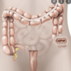
Where are GI neuroendocrine tumors located?

A

Can be located anywhere along the GI tract from the oesophagus to the rectum

13
Q

Squamous cell carcinomas tend to develop in which portion of the oesophagus? Explain why

A

Upper 2/3rds of the oesophagus

The type of epithelium lining the oesophagus above the Z-line is stratified squamous epithelium

14
Q

What is a common cause of squamous cell carcinoma oesophageal cancers?

A

Acetaldehyde pathway

[ACETALDEHYDE PATHWAY -EtOH – alcohol dehydrogenase (ADH) – oxidized acetaldehyde – aldehyde dehydrogenase (ALDH) – oxidized - acetate]

15
Q

Squamous cell carcinoma is a more common oesophageal cancer in which type of countries?

A

Countries in the less developed world

16
Q

Adenomacarcinomas are derived from which type of cells?

A

From metaplastic columnar epithelium

17
Q

Adenocarcinoma oesophageal cancers occur mainly in which part of the oesophagus and why?

A

Occur in distal third of the oesophagus

Below the Z-line, the epithelium is simple columnar and these cells can develop into adenocarcinomas

18
Q

Oesophageal adenocarcinomas are related to which condition?

A

Related to acid reflux

19
Q

Oesophageal adenocarcinomas is more common than squamous cell carcinomas in which types of countries?

A

Countries of the more developed world

20
Q

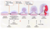
Briefly describe the transition of reflux into oesophageal cancer

A

Reflux leads to oesophagitis due to inflammation

Chronic inflammation can lead to metaplasia, resulting in presence of Barret’s Oesophagus

There is a chance overtime that metastatic epithelial cells present in Barret’s oesophagus patients can display neoplastic changes, resulting in formation of adenocarcinoma

N.B. Inflammation —> Metaplasia —> Neoplasia

21
Q

What percentage of the UK population experiencing oesophagitis is caused due to GORD?

A

30% of UK population experiencing oesophagitis

22
Q

What percentage of GORD population will end up developing Barret’s Oesophagus?

A

5% of GORD population

23
Q

What is the likelihood of someone with Barret’s oesophagus developing adenocarcinoma?

A

Barrett’s lifetime risk of cancer - 0.5-1%/ year.

24
Q

In comparison to the general population what is the fold risk of developing cancer in patient’s with Barret’s oesophagus?

A

30-100 fold risk of cancer

25
Q

State the Barret’s Surveillance Guidelines

A

BSGs refers to occasional endoscopic surveillance in barret’s oesophagus patients frequency changes depending on histological findings

No dysplasia: Every 2-3 years

Low grade dysplasia: Every 6 months

High grade dysplasia: Intervention required

26
Q

For both squamous cell carcinoma and adenocarcinomas, which age group is most likely to be affected?

A

Affects mainly the elderly

27
Q

How does squamous cell carcinoma rates differ between males and females?

A

Effects males more than females 10:1

28
Q

How common is squamous cell carcinomas in comparison to other cancers?

A

9th most common cancer

29
Q

What are two common late presentations of oesophageal cancers?

A

Dysphagia

Weight loss

30
Q

What percentage of patients diagnosed with oesophageal cancers end up being placed in palliative care?

A

65%

31
Q

What is the general morbidity and prognosis of oesophageal cancers?

A

Poor progonosis

Poor 5 year survival <20% (regardless of age its bad)

High morbidity and complex surgery

Providing adequate palliative care is difficult

32
Q

How are oesophageal cancers diagnosed?

A

Diagnosed by biopsy taken from endoscopy

33
Q

List 4 examples of imaging techniques used in the staging of oesophageal cancers

A

CT scan

Laproscopy (Keyhole surgery)

Endoscopic ultrasound (?)

PET scan (?)

34
Q

Depending on the prognosis of osophageal cancer, what are the two types of treatment plan options?

A

Curative

Palliative

35
Q

What is the curative treatment plan for oesophageal cancer?

A

Neo adjuvant chemotherapy followed by radical surgery

36
Q

What is the palliative treatment plan for oesophageal cancers?

A

Chemotherapy

Radiotherapy

Stent to keep oesophagus patent

37
Q

Briefly describe the Ivor Lewis approach to oesophagectomy.

A

Involves removal of tumar through abdominal incision and thoracotomy in which the upper portion of the stomach is removed and region of the oesophagus in which the tumor is located.

38
Q

What is the most common GI tract cancer in western societies?

A

Colorectal cancer

39
Q

Colorectal cancer is reponsible for how many portions of cancer deaths?

A

3rd most common cause of cancer deaths

40
Q

Colorectal cancer is more common in which gender and age groups?

A

Common in men (1 in 10 vs 1 in 14)

Common above age of 50 (pver 90% of cases)

41
Q

What are the 3 main forms of colorectal cancer?

A

Sporadic

Familial

Hereditary syndrome

42
Q

State the histopathology of colorectal cancers

A

Adenocarcinoma

43
Q

What are the 3 main signs that a colorectal cancer formed sporadically?

A

No family history

Developed at older age (>50)

A single lesion

44
Q

What are the 3 main signs that a colorectal cancer is in familial form?

A

Family history of colorectal cancer

Cancer occured in a close relative (1st degree relative)

Cancer developed at younger ages (<50)

45
Q

What are the 3 main signs that a colorectal cancer formed due to a hereditary syndrome?

A

Family history of cancer

Cancer developed at a young age

Evidence of specific gene defects

46
Q

Give 2 examples of hereditary syndromes which can result in the development of colorectal cancer.

A

.Familial adenomatous polyposis (FAP)

Hereditary nonpolyposis colorectal cancer (HNPCC or Lynch syndrome)

47
Q

Hereditary nonpolypososis colorectal cancer (HNPCC) is alternatively known as?

A

Lynch syndrome

48
Q

Describe the formation of a colon carcinoma

A

Begins due to APC mutation of normal epithelium which results in hyperproliferation.

Hyperproliferation leads to overexpression of COX2 leading to small adenoma formation

K-ras mutation in the small adenoma results in a large adenoma formation

p53 mutation followed by loss of 18q results in a colon carcinoma

49
Q

Name 5 medications/substances that can have protective effects against devolpment of colorectal carcinoma

A

Aspirin

NSAIDs

Folate

Calcium

Estorgen (perhaps why males more likely)

50
Q

What 4 past medical history risk factors should be checked for when taking a history of a patient suspected of having colorectal cancer?

A

Hx of colorectal cancer

Hx of any adenocarcinomas

Hx of Ulcerative colitis

Hx of radiotherapy

51
Q

What 2 family history risk factors should be checked for when taking a history of a patient suspected of having colorectal cancer?

A

1st degree relative (under 55) with history of colorectal cancer

Any identified hereditary syndromes such as FAP, HNPCC, Peutz-Jegher’s syndrome)

52
Q

What 4 dietary/lifestyle risk factors should be checked for when taking a history of a patient suspected of having colorectal cancer?

A

Carcinogenic foods

Smoking

Obesity

Socioeconomic status

53
Q

The clinical presentation of colon cancer depends on the location. 2/3rds of colon cancers are lcoated in which location?

A

Descending colon to the rectum

54
Q

The clinical presentation of colon cancer depends on the location. 1/2th of colon cancers are located in which location?

A

Sigmoid colon and rectum

55
Q

1/2th of colon cancers are located in the sigmoid colon and rectum, what is the significance of this in terms of being diagnosed?

A

Majority can be identified on a flexible sigmoidoscopy

56
Q

For more uncommon ceaceal and right-sided colon cancers, list 4 clinical presentations.

A

Iron-deficiency anaemia (most common)

Change in bowel habits (e.g. diarrhoea)

Distal ileum obstruction (late presentation)

Palpable mass (late presentation)

57
Q

What is the most common sign of right-sided/caecal colon cancer?

A

Iron-deficiency anaemia

58
Q

Give 2 examples of late presenting signs/symptoms of right-sided colon cancer.

A

Palpable mass

Distal ileum obstruction

59
Q

Give 2 examples of signs/symptoms of left-sided/sigmoid colon cancers.

A

Thin stool (late presentation)

Rectal bleed with mucus

60
Q

Give 2 examples of signs/symptoms of rectal colon cancers.

A

PR bleeding and mucus

Tenesmus

Anal, sacral, perineal pain (late presentation)

61
Q

What is a common late presentation of all colon cancers?

A

Bowel obstruction

62
Q

Give 2 examples of late presenting symptoms of colon cancer related to local invasion

A

Bladder symptoms

Femal genital tract symptoms

63
Q

List 4 examples of common areas of metasteses of colon cancer and resulting clinical presentations.

A

Liver (Hepatic pain and jaundice)

Lungs (Cough)

Regional lymph nodes

Peritoneum (Sister Mary Joesph Nodule)

64
Q

Metatstaic umbilical nodules indicative of abdominopelvic malignancies is known as?

A

Sister Mary Joesph Nodule

65
Q

List 4 common signs of primary colorectal cancer

A

Abdominal mass

Feeling of mass in digital rectal examination (DRE)

Rigid sigmoidoscopy

Abdominal tenderness and distention (indicative of large bowel obstruction)

66
Q

List 3 common signs of metastatic colorectal cancer

A

Hepatomegaly

Monphonic wheeze

Bone pain

67
Q

List 6 examples of investigations that can be performed if a patient is under suspicion of having colon cancer

A

Faecal occult blood test (FOB)

Blood tests

Colonoscopy

CT colonscopy/colonography

CT chest/abdomen/pelvis

MRI pelvis

68
Q

Give two examples of faecal occult blood test and explain what they detect.

Which one is more commonly used?

A

Guaiac test (aka Haemoccult test): looks at pseudoperoxidase activity of haematin

Feacal immunochemical test (FIT): Detects minute amounts of blood in the faeces

FIT more commonly used nowadays

69
Q

Give two examples of disadvantages of guaiac test (Hemoccult).

A

Has high specificity but is not very sensitive (40-80%)

Requires dietary restriction (e.g. red meat, melons, horse-radish, vitamin C) and certain medication such as NSAIDs

70
Q

Give two examples of useful blood tests which could be indicative of colon cancer but should not be used as a diagnostic tool

A

FBC: Can detect anemia, and low ferritin

Tumor marker tests such as detection of carcinoembryonic antigen test (CEA)

71
Q

What is a common use of a CAE blood test.

A

Can be used to monitor the spread of bowel cancers and can indicate whether chemotehrapy is effective

72
Q

Name the antigen most commonly associated with colon cancers

A

Carcinoembryonic antigen (CAE)

73
Q

Give 2 reasons why colonoscopy is an effective type of investigation into colon cancers.

A

Can identify lesions <5mm in size

Can involve identification and removal of small polyps which can reduce incidence of cancer

74
Q

Does colonoscopy require sedation?

A

Often requires sedation

75
Q

Compare CT colonoscopy to colonoscopy (2)

A

Can only identify lesions larger than 5mm whereas colonoscopy can identify smaller lesions

Is less invasive versus colonscopy and does not involve sedation

76
Q

If a lesion is idenitfied in a CT colonoscopy, what must be performed after?

A

Colonoscopy for diagnosis (need a biopsy)

77
Q

What would be the purpose/benefit of ordering an pelvis MRI for a patient diagnosed with rectal cancer?

A

Can help to identify depth of invasion and if there is mesorectal lymph node involvement

Can aid in decision to strat pre-operative chemotherapy or straight to surgery

78
Q

What is the purpose of pre-operative chemotehrapy?

A

Can help to reduce the margins if there is uncertainty, making it easier to completely remove the cancer in surgery with clearer margins

79
Q

What is the purpose of a CT chest/abdo/plevis in management of colorectal cancer?

A

Determines the staging of the cancer before treatment

80
Q

What is the primary management of colon cancer?

A

Surgery

81
Q

Besides surgery, what are 3 alternative management plans for patients with colorectal cancer?

A

Chemotehrapy

Radiotherapy

Stent

82
Q

What type of surgery is required for a obstructing colon carcinoma located in the right-sided or transverse section of the colon?

A

Resection and primary anastamosis (rejoining the proximal instestine with distal after portion is removed)

83
Q

What 3 types of surgery may be required for a obstructing colon carcinoma located in the left-sided section of the colon?

A

Hartmann’s procedure (Proximal end colostomy)

Primary anastamosis

Palliative stent

84
Q

What is Hartmann’s procedure

Is the procedure reversible or irreversible?

A

Type of surgical procedure characterised by resection with a proximal-endcolostomy

The procedure can be reversible after -/+ 6 months

85
Q

What is involved in primary anastamosis of the left-sided bowel?

What is the disadvantage of this procedure vs hartmanns?

A

Intraoperative bowel lavage with primary anastomosis with defunctioning ileostomy (would be on right side versus left liek hartmanns)

There is a 10% risk of leak so hartmanns is a bit safer

86
Q

Memorise the main blood supply of the colon

A
87
Q

The type of colon resection is dependant mainly on?

A

The blood supply of the colon

88
Q

Removal of the caecum and ascending colon is known as?

A

A right hemicolectomy

89
Q

In a right hemicolectomy, what type of anastamosis is performed?

A

Ileocolic anastamosis

90
Q

When would an extended right hemicolectomy be performed and what does it involve and why?

A

Would be needed if the tumor is located within most ascending portion fo the right colon/transvere portion.

Procedure involves removal of caecum, right colon and a portion of the transverse colon

Removing any less would disrupt the blood supply

91
Q

What does a left hemicolectomy involved?

A

Removal of the descending colon with anastomosis with the sigmoid colon

92
Q

What is the most common form of pancreatic cancer?

A

Pancreatic ductal adenocarcinoma

93
Q

What is the overall prognosis of rectal cancer?

What percentage of patietns would present late and what is the median survival rate of these patients?

A

Very poor

80-85% present late

Median survival rate <6 months and 5 year survival 0.4-5%

94
Q

15-20% of pancreatic cancer patients have resectable disease, what is the survival rate in comparison to late presenting

A

Median survival 11-20 months

5-year survival 20–25%

Virtually all pts dead within 7 years of surgery

95
Q

Pancreatic cancer is most common in which regions of the world, between which of the genders and of what age ranges?

A

Most common in western world

Affects men more than woman

Affetcts mainly 60-80 year olds (under 45 is rare)

96
Q

Pancreatic cancer is the _ most common cause of cancer deaths?

Pancreatic cancer is the _ most common cause of cancer deaths in the US?

A

4th most common

2nd most common

97
Q

How does the incidence and mortality rate compare in pancreatic cancer patients?

A

Incidence and mortality very similar e.g.

  • 9,921 new cases of PDA
  • 9263 deaths from PDA
98
Q

List 9 risk factors associated with pancreatic cancer

A

Chronic pancreatitis

Type II diabetes (1.8 fold risk)

Smoking

Family history

Diet (weak evidence)

Occupation

Cholelithiasis, previous gastric surgery & pernicious anaemia (weak evidence)

99
Q

What 4 risk factors have the biggest risk of developing pancreatic cancer?

A

Chronic pancreatitis (18 fold increase)

Smoking (cause of 25-30%) of cases of pancreatic ductal adenocarcinomas

Family history: 2, 6 & 30-fold with: 1, 2 & 3 affected first degree relatives respectively

T2D (1.8 fold risk)

100
Q

What percentage of PDA patients have a family history of the condition?

A

7-10%

101
Q

List 5 hereditary conditions which can cause pancreatic ductal adenocarcinoma alongside their lifetime risk

A

Hereditary pancreatitis (40%)

Familial atypical multiple mole melanoma (10-17%)

Familial breast- ovarian cancer syndrome (5-3.6%)

Peutz-Jeghers syndrome (11-36%)

HNPCC (Lynch syndrome) 3.7%

FAP 4.5%

102
Q

Describe the pathogenesis of pancreatic ductal adenocarcinomas

A

Most common pathological pathway involves Pancreatic Intraepithelial Neoplasias (PanIN)

PDAs evolve through non-invasive neoplastic precursor lesions

PanINs are microscopic (<5 mm diameter) & not visible by pancreatic imaging (hence difficult to diagnose early)

Acquire clonally selected genetic & epigenetic alterations along the way

103
Q

At least 2/3rds of PDA arise in which region of the body?

A

The head

104
Q

List 6 symptoms of PDA and explain why they present

A

Jaundice (with palpable gallbladder) [due to invasion or compression of common bile duct)

Weight loss (due to anorexia, malabsorption secondary to exocrine insufficiency and development of diabetes)

Pain in epigastrum which can radiate to the back (back pain indicative of posterior capsule invasion)

Atypical atatck of acute pancreatitis

Gastrointestinal bleeding (duodenal invasion or varices secondary to portal or splenic vein occlusion)

Persistent vomitting (if there is severe duodenal obstruction)

105
Q

Describe the typical presentation of PDA pain

A

Pain is located in epogastrum region and in 25% of cases this pain radiates to the back

This is a very common presentation at diagnosis

106
Q

How does cancer affecting the body and tail of the pancreas compare to that affecting the head in terms of symptoms and presentation?

A

Develop insidiously and are asymptomatic in early stages

There is marked weight loss with back pain in 60% of patients.

Jaundice is uncommon

Vomiting sometimes occurs at a late stage from invasion of the DJ flexure

Most unresectable at the time of diagnosis

107
Q

What symptoms of PDA suggests that the cancer is mainly in the head versus the body/tail?

A

Jaundice due to common bile duct obstruction (in >90% of cases)

108
Q

What is the tumor marker for pancreatic cancer and why should this not be used in diagnosis?

What ranges would increase the likelihood of it being a result of pancratic cancer?

A

CA19-9

Can be falsely elavated in pancreatits, hepatic dysfunction and obstructive jaundice

Concentrations > 200 U/ml confer 90% sensitivity

Concentrations in the thousands associated with high specificity

109
Q

List 3 useful investigations in the diagnosis of pancreatic cancer

A

Tumor marker CA19-9

Ultrasonography

Dual-phase CT

110
Q

Give 3 reasons why ultrasonography is useful in diagnosis of pancreatic cancer

A

Can identify:

Pancreatic tumours

Dilated bile duct

Liver metastases

111
Q

Explain why dual-phase CT in pancreatic cancer patients is useful

A

Accurately predicts resectability in 80–90% of cases by looking for:

Contiguous organ invasion

Vascular invasion (coeliac axis & SMA)

Distant metastases

112
Q

Why is a CT scan prefered over MRI when investigating for pancreatic cancer?

A

•MRI imaging detects and predicts resectability with accuracies similar to CT (MRI are expensive, noisy, take a while ect so may as well use CT of both have simialr accuracy)

113
Q
A