General neuro Flashcards
(66 cards)
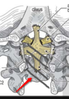
4 sutures of skull
Which suture closes first?

Metopic (in front), Lamboid (in back), Coronal, Saggital.
Metopic closes first - Male pattern baldness happens first

Which suture is most likely to close abnormally? What are french names for this? (2 names

Saggital (boat shaped head). Scaphocephaly, Dolichocephaly
What two canals are connected to this canal?

Pterygopalatine fossa (PTF) is the canal
Foramen rotundum (posterior), Spenopalatine foramen (medial)
Foramen Rotundum what runs in it? Relation to PTF?
Runs horizonal anteriorly to connect with the posterior aspect of PTF.
R2V2.
Foramen Ovale (relation to Spinosum)
Name these other canals

Shaped like oval on axial scan. Anteromedial.
(stilleto heel visual)

Foramen Spinosum (position, related to ovale)
posterolateral to foramen ovale
Stilleto heel visual
Relation of Vidians, ovale, and spinosum on axial. (visual to help remember Ovale and Spinosum)
From lateral to medial it goes SOV. On axial view, Ovale and Spinosum look like the imprint of a woman’s high heel shoe, which is pointed inwards. (ovale I ball of foot, spinosum is heel). The feet are pointed towards Viridans canal.
On Coronal plane, Anterior cliniod process is seen on slices which contain which foramen?

Foramen Rotundum. (and also likely Vidians canal)

Relationship of Rotundum and Vidians canal on coronal views

Rotundum is lateral, Vidians is medial. V in the middle

Juvenile angiofibroma starts where?
Sphenopalaine foramen. It then extends to Ptf.
JNA is fed by branches of which artery?
ECA
Fetal Pcomm frequencey and clinical significance
Also what is relation of PCOMM to CN3 normally, and what is in fetal pcomm?
30% prevalance. Stroke from ICA may hit both MCA and PCA distribution.
Normally fetal pcomm is medial/superior to CN3, but with Fetal Pcomm, it is lateral/superior
Most common persistent fetal connection b/w vertebrobasilar and carotid systems? Associated complication and associated sign?
Persistent trigeminal artery. This may be prone to aneurysm. Tau sign (seen as the vessel is coming off of the carotid)

Things that connect with pterygopalatine fossa
Rotundum, IOF, greater palatine canal, spenopalatine foramen, infratemporal fossa, viridans canal.
Superior ofbiral fissure contains lots of nerves (Need 3,4,6 for looking)
3,4,6, Lacrimal, frontal, nasocilliary (V1)
Cavernous sinus contains what? What are medial structure

CN2, 3, 4, 6, V1, V2. Internal carotid.
Abducens and internal carotid are medial

What passes through the optic canal?
CN2- opthalmic artery
What passes through the hypoglossal canal? Where is hypoglossal canal in relation to occipital condyle?
Hypoglossal nerve (CN12)
medial and superior to occipital condyle
What traverses the jugular foramen?
Pars vascularis?
Pars nervosa?
Pars vascularis:- Jugular vein- CNs 10 & 11 (go with the vessels)- posterior meningeal branch of ascending pharyngeal artery
Pars nervosa:- CN 9 (Nine is longer, goes alone in Nervosa)- inferior petrosal sinus venous return
What traverses the foramen spinosum?
Middle meningeal artery
What traverses the foramen rotundum?
CN V2 “R2V2”
What traverses the superior orbital fissure?
CNs V1, 3, 4, 6
What traverses the foramen ovale?
CN V3, accessory meningeal artery
Stylomastoid canal
CN 7