GI Anatomy and Physiology Flashcards
(77 cards)
1
Q
Gastrointestinal Tract Functions Overview:
A
- Breaks down ingested food and prepares food for uptake by the body’s cells
- Water absorption
- Eliminates wastes
- Controlled by hormones and the autonomic nervous system (except for chewing, swallowing, and defecation)
2
Q
Pancreatic/Small Intestine Digestive Enzymes
A

3
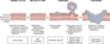
Q
Slide 5
A
Slide 5 GI PPT
4
Q
Intestinal Bacteria
A
- Stomach enviornment is relatively sterile because of secreted stomach acid
- Bile acid secretion, motility, and antibody production keep bacterial numbers in the duodenum to a minimun
- A low concentration of aerobes in the jejunum
- Anaerobic bacteria distal to the ieloceceal valve
- Anaerobes are 95% of the fecal flora in the colon
- Bacteroids, clostridia, anaerobic lactobacilli and coliforms are the most common microogranism found from the ileum to the cecum
- Intestinal tract is sterile at birth and is colonized within a few hours
- Normal flora are non-pathogenic and these microorganisms are immune tolerant
5
Q
GI Disorders
A
- Rank 3rd in total economic burden
- 60-70million Americans have digestive disease
- Many could be prevented or minimized by proper nutrition and changes in health practices
6
Q
Esophageal Disorders
A
- Congenital Anormalies
- Dysphagia
- Tears (Mallory Weiss Syndrome)
- Hiatal hernia
- Gastro-esophageal reflux disease (GERD)
- Barrett esophagus
- Esophageal carcinoma
7
Q
Congenital Anomalies
A
-Esophageal Atresia & Tracheoesophageal Fistula
8
Q
Dysphagia
A
- Dysphagia is diffculty in swallowing
- Mechanical or Functional
- Mechanical Obstructions
- Intrinsic
- Tumors, strictures or diverticular herniations
- Extrinsic
- Tumors
- Intrinsic
- Functional dysphagia: Neural or muscular disorders that interfere with voluntary swallowing or peristalsis
- Aschalasia
- Failure of the lower esophageal sphincter to relax due to denervation of smooth muscle in the esophagus
9
Q
Mallory-Weiss Tears
A
- Longitudinal tears in the esophagus
- Most common at the gastroesophageal junction
- Associated with severe retching or vomiting
- Usually heal on their own
10
Q
Esophageal Varices

A
- Caused by portal hypertension
- 50% of cirrhotic patients
- Hepatic schistosomiasis
- Often asymptomatic
- Rupture can lead to massive hemorrhage and death
11
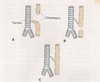
Q
Hiatal Hernia
A
- A defect in the diaphragm that allows protrusion of the upper part of the stomach through the diaphragm (hiatus) into the chest cavity
- Two basic types
- Sliding hiatal hernia (most common 95%)
- Para-esophageal hiatal hernia

12
Q
Gastroesophageal Reflux Disease (GERD), Predisoping factors, manifestations and treatment
A
- A failure of the lower esophageal sphincter (LES) to close fully
- Predisposing factos
- Transient relaxation or weakness of the LES
- An increase in abdominal pressure
- High food volume, alcohol, smoking and drugs
- Hiatal hernia
- Children under 2, especially 0-6 months
- Manafiestations
- Heartburn, regurgitation of chyme, and upper abdominal pain within 1 hour of eating
- Treatment
- Most effective treatment is proton pump inhibiotrs
- Major consequence of GERD is Esophagitis

13
Q
Barrett Esophagus
A
- Chronic irration results in the replacement of normal squamous epithelium of the lower esophagus with columnar epithelial tissue (metaplasia).
- Occurs in males more often than females (4:1)
- Barrett esophagus is associated with an increased risk of developing esophageal cancer
- These individuals must be monitored closesly

14
Q
Two types of Esophageal Carinomas & Incidences
A
- Two main types
- Adenocarcinomas
- Squamous cell carcinomas
- Incidences
- Worldwide, squamous cell carinomas accounts for about 90% of esophageal cancer
- However adenocarcinomas are more commin in US due to the association with Barrett esophagus
- US incidence is 6 per 100,000
- 1-2% of all cancer deaths
- Parts of Asia and Iran
- Pervalence is 100 per 100,000
- 20%
15
Q
Esophageal Adenocarcinomas
A
- Only recognized precursor is Barrett esophagus
- Multistep process that takes many years to develop
- Degree of dysplasia is the strongest predictor to the progression of cancer
- Mutations in p53 are common
16
Q
Esophageal Squamous Cell Carcinoma
A
- Predisposing factors
- Retarded passage of food through the esophagus (achalasia)
- Potential carcinogens (smoking and alcohol)
- Diet
- High levels of nitrites
- Fungal contamination
- Most cases are abnormalities in P16/INK4
17
Q
Diseases of the Stomach
A
- Congential abnormalities
- Gastritis
- Peptic Ulcer Disease
- Gastric Carcinomas
18
Q
Congenital Abnormalities
A
- Pyloric Stenosis
- 1-300 to 900 live births
- 4:1 (males to females)
- Muscular hypertrophy of pyloric smooth muscles
- Diaphragmatic hernia
- Herniation of stomach and other abdominal contents (intestine) into thorax through diaphragmatic defect
- Cause strangulation of intestine
19
Q
Gastritis & Potential Causes
A
- Normally gastric mucosal barrier protects the stomach from digestive enzymes
- Tight cellular junctions
- A protective mucus layer
- High prostaglandin level
- Disruption of these defense mechanism leads to inflammation of gastric mucosa
- Can be Acute or Chronic
- Potential Causes
- Aspirin ingestion (and other NSAID’s)
- Ingestion of toxic chemicals
- Excessive alcohol intake
- Stress/caffeine
- Heliobacter pylori- thrives in acid environment, disrupts mucosal barrier
20
Q
Gastritis: Manifestions
A
- Manifests itself by the possible presence of nausea, vomiting, anorexia
- Blood may occasionally appear in vomit (Hematemesis) due to gastric mucosal damage
- Usually improves after removal of causative agent(s) with in a week
- Antacids and proton pump inhibitors help
- Can progress to a chronic form
21
Q
Immune/Chronic Gastritis
A
- Least common
- Limited to the fundus of stomach
- Autoantibodies to parietal cells
- Gland destruction and mucosal atrophy
- Decrease acid and intrinsic factor production
22
Q
Non-Immune Chronic Gastritis
A
- Usually only involves the antrum
- Major cause is infection with H. pylori
- Present in 90% of individuals with chronic gastritis
- Chronic Inflammatory changes
- Mucosal atrophy and epithelial metaplasia
- More common in older individuals
- 50% of individuals over 50 are colonzied with H. pylori
23
Q
Peptic Ulcers
A
- Caused by an injury to the protective mucosal lining anywhere along the GI tract
- 98% of peptic ulcers occur in the first portion of the duodenum or the stomach (4:1)
- 3 times more common in males than females
- May lead to tissue erosion (ulceration) by stomach hydrochloric acid/pepsin into the underlying tissue layers
24
Q
Peptic Ulcers: Main predisposing factors
A
- Main predisposing factors
- chronic use of NSAIDs
- infection with Helicobacter pylori (#1)
- Other less commoon factors
- Smoking and alcohol use
- Chronic Stress?
- Zollinger Ellision Syndrome
25
*Helicobacter pylori*
* Can colonize the gastric epithelial cells
* Can survive in low pH
* Have flagella
* Secrete urease that produces ammonia
* Produce a vacuolating toxin (VacA) that promtoes bacterial survival and causes epithelial injury
* Presence of CagA gene

26
Recap/ Gastritis/Ulceration

27
Gastric Carinomas
* 90-95% of malignant tumors of the stomach are gastric carinomas
* 2 leading cause of cancer related deaths in the world
* One of the leading killers among cancers
* low 5 year survival rate of less than 20%
28
Risk Factors of Gastric Carcinomas
* Infection with *H. pylori*
* Food additives such as nitrates
* High levels can lead to the conversion of carcinogenic nitrosamines in the stomach
* High salt intact
* Enhances conversion to nitrates
* Caustic to stomach
* Low intake of fruits and vegetables
* Vit C and carotenoids thought to be protective
29
Gastric Carincoma: Pathogenesis
* Atropic gastritis and intestinal metaplasia are strongly linked to the association with gastric cancer
* Leads to insufficent acid production which allows bacteria colonization
* Bacteria convert nitrates to nitrosamines
* Cause DNA damage to mucosal epithelium
* Leads to further damage
30
Diseases/ Disorders of the Intestine
* Lactose Intolerance
* Malabsorption syndrome
* Celiac sprue
* Inflammatory bowel disease (IBD)
* Diverticulosis/diverticulitis
* Bowel Obstruction
* Appendicitis
* Diarrhea
* Irritable Bowel
* Colon cancer
31
Lactose Intolerance
* A deficiency in the intestinal mucosal enzyme lactase
* Leads to the inability to hydrolyze lactose into glucose and galatose
* Undigested and unabsorbed lactose is metablized by bacteria in the colon

32
Malabsorption Syndrome
* Syndrome implies a common constellation of symptoms arising from multiple causes
* Inadequate gastric mixing
* Insufficient digestive agents
* Improper milieu
* Damage to intestinal epithelium
* Impaired transport
33
Celiac Sprue
* Immunological sensitivity to gluten (gliaden peptide)
* Low absorptive villi (flattened villi)
* Improves when gluten is removed from diet
* Large genetic component (HLA-DQ2, HLA-DQ8)
34
Celiac Sprue: Mechanism
* Gliadin induces epithelial cells to produce IL-15
* IL-15 triggers activation and proliferation of CD8+ intraepithelial lymphocytes that express NKG2D and MIC-A receptor
* These CD8+ lymphocytes destroy enterocytes that express MIC-A
35
Inflammatory Bowel Disease (IBD)
* Crohn's disease and ulcerative colitis are chronic relapsing inflammatory disoders of unknown origin
* 1 million Americans have IBD
* Likely a combination of the following
* Inappropriate host interactions with intestional flora
* Intestinal epithelial dysfunction
* Abnormal mucosal immune response

36
IBD- Proposed Mechanism
* 1-Genetics
* More apparent in Crohn's Disease
* NOD2, ATG16L1, IRGM
* 2-Mucosal Immune Response
* Immunosuppression therapy improves symptoms
* IL-23 receptor
* 3-Epithelial barrier defects
* Dysfunction of tight junction (Crohn's)
* Allow organism to invade and active the immune response
* 4-Microbiota
* Antibodies against flagella proteins
* Probiotics help?

37
Crohn's Disease & Incidence
* Transmural inflammation of the GI tract
* Generally affects the proximal colon and the terminal ileum
* 30% are small bowel only
* 50% are small and large bowel
* 15-20% are large bowel only
* Noncontiguous "skip" lesions
* Appears to begin in the intestinal submocsa and spreads to the mucosa and serosa
* Obstruction of lymphatics within the GI tract
* Inflammatory reaction leads to deep penetrating ulcers in the intestinal wall
* Abscesses, fistulas, and microperforations
* Pain is often constant with diarrhea and malabsoprtion syndrome
* Incidence
* 1-10cases/100,000
* Males and females are equally affected
* Higher incidence in European ancestry and Ashkenazi jews
* Genetic factors
* NOD2, ATG16L1, IRGM
38
Crohn's Disease: Treatment
* Cortiosteroids, immunosuppresive drugs
* Infliximab (monoclonal antibody thta targets TNF-A) and ultimately surgery
39
Ulcerative colitis (UC) & Assess UC disease Severity
* Ulcerative colitis is an inflammatory disease of the mucosa of the colon and rectum.
* The inflammation involves the mucosal surface and not the underlying tissue layers
* UC presents with abdominal pain, diarrhea and rectal bleeding.
* Results in inflammation (neutrophilic/mono-cytic inflitration) and eventual abscess formation
* Both B and T cells seems to be involved with large amounts of IgG antibodies produced
* Tends to be more chronic relapsing than Crohns
* Increased risk of colon/rectal cancer
* Assess UC disease severity
* Examining the Number of stools per day
* Hematocrit
* Albumin level
40
Compare Cronh's Disease & Ulcerative Colitis in the following areas:
* Region,
* Distribution
* Inflammation
* Ulcers
* Malignat potential
* Reucrrence after Surgery
* Bloody Stools
* Strictures and Fistula
* Toxic Megacolon

41
Diverticulosis
* Small herniations of the mucosal of the mucosal and sub-mucosal layers of the colon wall
* Usually occurs in the large intestine due to the structural differences
* Occur as a result of increased intraluminal pressure and decrease in luminal diameter
* Age
* High density low bulk (low fiber)
* Straining during defection
* An inflamed or infected diverticulum
* This inflammation can result in severe abdominal pain, abscess and possible rupture into the peritoneal cavity causing life-threatening peritonitis
* Rx: Increase fiber (increase stool weight and decrease intraluminal pressure) and antibiotics

42
Bowel Obstruction
* Small intestine more commonly obstructed
* Causes
* Tumors
* Herniation
* Torsion (volvulus)
* Intussusception
* Adhesion
* Paralytic ileus
* Neuropathies

43
Diarrheal Disease
* Affects 500 million children throughout the world and is the leading cause of death of children under 4
* About 220,000 Americans children hospitalized for gastroenteritis
* Two-types
* Large-volume
* Small volume
44
Diarrhea: Mechanism
* Osmotic
* Non absorbale substance in the intestine draws water into the lumen by osmosis
* Secretory
* Excessive mucosal secretion or lack of Na+ reabsorption
* Motility
* Increased motility
45
Viral Enterocolitis
* Tend to affect the superificial epithelial of the small intestine leading to destruction of these cells
* Repopulation of epithelial cells result in immature enterocytes that leads to a lack of absorption of water and nutrients
* Norwalk Virus and Rotavirus are classic examples
46
Bacterial Enterocolitis
* Tend to be more severe than viral forms
* Massive fluid lsos, destruction of intestinal mucosa, sepsis and perforation
* Can be caused by several mechanism
* Ingestion of preformed toxins
* Infection by toxigenic organisms that proliferate and produce toxins
* Infection be enteroinvasive organism which invade and destroy the epithelial mucosa
47
Irritable Bowel Syndrome (IBS)
* Functional disorder with no identifiable pathology
* Fluctuations in stool frequency and consistency
* Characterized by a variable combination of chronic and recurrent intestinal symptoms
* Hallmark is abdominal pain/bloating alleviated by defecation
48
Potential Causes of IBS
1. Abnormal GI motility and secretion
2. Post infectious IBS
3. Overgrowth of Intestinal flora
4. Food allergy or food intolerance
5. Psychoscial factors
6. Hormones
It is not IBS, if fever, bloody stools, nocturnal diarrhea, or weight loss are present
49
Tumors of the SI & LI
* Colon/rectum is host to more primary neoplasms than any other organ in the body
* Colorectal carcinoma is the 2 leading cancer in the US
* Adenocarcinomas constitute the vast majority of the colorectal cancers
50
Adenomatous Polyps
* Make up about 70% of intestinal neoplasms
* A polyp is a mass that protrudes into the gut lumen
* Begin as benign neoplasms that arise from the mucosal epithelium due to alterations in the replication of the crypt epithelial cells
* Maligant risk with adenomatous polyps is correlated with the size of the polyp and the degree of dysplasia
* Familial Adenomatous Polyposis (AD)
* Mutations in the adenomatous polyposis coli (APC) gene
* Develop 500-2500 colonic adenomas
* Development of adenocarcinoma is nearly 100% by midlife unless a prophylactic colectomy is performed
51
Adenocarcinomas
* 2nd leading cause of cancer related deaths in US
* Risk Factors
* Family history
* UC
* adenomatous polyps
* perhaps diet
* Aspirin may protect
* May be asymptomatic for a long time
* Bleeding often first symptom
* Believed to be two distinct pathways involved in colorectal carcinogenesis
* 1) APC. B-catenin pathway
* 2) Mismatch Repair Pathway
52
APC/B-catenin pathway
53
Mismatch Repair Pathway

54
Modified Dukes Classification for colorectal cancers: Define
1. A
2. B1
3. B2
4. C1
5. C2
6. D
As well as survial % after treatment
1. A-Cancer limited to the mucosa or submucosa. 90% surival
2. B1- Cancer pernetrates into but not through the muscularis propria. 80% surivival
3. B2-Cancer penetrates through the muscularis. 70%
4. C1-Same, as B1, plus lymph node metastases 50%
5. C2-Same as B2, plus lymph node metastases. 50%
6. D-distant metastases are present, x\<30%
55
Gallbladder Disorders
* Cholelithiasis
* Cholecystitis
* Choledocholitiasis
* Cholangitis
56
Cholelithiasis (Ga
llstones)
* A condition in which cholesterol stones form within the gallbladder
* The formation of gallstones is the result of bile that is supersaturated with cholesterol
* Supersaturation sets the stage for cholesterol crystal formation which leads to "microstones"
* Overtime microstones aggregate and grow to form "macrostones"
* Several factors
* Enzyme defect; increases cholesterol synthesis by hepatocytes
* Decreased secretion of bile acids, which normally promote cholesterol solubility
* Decreased reabsorption of bile acids from the ileum
* Gallbladder smooth muscle hypomotility, stasis
57
Cholecystitis
* **_Cholecystitis:_** is an inflammatory disease of the gall bladder
* Typically a result of cholelithiasis and obstruction of the cystic duct
* If left untreated the gallbladder may perforate and rupture possibly resulting in empyema
58
Choledocholithiasis/Cholangitis
* Choledocholithiasis
* Blockage and inflammation of the common bile duct
* Typically due to a stone
* May result in an secondary pancreatitis
* Cholangitis
* Bacterial infection of the bile duct
59
Acute Pancreatitis
* Acute pancreatitis is an inflammatory condition of the pancreas which alters the exocrine and endocrine functions of the pancreas
* General precipiating factors
* pancreatic duct obstruction (stone, tumor)
* edema (leads to ischemic injury)
* acinar cell injury (alcohol, drugs, virus)

60
Chronic Pancreatitis
* Chronic inflammatory lesions within the pancreas along with persistent pancreatic insufficiency over long periods.
* Eventually, fibrosis adversely affects the exocrine and endocrine tissue and calcification develops
* Almost all cases of chronic pancreatits are associated with alcoholism
61
Pancreatic Cancer
* One of the most deadly malignancies with a 5 year survival rate of about 4-5%
* Age (50+), smoking, alcohol, chronic pancreatitis and diabetes mellitus
* 95% are adenocarcinomas of the ductal epithelium
62
Disorders of the Liver
* Jaundice (symptom)
* Hepatitis
* Cirrhosis
* Toxic liver disorders
* Liver tumors
63
Bilirubin Metabolism and Elimination

64
What is Jaundice?
* Jaundice (Icterus) is a yellow pigmentation of the skin caused by hyperbillirubinemia
* Can result from
* Extrahepatic obstruction to bile flow (gallstones)
* Intrahepatic obstruction (hepatitis or cirrhosis)
* Excessive production of billrubin (excessive hemolysis of RBCs)
65
Pathways of Jaundice

66
Viral Hepatits
* Hepatitis refers to inflammation of the liver parenchyma. This inflammation may be due to an infectious agent or it may be chemically induced
* There are many viruses capable of infecting the liver. The types we conser are Hepatitis A-E
67
Hepatitis A
* HAV: RNA virus
* Spread by fecal-oral route
* Drinking contaminted milk, or water, or eating shellfish from infected waters
* 2-7 week incubation period
* May be asymptomatic or mild (jaundice variable)
* Malaise, aneroxia, nasuea, fever, upper right pain are symptoms
* Clinical course usually self limiting
* Usually does not result in chronic hepatitis
* Vaccine available
68
Hepatitis B
* HBV: DNA virus
* Prevalent worldwide (5% of population)
* Spread by blood/body fluids, sex perinatal
* Incubation period: 2-6months
* May be silent, mild hepatisis or fulminant
* May procedd to chronic hepatits...cirrhosis
* Increased risk of liver cancer
* Vaccine available (Recombivax)
69
Hepatitis C
* HCV: RNA virus
* Prevalent worldwide (3% of population)
* Most common cause of chronic hepatits
* Incubation 15-150 days (Avg 50)
* Spread: same as HBV (sex, perinatal less likely)
* Acute infection usually asymptomatic
* Increased risk of liver cancer
* No vaccine available
70
Hepatitis D
* Hepatitis Delta virus-defective RNA virus
* Two forms
* Coinfection with acute Hep B
* Super-infection which Hep D is imposed on chronic Hep B
* HDV can increase severity of HBV
* Transmission is similar to HBV
71
Hepatitis E
* Uneveloped, single stranded RNA
* Transmitted by fecal oral route
* Manifestations similar to Hep A
* Does not cause Chronic Hep or carrier state
* High mortality rate-20% in preg women
* Occurs primarily in developing areas
* US cases only from those who traveled to edemic areas
72
Chronic Hepatitis
* Chronic persistent or chronic active liver inflammation of more than six months duration.
* Progressive and destructive inflammatory disease that involves necrosis of the hepatic lobule
* HBV/HCV
* Maybe autoimmune in nature
* Chief reason for liver transplants
73
Cirrhosis of the Liver
* Cirrhosis of the liver is an irreversible state that represents the end stage of a disease process (e.g. hepatitis, alcoholic liver disease). It is represented by widespread hepatic fibrosis
* This fibrosis alters liver blood flow, bile flow and hepatocellular biochemical functions (e.g. protein synthesis, urea synthesis).
* The eventual end result is liver failure
74
Pathway of Cirrhosis

75
Alcoholic Liver Disease
* Chronic abuse of alcohol commonly leads to liver disorders such as: fatty liver, hepatitis and cirrhosis.
* Fatty liver (steatosis) is an accumulation of triglycerides in the hepatocytes
* Decreased ability to metabolize them
* Defective transport mechanism.
* Results in an enlarged liver and is usually asymptomatic
76
Metal Storage Diseases
* Hemochromatosis: iron overload due to an increase in iron absorption in the small intestine.
* Iron is deposited in many tissues in excess including the liver. Inherited disorder (AR).
* Wilson’s Disease: Copper deposition in the liver as a result mutation in the (ATP7B) gene which there is decreased production of ceruloplasmin, the protein carrier of copper in the blood. (AR)
77
Cancer of the Liver
* Relatively rare – 0.5-2% of all cancers
* 5-yr survival – 1%- most die within 6 months
* Two main types
* Hepatocellular carcinoma – etiologic agents include chronic viral Hep, cirrhosis, arsenic contaminated water, chronic aflatoxin (molds) exposure
* Serum alpha-fetoprotein found in 60-70%
* Cholangiocarcinoma –
* Risk factors include chronic inflammation and injury to bile duct epithelium
