GI Imaging Pictures Flashcards
(21 cards)
What does this image show?

Displacement of stomach by hepatic mass?
What does this image show?
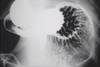
Normal dog stomach mucosa using double contrast gastrogram
How is focal dilatation of SI defined?

1.6x depth of L5 or 2x rib
What does this image show?

Dilated uterus (no foetuses will be present until day 44)
What does this image show?

General dilation
What does this image show?

Ball FB with local dilation
What does this image show?

Gravel sign of partial obstruction
What does this image show?

Linear FB - plicated SI moved to one side of the body
What does this image show?

FB consistently seen between views
What does this image show?

Mass in lumen
What does this image show?

Intussecption - long filling defect with contrast around it
What does this image show?

Duodenal mucosal changes due to pancreatitis
Does this kitten have a complete obstruction? Images taken immediately and 20 mins later.

20 mins later contrast can be seen in rectum so not an obstruction
What does this image show?

Intestine
Which is the lumen?

Which is the mucosal layer?


Which is the submucosa layer?

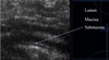
Which is the muscularis layer?


Which is the serosal layer?


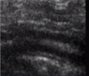
What does this image show?

Corrugated intestine shows hypermotility. Kidney can be seen below.
What does this image show?

Descending duodenum. Whiter (hyperechoic or more echogenic) line = R limb of pancreas
