Glomerular Diseases Flashcards

(88 cards)

1
Q

What are the characteristics of nephrotic syndrome?

A

Hypoalbuminemia, edema, hyperlipidemia, lipiduria, and SEVERE proteinuria (> 3.5 g/day)

2
Q

What is the most common cause of secondary nephrotic syndrome in adults?

A

Diabetic glomerulosclerosis

3
Q

What is another name for glomerulonephritis?

A

Nephritic Syndrome

4
Q

What is Nephritic Syndrome characterized by?

A

Hematuria, Proteinuria, decreased GFR

5
Q

What conditions does Nephrotic Syndrome lead to?

A

Elevated BUN, Elevated Serum Creat., oliguria, salt/water retention, hypertension, edema

6
Q

What causes decreased GFR in Nephritic Syndrome?

A

Inflmmatory damage that may impair glomerular flow and filtration

7
Q

What is the only means of definitive Dx for most glomerular diseases?

A

Renal biopsy

8
Q

Describe the differences betwixt NephrItic and NephrOtic syndrome

A

Nephritic vs Nephrotic Syndrome Table

9
Q

What findings are observed (uscope, IF, Electron) in minimal-change nephropathy?

A

No lesions, no ICs, no ICs

10
Q

What findings are observed (uscope, IF, Electron) in Focal Segmental Glomerulosclerosis?

A

Focal and Segmental Glomerular Consolidation, no ICs, no ICs

11
Q

What findings are observed (uscope, IF, Electron) in Membranous Glomerulopathy?

A

Diffuse global capillary wall thickening, diffuse capillary wall Igs, and Diffuse subepithelial dense deposits

12
Q

What findings are observed (uscope, IF, Electron) in Membranoproliferative Glomerulonephritis?

A

Capillary wall thickening and endopaillary hypercellularity, diffuse capillary wall complement and subendothelial (type I) dense deposits; intramembranous (type II) dense deposits

13
Q

What are the 3 major pathogenetic forms of glomerulonephritis?

A

In situ IC formation, Deposition of ICs, and ANCA

14
Q

What is IC formation in situ glomerulonephritis?

A

Abs bind to intrinsic antigens or foreign antigens within glomeruli

15
Q

What is circulating IC glomerulonephritis?

A

Circulating IC deposit in glomeruli and incite inflammation

16
Q

What is the mechanism of ANCA glomerulonephritis?

A

Circulating autoAbs to antigens within neutrophil cytoplasm (myeloperoxidase or proteinase-3). This activates the neutrophils causing endothelial damage especially in glomerulus

17
Q

What lab tests are needed to diagnose glomerular disease?

A

Light uscopy, electron uscopy, and Immunofluorescence

18
Q

What histologic finding is associated with a rapidly progressive course?

A

Glomerular Crescent Formation

19
Q

What is Minimal-Change Glomerulopathy characterized by?

A

Effacement of podocyte foot processes

20
Q

A biopsy of MCG is shown below. What are characteristic light microscopic findings?

A

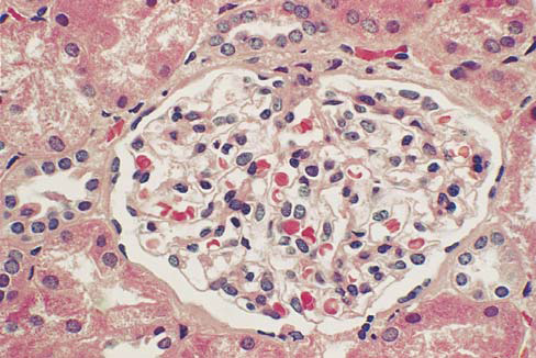
No changes are observed on light uscopy

21
Q

What does electron uscopy show in a patient with MCG?

A

Podocyte foot process obliteration

22
Q

What condition does MCG cause?

A

Nephrotic Syndrome

23
Q

A patient Dx with MCG has azotemia. What is the likely to true Dx?

A

Focal Segmental Glomerulosclerosis

24
Q

What parts of the glomeruli are affected in Focal Segmental Glomerulosclerosis?

A

(Focal) Some glomeruli and initially only affects the glomerular tuft (segmental)

25
A biopsy of a patient with Focal segmental glomerulosclerosis is shown below. What are the characteristic findings?
Periodic acid-schiff staining shows perihilar areas of segmental sclerosis and adjacent adhesions to Bowman Capsule
26
What is believed to be the general cause of FSGS?
Injury to or dysfunction of podocytes
27
What are agents of podocyte injury that can lead to FSGS?
Viruses, drugs, serum factors, congenital abnormalities
28
What are typical findings of a kidney biopsy of a patient with FSGS?
Segmental obliteration of capillary loops by increased matrix/cells, glassy hyalinosis (insudation of plasma proteins and lipids)
29
In patients with obesity or reduced renal mass what type of FSGS is observed?
Perihilar
30
In a patient with HIV, what type of FSGS is observed?
Collapsing sclerosis
31
T or F: Immune complexes are observed in FSGS
FALSE
32
What type of FSGS patient has the worst prognosis?
HIV and Idiopathic collapsing patients
33
What is HIV associated nephropathy?
Severe rapidly progressive collapsing for of focal segmental glomerulosclerosis
34
A biopsy of a patient with HIV1 associated nephropathy is shown. What are the characteristic findings of this biopsy?
Segmental or global collapsing pattern of focal sclerosis
35
What are typical causes of Membranous Glomerulopathy?
Accumulation of immune complexes in the subepithelial zone of the glomerular capillaries
36
What can membranous glomerulopathy cause?
Nephrotic Syndrome
37
In MG, where do Immune Complexes localize?
Betwixt the Podocyte and the GBM in the subepithelial zone
38
What are the general causes of primary membranous glomerulopathy?
Anti-PLA2R autoAbs, Anti-neutral endopeptidase alloantibodies
39
What are some general causes of secondary membranous glomerulopathy?
Autoimmune disease, infectious disease, drugs, cancer
40
A patient with Membranous glomerulopath is shown. What are the characteristic findings in this biopsy?
Slightly enlarged glomerulus, diffuse thickening of capillary walls, no hypercellularity
41
A biopsy of a patient with membranous glomerulopathy is shown below. A silver stain was performed. What are the lesions observed? What stage of MG is shown?
Spikes in the GBM, Stage II
42
A patient with membranous glomerulopathy is shown below. What markers are observed in IF testing?
IgG and C3
43
What is the most common primary glomerular cause of nephrotic syndrome in white and Asian adults in the US?
Membranous glomerulopathy
44
What is the most common secondary cause of nephrotic syndrome?
Diabetic glomerulosclerosis
45
A biopsy of a patient with diabetic glomeruloscerlosis is shown below. What are typical findings observed?
Prominent increases in mesangial matrix, dilation of glommerular capillaries, and thickening of capillary BM. Kimmelstiel-Wilson nodules also form
46
What is the leading cause of end-stage renal disease in the US?
Diabetic glomerulosclerosis
47
From what is AA amyloid derived? What is it associated with?
Serum amyloid A protein (SAA); Chronic inflammatory states
48
From what is AL amyloid derived? What is it associated with?
Either Lambda or Kappa Ig light chains, associated with multiple myeloma and B cell neoplasms
49
A biopsy of a patient with amyloid nephropathy is shown. What are the characteristic findings?
Amorphous acellular material epands the mesangial areas and obstructs the glomerular capillaries.
50
What type of stains identify amyloid?
Congo red stain
51
What type of disease leads to monoclonal Ig deposition disease?
B-Cell Neoplasia
52
What is the most common type of Ig that causees MIDD?
Kappa light chains
53
What is Alport Syndrome and what causes it?
Hereditary Nephritis caused by abnormal glomerular basement membrane type IV collagen
54
What type of inheritance is 85% of Alport Syndrome?
X linked and caused by a mutation in the gene for Alpha5 chain of IV collagen
55
What is used to make the Dx of Hereditary Nephritis?
Electron Microscopy
56
A biopsy of a patient with Hereditary Nephritis is shown. What are the key characteristics?
Lamina densa of the GBM is laminated rather than forming a single dense band
57
What does Thin Glomerular Basement Membrane Nephropathy lead to?
Benign Hematuria
58
What are the agents that typically cause Acute Postinfectious Glomerulonephritis?
Group A Strept (Beta Hemolytic) or Stapylococcus infection
59
What is believed to be the cause of postinfectious glomerulonephritis?
Deposition of immune complexes of Ab plus bacterial antigens in glomeruli
60
What is the general mechanism leading to postinfectious glomerulonephritis?
Immune complexes w/I glomeruli initiate inflamm response by activating complement and other humoral and cellular inflammatory mediators
61
Shown below is the biopsy of a patient with acute poststreptococcal glomerulonephritis. What are characteristic findings?
Distinctive subepithelial dense deposits shaped like humps.
62
A biopsy of a patient with type I membranoproliferative glomerulonephritis is shown below. What are characteristic findings?
Hypercellularity and capillary wall thickening, mesangialproliferation and extension into the subendothelial zone
63
What was the segmentation of the glomerulus in Type I membranoproliferative glomerulonephritis called?
Lobular glomerulonephritis
64
A renal biopsy of a patient with type II membranoproliferative glomerulonephritis is shown below. What are the typical findings?
Electron-dense deposits in the glomerular basement membrane
65
What is the complement factor that plays a role in type II MPGN?
C3 nephritic factor
66
What is the Dx? Type I or Type II MPGN?
Type II
67
What is the Dx? Type I or Type II MPGN? What describes the fluorescence pattern?
Type I; Granular to band-like staining for C3
68
Shown below is an EM of a patient with Type II MPGN. What is another name for this disease?
Dense deposit disease
69
What is class I of SLEG? Findings?
Minimal mesangial lupus glomerulonephritis; No findings on light uscopy
70
What is class II of SLEG? Findings?
Mesangial proliferative lupus glomerulonephritis; Immune complexes are confined to mesangium and cause varying degress of mesangial hypercellularity and matrix expansion
71
What is class III of SLEG? Findings?
Focal Lupus Glomerulonephritis; ICs w/I subendothelial zone, mesangium resulting in inflamm and proliferation of mesangium and endothelium
72
What is class IV of SLEG? Findings?
Diffuse lupus glomerulonephritis; Class III but involves \> 50% of glomeruli
73
What is class V of SLEG? Findings?
Membranous lupus glomerulonephritis; ICs primarily in the subepithelial zone
74
What is class VI of SLEG? Findings?
Advanced sclerosing lupus glomerulonephritis
75
Granular staining along tubular basement membranes and interstitial vessels is seen below. What is the Dx?
Diffuse proliferative lupus glomerulonephritis\
76
What type of renal conditions are caused by Berger Disease?
IgA Nephropathy due to IgA renal complexes
77
What type of IgA is most commonly deposited in Berger disease?
IgA1
78
An IF for IgA is shown below. What is the Dx?
Immunoglobulin A Nephropathy
79
What is anti-GBM glomerulonephritis mediated by?
Autoimmune response against type IV collgen in the GBM
80
What symptoms are often associated with anti-GBM Glomerulonephritis? Why?
Pulmonary hemorrhage and hemoptysis; Target antigens are also expressed in alveolar BM
81
An IF of anti-GBM glomerulonephritis is shown. What are the key characteristics?
Diffuse linear GBM immunostaining for IgG on basement membrane
82
What other condition is observed in patients with anti-GBM glomerulonephritis?
Glomerular Crescents
83
What is a subtype of anti-GBM glomerulonephritis that is rapidly progressive?
Crescentic glomerulonephritis Crescentic glomerulonephritis
84
What is ANCA glomerulonephritis characterized by?
Glomerular necrosis and crescents
85
What are ANCAs specific for in ANCA glomerulonephritis?
Myeloperoxidase and proteinase-3
86
What condition do three quarters of those with ANCA glomerulonephritis have?
Systemic small vessel vasculitis
87
What does ANCA glomerulonephritis and pulmonary vasculitis lead to?
Pulmonary-renal syndrome
88
A patient with ANCA glomerulonephritis is shown. What does the silver stain reveal?
Focal disruption of glomerular basement membranes and crescent formation within the Bowman space