GN with pictures Flashcards
(25 cards)
how does the basement membrane manage to control what is filtered?
see image
recall that flow is from the endothelium TOWARDS the podocytes

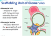
what is the role of the mesangium?
see pic
What do we look at in a renal biopsy?
see image

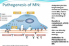
what is the pathogenesis of membranous nephropathy?
There is some sort of anti-podocyte antibody that leads to clustering of complement on the podocyte. This leads to C5b-C9 on the podocyte surface.
This leads to change in the podocyte behaviour
There is also detection of an antibody against “PHOSPHOLIPID A2 RECEPTOR”
What does the histopath slide look like in membranous nephropathy?
When you look at the histo slide you can see that the basement membrane is much thicker than adjacent tubular membrane. There is not increased cellularity. It is all about the membrane

How do we treat membranous nephropathy?
We first have to stratify the patients.
Low risk: normal renal function and < 4gm proteinuria/day
Medium risk: normal RF and 4 – 8 gm/day
High risk: abnormal RF and >8gm/day
Low risk is treated with ACEi and bring down BP to <125/75
Medium risk: trial ACEi (many will spontaneously resolve) – if no response after 6 months of therapy, probably steroids and cytotoxics
If high risk – 3 month trial with ACEi, then steroids + cytotoxics or calcineurin inhibitor
ALSO MUST PAY ATTENTION TO ADJUVANT THERAPY
That is,
Control lipids
Manage fluid balance
No smoking or NSAID
Control BP (<125/75)
ACEi
Diet
ANTICOAGULATION IF SERUM ALBUMIN < 20g/dL
What is the pathology of mesangiocapillary GN/membranoproliferative GN (MPGN)?
There are discrete deposits in the subendothelial space and in the mesangial space
There are several subtypes:
Type 1: subendothelial deposits
Type 2: dense desposits in GBM
Type 3: subepithelial and subendothelial deposits
HOWEVER, we no longer use this classification
Currently we use:
- Imune complex mediates – increased levels of immune complexes
- immunoglobulin negative, complement positive GN (this occurs through dysregulation of the alternative pathway of complement) – this can then be differentiated into dense deposit disease and C3GN
What is the pathology of rapidly progressive glomerulonephritis (RPGN)?
This condition is also called crescentic nephritis.
This can be broken up into:
type I – anti-GBM
type 2 – pauci-immune
type 3 – immune complex
Type 3 RPGN can present as:
IgA immune complex:
- IgAN
- HSP
the full house of immune complexes:
- SLE
IgG and complement associated:
- postinfectious
- cryoglobulinaemia
- endocarditis
What are some of the antibodies we use in post-infective GN?
Post streptococcal antibodies include:
- ASOT
- anti-DNAase B
- anti-hyaluronidase Ab
these can be present, but it does not predict the severity of disease
What do we use for management of post-infective GN?
There is no role for immunosuppression. It is all supportive.
What is the meaning of ANCAs?
How do they cause their pathology?
The attached slide gives the associations between these diseases and the ANCAs.
They are pauci-immune conditions

How do we manage ANCA vasculitis?
It is similar to oncology - we need to complete remission, consolidation and then remission.

What is the staging of lupus nephritis?
See the attached image. Class I and II are early days. III and IV are active mesangial disease
V is membranous
VI is burnt out

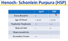
What are some features you would use to differentiate IgAN and HSP?
.
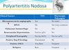
PAN v polyarteritis nodosa in renal disease?
.
What are the things you might see on a renal biopsy of someone with diabetic nephropathy?
.

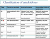
What is the stain that we use in amyloid nephropathy?
In this condition we use the Congo red stain and we can see apple-green birefringence
This is amyloid deposition. An important tid-bit of info
What is Alport syndrome?
What is thin basement membrane disease?
Alport is a genetic problem with collage and we get “splitting” of the basement membrane
in thin basement membrane disease, we often get isolated haematuria. Renal biopsy demonstrates a basement membrane that is about half the thickness of normal.
What GNs are associated with which malignancy?
probably low yield for the amount of work needed to get this info in.
MCN with Hodgkin’s
bowel/GI malig more assoc with membranous
CLL (often Hep C assoc) can cause cryoglob GN
MM and waldenstrom’s can cause systemic amyloidosis
Which of the nephrotic syndromes have the best evidence for anticoagulation when hypoalbuminaemic?
Membranous nephropathy seems to be the strongest (according to QLD lecture)
Which of the following is most strongly associated with a PERSISTENT reduction of C3?
a. mesangiocapillary (mesangioproliferative GN)
b. diffuse SLE
c. idiopathic membranous
d. post-streptococcal
e. mesangial IgA disease
The answer is A. This is associated with persistent decrease
Both diffuse SLE and post-strept will have low C3, but only when disease is active.
How does IgA nephropathy usually present?
It usually presents with recurrent episodes of isolated haematuria.
On a renal biopsy from patient with SLE, what feature would suggest poor response to therapy?
a. wire loops
b. crescents
c. advanced glomerulosclerosis
d. immunoglobulin deposits in the tubules
e. diffuse glomerular involvement
The finding of advanced glomerulosclerosis in SLE suggests burnt out lupus. That’s bad news bears for treatment.