Gynae + testicular pathology Flashcards Preview

*Medicine: Cellular Pathology > Gynae + testicular pathology > Flashcards

Flashcards in Gynae + testicular pathology Deck (52)
Loading flashcards...
1
Q

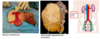
What is the epithelium type of the cervix?

A
  • endocervix lined by columnar (glandular) epithelium
  • ectocervix lined by squamous epithelium

the two types of cells meet at the squamocolumnar junction

2
Q

How does the cervix histology change during puberty and pregnancy?

A

During puberty and pregnancy, hormonally-induced eversion of cervix occurs: lower pH of vagina results in formation of a physiological transformation zone (TZ):

  • the columnar epithelium undergoes physiological metaplasia to tougher + more resistant squamous epithelium
3
Q

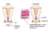
Following the physiological metaplasia, what other changes can occur in the transformation zone?

A
  • cells undergoing metaplasia are predisposed to develop dysplasia
  • so it is in transformation zone that virtually all cervical dysplasia arises
  • in the cervix, the agent inducing dysplasia is HPV
  • dysplasia occuring in the cervix is called cervical intraepithelial neoplasia (CIN) - usually asymptomatic
  • left untreated, CIN may progress to invasive squamous cell carcinoma
4
Q

How likely is CIN progression to invasive carcinoma?

A
  • ~ 11% of CIN 1 will progress to CIN 3 (other 89% stays as CIN 1 or regresses)
  • ~ 12% of CIN 3 will progress to invasive cancer (other 88% stay as CIN 3 or regresses)
5
Q

What is the major risk factor for development of CIN and cervical cancer?

A
  • persistent HPV infection
  • over 99.5% of cervical cancers associated w/ high risk of HPV infection
  • more than 130 HPV genotypes identified
  • sexually transmitted
6
Q

HPV infections of the genital tract have been subclassified into those associated w/ benign (low risk) and malignant (high risk) genital tract disease.

What are the high risk subtypes?

A
  • HPV types 16 + 18
  • associated w/ 70% of cervical cancers
7
Q

What are the low-risk HPV subtypes?

A
  • HPV types 6 + 11
  • subtypes associated w/ anogenital warts (condyloma acuminata) but not CIN and cervical cancer
8
Q

What is the pathophysiology of the HPV virus and how it infects?

A
  • HPV viral DNA integrates into host cell DNA in the cervical squamous epithelium
  • the virus preferentially infects cells in the TZ since these cells are undergoing metaplasia and altered gene expression
  • the viral E6 + E7 gene products interact with and inhibit tumour suppressor gene products: P53 and retinoblastoma protein
  • these proteins are important for cell cycle control + apoptosis
  • inactivation of these genes -> uncontrolled cell proliferation
  • HPV is a prerequisite for cervical cancer but only small proportion of HPV infections progress to either high-grade CIN or cancer
  • progression to malignancy requires one or more cofactors eg. smoking, immunosuppression
9
Q

Cervical cancer is the most common cause of cancer in women aged 18-35yrs. How does cervical cancer usually present?

A
  • vaginal bleeding
  • most commonly post-coital bleeding
  • post-coital bleeding is due to cervical cancer until proven otherwise
  • there may be an offensive vaginal discharge
  • pain is not an early feature
  • speculum exam usually reveals an ulcer or a mass on cervix
10
Q

Following examination, what investigations can be carried out for suspected cervical cancer?

A
  • biopsy -> to confirm diagnosis, give the type and grade
  • staging -> examination under anaesthesia, abdo/pelvis CT
    • staged using FIGO system

NB. a smear is a screening test for asymptomatic women and so is not appropriate in a symptomatic woman

11
Q

What is the pathology/type of most cervical cancers?

A
  • invasive squamous cell carcinomas (80%)
  • precursor lesion is CIN
12
Q

What are the remaining 20% of cervical cancers?

A
  • adenocarcinomas
  • arising from endocervical epithelium in cervix
  • high risk HPV also important in causing cervical adenocarcinoma
  • precursor lesion is CGIN (cervical glandular intraepithelial neoplasia)

CGIN beyond scope of T year syllabus

13
Q

Describe the cervical screening programme

A
  • to detect + treat premalignant lesions (ie. CIN)
  • therefore to reduce incidence + mortality of squamous cell carcinoma
  • cervical smears are basis of programme
  • women screened every 3 years from 25-49yrs and every 5yrs from 50-64yrs
  • test involves opening up vagina w/ speculum and using a brush to take sample of cells from the transformation zone
  • the brush, where cells are lodged, rinsed directly into preservative fluid
  • this is sent to cytology lab for prep as a slide
  • stained slides scrutinised for squamous epithelial cells showing dyskaryosis
14
Q

What is dyskaryosis?

A
  • refers to abnormalities of the cell nucleus
  • eg an irregular shape, an increased size
  • term that is only used in cervical smear reports
  • dyskaryosis is graded depending on severity of nuclear abnormalities:
    • low grade - mild
    • high grade - moderate or severe
15
Q

What is the difference between dyskaryosis and CIN regarding terminology?

A
  • dyskaryosis - diagnosis rendered on cervical smear (screening)
  • CIN - diagnosis only rendered on cervical biopsy (gold-standard)
16
Q

A smear showing dyskaryosis is a good predictor of the presence of CIN in the cervix. What dyskaryosis grades correlate with the level of CIN?

A
  • low grade (mild) dyskaryosis - CIN 1
  • high grade (mod) dyskaryosis - CIN 2
  • high grade (severe) dyskaryosis - CIN 3

These are all PREDICTIONS - like all screening tests, the cervical smear is not perfect + sometimes is wrong. A definite diagnosis of CIN can only be made on biopsy (or LLETZ) taken at colposcopy.

17
Q

What is meant by ‘borderline nuclear change’?

A
  • a reporting category which is best thought of as a holding category
  • used when the pathologist is uncertain whether the smear is normal or shows dyskaryosis
  • does not correspond to a particular disease process
  • borderline category is necessary bc the cervical smear is an imperfect screening test which doesn’t always give a clear result
18
Q

What are the national guidelines for management of abnormal cervical smears?

A
  • high-grade dyskaryosis (mod/severe) -> refer pt to colposcopy
  • low-grade dyskaryosis (mild) -> HPV testing using PCR performed on smear sample to see if high risk HPV subtypes (16+18) are present in sample:
    • high-risk HPV positive -> refer pt to colposcopy
    • high-risk HPV negative -> return pt to routine recall (3-5yrs)
  • borderline nuclear change managed in same way as low-grade dyskaryosis (ie HPV triage)
19
Q

Why is it reasonable to “return to routine recall” if the high-risk-HPV test is negative?

A
  • in absence of high-risk HPV, CIN is very very unlikely
  • therefore it is safe to assume the ‘mild dyskaryosis’ seen in smear is actually a false positive
  • reflects imperfections of the screening test
20
Q

What happens at colposcopy?

A
  • cervix exposed using a speculum
  • colposcopist carefully examines cervix using colposcope
  • both before and after application of acetic acid
  • certain abnormal colposcopic appearances are associated w/ CIN eg. acetowhite epithelium
  • however, CIN is a histological diagnosis so biopsy required to confirm diagnosis
21
Q

What is the management of CIN?

A
  • CIN 1 - observation + regular follow-up smears
  • CIN 2 + 3 - excision of transformation zone w/ cutting diathermy under local anaesthesia - ‘large loop exicison of transformaton zone’ (LLETZ)
    • specimen sent to lab to be carefully examined histologically by pathologist
    • pts who have had a LLETZ for CIN are offered a repeat smear + high risk-HPV testing 6 months later
22
Q

What is the endometrium composed of?

A
  • endometrial glands
  • endometrial stroma
  • in normal endometrium, the glands are widely spaced in the stroma
23
Q

What is endometrial hyperplasia?

A
  • increase in the number of endometrial glands relative to the endometrial stroma
  • glands become more closely packed
  • results in a thickening of endometrium
  • can be seen at hysteroscopy or on imaging (transvaginal ultrasound)
24
Q

How does endometrial hyperplasia usually present?

A
  • abnormal vaginal bleeding
  • intermenstrual, polymenorrhoea (unusually frequent periods)
  • or postmenopausal
25
Q

Endometrial hyerplasia is caused by high levels of unopposed oestrogen (ie. a high oestrogen:progesterone ratio).

What are causes of high levels of unopposed oestrogen?

A
  • polycystic ovaries syndrome (common cause in young women)
  • obesity (peripheral aromatisation of androgens -> oestrogens)
  • tamoxifen therapy - tamoxifen is a selective oestrogen receptor modulator (SERM) - an antagonist in the breast + used in treatment of breast carcinoma, it is an agonist in endometrium
  • anovulatory cycles in perimenopause - falling numbers of follicles in ovaries -> failure of ovulation. Anovulatory cycles do not produce a corpus luteum so there is no progesterone to stimulate a switch from proliferative to secretory phase
  • unopposed oestrogen HRT (combined oestrogen-progesterone HRT and OCP are protective)
26
Q

What are the two types of endometrial hyperplasia?

A
  • non-atypical hyperplasia (‘endometrial hyperplasia without cytological atypia’)
    • left untreated, risk of progression to cancer is very low (<2%) so this is generally not regarded as premalignant
  • atypical hyperplasia (‘endometrial hyperplasia with cytological atypia’)
    • left untreated, risk of progression to cancer is up to 50% and so this is regarded as premalignant
    • patients are usually recommended to have a hysterectomy

The terms ‘atypia’ / ‘atypical’ equate to the use of terms dysplasia or intraepithelial neoplasia at other anatomical sites ie. atypical hyperplasia is premalignant/precancer

Dysplasia in the endometrium is called atypical hyperplasia

27
Q

How common is endometrial cancer?

A
  • endometrial carcinoma = most common gynae malignancy
  • most endometrial cancers (>90%) occur in women aged >50yr
  • majority are adenocarcinomas arising from atypical hyperplasia
28
Q

Unopposed oestrogen is the major cause of endometrial hyperplasia and so this is the most important risk factor for endometrial carcinoma.

What is Lynch syndrome and its link with endometrial carcinoma?

A
  • confers an increased risk of developing a wide variety of cancers
  • in particular colorectal cancer and endometrial cancer
  • in fact, women with LS, endometrial cancer is more common than colorectal cancer + it is more often the first (sentinel) cancer they develop
29
Q

How does endometrial cacrcinoma most commonly present as?

A
  • post-menopausal bleeding
  • ‘post-menopausal bleeding is due to endometrial carcinoma until proven otherwise’
  • this symptom permits detection at an early stage + accounts for the better prognosis of endometrial cancer (compared to ovarian cancer)
30
Q

What are the gold standard investigations for post-menopausal bleeding (suspected endometrial cancer)?

A
  • hysteroscopy and endometrial biopsy
  • gynaecologist is able to visualise the endometrium + biopsy suspicious areas
  • biopsy will establish diagnosis of carcinoma + also provide grade of tumour (1, 2 or 3)
  • once cancer dx established, tumour needs to be staged - done by MRI, staged using FIGO system
31
Q

Ovarian tumours are a common cause of ovarian masses. The ovary can give rise to a bewildering array (over 50) of tumour types. How do ovarian masses come to attention?

A
  • ovarian masses generally often clinically silent
  • produce no symptoms
  • they may come to attention when v large + cause abdo distension
  • or when USS/laparoscopy/laparotomy is performed for unrelated reasons
32
Q

An ovarian mass may present acutely. What one of three things might it be associated with in a patient?

A
  • torsion of pedicle of the mass -> impairment to blood supply, initially causes ischaemia + abdominal pain. If torsion isn’t corrected, the mass will undergo infarction + patient’s clinical condition will worsen
  • rupture of a cystic mass with a release of cyst contents into the peritoneal cavity -> intense pain, this is particularly associated w/ a mature cystic teratoma
  • haemorrhage of a cystic mass into itself or into peritoneal cavity often causes pain, peritoneal cavity haemorrhage can be so severe as to cause hypovolaemic shock
33
Q

What is a mature cystic teratoma (‘dermoid cyst’)?

A
  • common benign tumours
  • typically occurring in the pre-menopausal age group
  • often asymptomatic but rupture is painful
34
Q

What is a teratoma?

A
  • germ cell tumours which form normal tissue structures
  • most teratomas contain elements dervied from all three embryonic layers (ectoderm, mesoderm and endoderm)
  • teratomas arising in the ovary are typically cystic tumours lined by skin w/ underlying sebaceous glands + hair follicles
  • the cyst becomes filled w/ thick greasy sebaceous material + hair
35
Q

What is the most common type of malignant ovarian tumour?

A
  • high grade serous carcinoma (HGSC)
  • on naked eye examination, HGSC may be purely solid but more commonly it has solid + cystic components
  • over 90% of ovarian cancers are carcinomas (ie. tumours arising from epithelial cells)
  • HGSC accounts for ~70% of all ovarian carcinomas
36
Q

What are risk factors for developing ovarian carcinomas?

A
  • number of ovulations is important: the more ovulations, the higher the risk. Therefore, pregnancy + use of OCP is protective (they reduce the number of ovulations) whilst nulliparity increases risk
  • family history - inherited gene mutations confer inc risk of developing ovarian cancer in ~20% women
    • pts w/ BRCA1 and BRCA2 germline mutations have a 60% + 25% lifetime risk respectiveley of developing HGSC
    • pts w/ Lynch syndrome have a 10% lifetime risk of developing ovarian cancer (typically endometrioid or clear cell subtypes; not HSC)
37
Q

The symptoms and signs for ovarian cancer are non-specific and often only manifest when the tumour is large.

What is the clinical presentation of ovarian cancer?

A
  • abdo distension/bloating, feeling of fullness, anorexia
  • pelvic/abdo pain, inc urgency/frequency
  • unexplained weight loss, fatigue + change in bowel habit
  • signs: pelvic/abdo mass, ascites + pleural effusion

Patients usually present at a high stage due to the diverse and non-specific symptoms + signs -> poor prognosis

Often also a delay in diagnosis bc of the above symptoms are common, whilst conversely ovarian cancer is fairly uncommon - an avg GP will see 1 woman w/ ovarian cancer over 3-5yrs. It is not uncommon for a patient w/ a subsequent diagnosis of ovarian cancer to have presented to their GP with symptoms 3+ times before diagnosis is made.

38
Q

What are the investigations for suspected ovarian cancer?

A
  • RMI (risk of malignancy) should be calculated
  • RMI = ultrasound score x menopausal score x serum CA125 level
    • ultrasound score - 1 or 3 (depending on presence of certain features on ultrasound)
    • menopausal score - 1 (pre) or 3 (post)
    • serum CA125 level - tumour marker level in iu/ml

RMI aids in differentiation of benign + malignant lesions: women w/ an RMI >250 have an approx 75% risk of having cancer + so should be referred to the gynaecology oncology service. If index of suspicion is high, then CT/MRI can be used to examine the pelvis + look for peritoneal spread, ascites, liver mets etc.

Most women w/ ovarian cancer will require surgery to establish dx, formally stage the extent of disease (FIGO) and to de-bulk tumour

39
Q

How common is testicular cancer?

A
  • uncommon (not in top 20 most common cancers in men overall)
  • however, in male 15-49yo age group, it is the most common type of cancer
  • it is also highly responsive to treatment
  • therefore, important to recognise + diagnose
40
Q

How do testicular tumours usually present?

A
  • as painless lump
  • NICE recommend any patient w/ swelling or mass in body of testis should be referred urgently (2 week suspected cancer referral pathway) to a urologist for investigation
41
Q

What is the work-up by the urologist for suspected testicular cancer?

A
  • ultrasound of testicles
  • serum tumour markers
42
Q

What are the majority of testicular cancers?

A
  • >90% germ cell tumours (GCTs)
  • germ cell tumours are so called bc they are derived from germ cells of testis which normally develop into mature spermatozoa
43
Q

There are many types of germ cell tumours, they are divided into two groups. What are these two groups?

A

Seminoma and non-seminomatous GCTs

For T year, we only expect you to know about seminoma. You are not expected to know about the subtypes of NSGCTs but you should be aware of the existence of the NSGCT group of tumours.

44
Q

Why is the classification of germ cell tumours useful?

A
  • prognosis - NSGCTs generally behave more aggressively than seminoma
  • management - bc of their aggressiveness, NSGCTs often require chemotherapy in addition to radical orchidectomy. Seminoma is usually managed w/ radical orchidectomy alone as is less likely to require chemotherapy
45
Q

What is meant by ‘pure seminoma’?

A
  • pure seminoma = seminoma without other germ cell components is classified as seminoma
  • however, a tumour containing seminoma mixed with one or more germ cell components listed in the table (prev) above is classified as a ‘non-seminomatous germ cell tumour’. At first glance, this may seem confusing but it makes sense bc the non-seminomatous component behaves more aggressively + so its presence trumps the seminomatous component for the purposes of prognosis/management + so determines the classification
46
Q

Seminoma has a peak incidence in the 30-40yr age group. Where does it arise?

A
  • type of germ cell tumour
  • arises in the seminiferous tubules of testis
  • remember that the seminiferous tubules are site of spermatogenesis:
    • germ cell -> spermatogonia -> spermatocyte -> spermatid -> spermatozoa
47
Q

What is the primary treatment for seminoma?

A
  • radical orchidectomy
  • seminoma usually spreads via lymphatics to para-aortic retroperitoneal lymph nodes; chemo to treat involved retroperitoneal nodes may be employed (if required)
48
Q

Measuring certain tumour markers is part of the work up of testicular masses. They ar euseful for initial assessment, monitoring response to therapy and in the long-term follow-up for recurrence.

What does alpha-fetoprotein (AFP) indicate?

A
  • marked elevation of serum AFP is typical of yolk sac tumours
  • not elevated in pure seminoma
49
Q

What does human chorionic gonadotrophin (HCG) indicate?

A
  • HCG normally synthesised by syncitiotrophoblast cells of placenta
  • choriocarcinomas often produce high levels of HCG
  • not usually elevated in pure seminomas (but may be)
50
Q

What does high levels of lactate dehydrogenase (LDH) indicate?

A
  • degree of LDH elevation corresponds well w/ bulk of tumour
  • therefore useful in assessing tumour burden
51
Q

What comparisons can be made between seminomas and non-seminomatous germ cell tumours?

A
  • testicular germ cell tumours are uniquely chemo- and radiosensitive
  • consequently, cure rates for these tumours are more than 80% even in those w/ metastases
  • however, treatment can be less intensive when disease is diagnosed at an earlier stage
52
Q

Compare and contrast the features of the most common ovarian and testicular tumours

A