Hand and Wrist (Session 9) Flashcards

(60 cards)

1
Q

What percentage of carpal bone fractures do scaphoid fractures account for?

A

70-80%

(10% of all hand fractures)

2
Q

What is the most common mechanism for a scaphoid fracture?

A

FOOSH (young adults)

=Hyperextension and impaction of scaphoid against rim of radius

3
Q

Where do patients usually complain of pain if they have a scaphoid fracture?

A

Anatomical snuffbox

  • Pain=exacerbated by moving wrist*
  • Swelling around radial and posterior aspects of wrist*
4
Q

Where and how commonly do fractures occur in the scaphoid (%)?

A
  1. Waist: 70-80%
  2. Proximal pole: 20%
  3. Distal pole: 10%
5
Q

Why are follow-up x-rays sometimes required for a scaphoid fracture? (10-14days after)

A
  • May not show up initially
  • Fracture line may be more visible after some bone reabsorption

(In the meantime- patient should be treated as if they have a fracture if it is suspected)

6
Q

If a suspected scaphoid fracture still doesn’t show up on an x-ray after 10-14 days and the patient is still symptomatic what should be done?

A

CT/MRI

7
Q

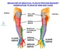
Describe the blood supply to the scaphoid.

A
  • Mainly retrograde (from distal to proximal pole)
  • Blood supply to proximal pole=tenuous
8
Q

What type of scaphoid fracture can result in avascular necrosis?

A

Waist of scaphoid

9
Q

What complications can arise from a fracture in the waist of the scaphoid?

A
  1. Non-union (8-10%)
  2. Malunion
  3. Avascualr necrosis
  4. Carpal instability
  5. Secondary osteoarthritis (non-union, malunion, avascular necrosis)
10
Q

What is a Colles’ fracture?

A
  • Extra-articular
  • Distal radial metaphysis
  • Dorsal angulation and impaction
11
Q

What other fracture is associated with a Colles’ fracture in 50% of cases?

A

Ulnar styloid fracture

12
Q

In which patients are Colles’ fractures common?

(colles’ fracture= most common type of wrist fracture)

A
  • Patients w./ Osteoporosis
    • Post menopausal women
13
Q

What is the usual mechanism of injury for a Colles’ fracture?

A

FOOSH

14
Q

How will a patient with a Colles’ fracture present?

A

Wrist=

  • Painful
  • Deformed
  • Swollen
15
Q

How are most Colles’ fractures treated?

A

Reduction

Immobilisation in cast

16
Q

What complications can arise following a Colles’ fracture?

A
  • Malunion (dinner fork deformity)
  • Median nerve palsy
  • Post traumatic carpal tunnel syndrome
  • Secondary osteoarthritis
  • Tear of extensor pollicis longus tendon
17
Q

What is a Smith fracture?

A
  • Distal radius
  • Palmar (volar) angulation- of distal fragment
18
Q

What % of smith fractures are extra-articular?

A

85%

19
Q

What % of fractures of the radius and ulna to smith fractures account for?

A

<3%

20
Q

WIn which patients are Smith fractures common?

A
  • Young men
  • Elderly women
21
Q

What are the 2 typical mecahnisms for a Smith fracture?

A
  1. Fall onto flexed wrist
  2. Direct blow to back of wrist
22
Q

What is the ‘garden spade’ deformity?

A
  • Malunion of Smith fracture
  • Residual volar displacement of distal radius
23
Q

What complication can follow the ‘garden spade deformity’?

A

Deformity narrows-distorts carpal tunnel

=Carpal tunnel syndrome

24
Q

What is Rheumatoid arthritis? (include the mechanism of its pathology)

A
  • Autoimmune disease
  • Autoantibodies= rheumatoid factor
  • Attack synovial membrane
  • Inflamed synovial cells- proliferate
  • Form pannus - penetrate through cartilage and adjacent bone
  • Causes erosion and deformaties
25
Name 3 joints which are commonly affected by **rheumatoid arthritis.**
1. Metacarpophalangeal joints (MCPJ) (Hands and feet) 2. Proximal interphalangeal joints (PIPJ) (Hands and feet) 3. Cervical spine
26
Rheumatoid arthritis is often described as 'symmetrical polyarthritis'. What does this mean?
1. Affects multiple joints 2. Symmetrical distribution
27
If a patient has rheumatoid arthritis, how might they present? (7)
1. Pain and swelling of joint 2. Erythema (redness) overlying joints 3. Stiffness- worst in morning/inactvity 4. Carpal tunnel syndrome (synovial swelling) 5. Fatigue/flu-like symptoms (systemic nature of disease) 6. Rheumatoid nodules - in fingers/elbows (late feature) 7. Deformities
28
What are the X-ray features of rheumatoid arthritis? (LESS)
* Loss of joint space * Erosions * Soft tissue Swelling * Soft bones (osteopenia)
29
Name 2 deformities that can be seen in patients with advanced rheumatoid arthritis?
1. Swan neck deformity 2. Boutonniere deformity
30
Describe the 'swan neck deformity'.
1. PIPJ hyperextends 2. MCPJ flexed 3. DIPJ flexed
31
Explain the 'swan neck' deformity
* PIPJ- laxed as adjacent synovitis * Imbalance between muscles acting on PIPJ * DIP- elongation/rupture at insertion of extensor digitorum
32
Describe the Boutinniere deformity.
* MCPJ= hyper extended * DIPJ= hyper extended * PIPJ= flexed
33
Explain the Boutinniere deformity
* Inflammation in PIPJ * Lengthening/rupture of extensor digitorum on dorsal surface of finger * Lateral band slips- acts on palmar surface- act as flexor rather than extensor
34
What is psoriasis?
* Skin condition * Causes: * red, flaky patches of skin- covered with silvery scales
35
Where does psoriasis characteristically occur?
* Elbows * Knees * Scalp * Lower back * *Can occur anywhere*
36
What % of the population has psoriasis?
1-2%
37
Does psoriatic arthritis usually develop in a symmetrical of assymetrical manner?
Assymetrical
38
How do patients with psoriatic arthritis usually present?
* Fusiform (sausage shaped) swelling of digits (*dactylitis)* * Joints stiffen Can develop into **Arthritis mutilans** (widespread joint destruction
39
Rheumatoid arthritis most commonly affects MCPJs and PIPJs. Which joint does psoriatic arthritis most commonly affect?
DIPJs
40
What other symptoms do 80% of patients which are affected by psoriatic arthritis have?
* Nail lesions: * Pitting * Onycholyis (separating nail from nail bed)
41
Name 2 other conditions where Onycholyis (separating nail from nail bed) is seen.
* Hyperthyroidism * Fungal nail infection
42
Which joint in the hand is most commonly affected by osteoarthritis?
1st Carpo Metacarpal joint *(between trapezium and first metacarpal)*
43
In what population is osteoarthritis in the first carpometacarpal joint more common?
Women (1/3 of women over 40 yrs will have x-ray changes)
44
How will a patient with osteoarthritis in the first carpometacarpal joint present?
* Pain at base of thumb * Pain exacerbated by movement, relieved by rest * Stiffness increased following periods of rest * Swelling may be evident at base of thumb * *Squaring of hand* (Later) * First metacarpal subluxes- ulnar direction * Loss of normal contour
45
During which decades of life are patients likely to acquire osteoarthritis in their fingers?
5th/6th
46
What are Heberden's nodes?
* Classic sign of osteoarthritis * Typically develop-middle age * Tend to run in families * More common in women * Affect **DIPJ** * Chronic swelling of affected joint * Sudden onset of: * Pain * Swelling * Loss of manual dexterity * Cystic swelling- contains gelatinous hyaluronic acid-osteophyte left when inital pain and inflammation subsides
47
What are Bouchard's nodes?
* Classic sign of osteoarthritis * Typically develop-middle age * Tend to run in families * More common in women * Affect **PIPJs** * Chronic swelling of affected joint * Sudden onset of: * Pain * Swelling * Loss of manual dexterity * Cystic swelling- contains gelatinous hyaluronic acid-osteophyte left when inital pain and inflammation subsides
48
What is carpal tunnel syndrome?
* Compression of median nerve *as is passes through carpal tunnel* * Most common site of _nerve entrapment_
49
Name some risk factors for carpal tunnel syndrome. (5)
1. Obesity 2. Repetitive wrist work 3. Pregnancy 4. Rheumatoid arthritis 5. Hypothyroidism
50
Name some complications which may follow nerve compression in carpal tunnel syndrome. (4)
1. Ischaemia 2. Focal demyelination 3. Decrease in axonal calibre 4. Axonal loss
51
How will a patient with carpal tunnel syndrome present?
* Paraesthesia in distribution of median nerve * Symptoms=worse at night (wrist drifts into flexion- narrows carpal tunnel further) * Daily activities can aggravate parasthesia *eg driving, combing hair, holding phone* * Manual dexterity= diminished and difficulty with daily actvities eg buttoning clothes * Pain in: forearm, elbow, shoulder, neck (up to 1/3 patients)
52
Why is sensation to the palm spared in carpal tunnel syndrome?
Palmar cutaneous branch of median nerve branches proximal to carpal tunnel and passes superficial to it
53
What may happen to the thenar muscles as a result of carpal tunnel syndrome? * *Flexor pollicis brevis (superficial head)* * *Abductor pollicis brevis* * *Opponens pollicis*
* Muscle weakness * Atrophy Motor branch of **median nerve** exits distal to carpal tunnel (supplies thenar muscles)
54
If a patient has long standing carpal tunnel syndrome, will they still be able to flex and adduct their thumb?
**Yes:** ## Footnote * **_Flexor pollicis longus_** innervated by **anterior interosseous branch of median nerve** * **_Flexor pollicis brevis (deep head)_** innervated by **ulnar nerve** * **_Adductor pollicis_** innervated by ulnar nerve
55
What is another name for Ulnar nerve compression in Guyon's canal?
1. Ulnar tunnel syndrome 2. Guyon's canal syndrome 3. Handlebar palsy
56
How will a patient with 'Guyon's canal syndrome present'?
* Parasthesia -ring and little fingers * Weakness in intrinsic muscles of hand supplied by ulnar nerve * *Adductor pollicis* * *Palmar and dorsal interossei* * *Lumbricals to ring and little fingers* * *Deep head of flexor pollicis brevis*
57
What is **Dupuytren's contracture**? (Common condition)
* Localised thickening+contracture of palmar aponeurosis * Causes _flexion_ and _deformity_ 1. Thickening/nodule in palm (painless/painful) 2. Myofibroblasts in nodule contract 3. Tight bands (cords) form 4. Overlying skin tightly adherent to palmar aponeurosis-now involved 5. Fingers stuck in flexed position (fixed flexion)
58
Which digits are commonly affected by Dupuytren's contracture?
1. Ring finger 2. Little finger 3. Thumb (may be involved)
59
What population does Dupuytren's commonly occur in?
40-60 years 70% cases have family history- autosomal dominant More common in males + northern european origin
60
Name 4 examples of conditions that increase a persons risk of developing Dupuytren's contracture:
1. Type 1 diabetes 2. Smoking 3. Hypercholesterolaemia 4. Heart disease 5. HIV 6. Hypo/hyperthyroidism 7. Trauma to hand/fingers