Head and Neck Muscles Flashcards
(63 cards)
1
Q
- What is this muscle?
- What group of muscles does it belong to?
- What is its origin?
- What is its insertion?
- What is its function?
- What is its innervation?

A
- Digastric Muscle
- Suprahyoid Muscles
- Anterior belly - digastric fossa of mandible
Posterior belly - mastoid notch of temporal bone - Intermediate digastric tendon (body of hyoid bone)
- Depresses mandible
Elevates hyoid bone when swallowing and speaking - Anterior belly - nerve to mylohyoid of inferior alveolar nerve (CNV3)
Posterior belly - digastric branch of facial nerve (CNVII)
2
Q
- What is this muscle?
- What group of muscles is it part of?
- What is its origin?
- What is its insertion?
- What is its function?
- What is its innervation?

A
- Corrugator supercili muscle
- Orbital group
- Frontal bone (medial end of superciliary arch)
- Middle of the eyebrow
- Draws the eyebrows medially and inferiorly (i.e. when squinting)
- Facial nerve - temporal branch
3
Q
- What is this muscle?
- What is its origin?
- What is its insertion?
- What is its function?
- What is its innervation?

A
- Middle scalene
- Posterior tubercles of transverse processes of vertebrae C3-C7
- Superior border of rib 1 (posterior to subclavian groove)
- Bilateral - neck flexion
Bilateral - elevation of rib 1
Unilateral - neck ipsilateral flexion
Unilateral - neck contralateral rotation - Anterior rami of spinal nerves C3-C8
4
Q
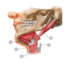
Identify the following structures in this image:

A
- Mylohyoid muscle
- Hyoglossus muscle
- Posterior belly of digastric muscle
- Stylohyoid muscle
- Anterior belly of digastric muscle
5
Q
- What is this muscle?
- What is its origin?
- What is its insertion?
- What is its function?
- What is its innervation?

A
- Auricularis anterior muscle
- Epicranial aponeurosis
- Spine of helix
- Draws auricle anteriorly
- Facial nerve - temporal branches
6
Q
Identify the following structures in this image:

A
- Sternocleidomastoid muscle
- Posterior belly of digastric muscle
- Mylohyoid muscle
- Anterior belly of digastric muscle
- Stylohyoid muscle
- Platysma muscle (partially cut)
7
Q
- What is this muscle?
- What is its origin?
- What is its insertion?
- What is its function?
- What is its innervation?

A
- Trapezius
- External occipital protuberance
Medial third of superior nuchal line (of occipital bone)
Ligamentum nuchae (nuchael ligament)
Spinous processes of C7-T12 - Superior fibres - posterior border of lateral third of clavicle
Middle fibres - medial margin of acromion and posterior border of scapula spine
Inferior fibres - converge at an aponeuosis inserted into the scapula spine - Upper fibres - scapulothoracic joint - draws scapula superiomedially
Upper fibres - atlantooccipital joint/upper cervical vertebrae - extension of head and neck and lateral flexion of head/neck
Upper fibres - atlantoaxial joint - contralateral rotation of head
Transverse/Central fibres - scapulothoracic joint - draws scapula medially
Lower fibres - scapulothoracic joint - draws scapula inferomedially - Motor - Accessory nerve (CNXI)
Sensory - Anterior rami of spinal nerves C3-C4 (via cervical plexus)
8
Q
- What is this muscle?
- What group of muscles is it part of?
- What is its origin?
- What is its insertion?
- What is its function?
- What is its innervation?

A
- Levator labii superioris alaeque nasi muscle
- Oral group
- Maxilla (near the bridge of the nose)
- Alar cartilage of the nose
Lateral upper lip (blending with orbicularis oris and levator labii superioris muscles) - Elevates the upper lip
Dilates the nostril - Facial nerve - zygomatic and buccal branches
9
Q
Identify the following structures in this image:
A
- Stylohyoid muscle
- Thyrohyoid muscle
- Sternohyoid muscle
- Omohyoid muscle (superior belly)
- Fibrous loop for intermediate digastric tendon
10
Q
- What is this muscle?
- What is its origin?
- What is its insertion?
- What is its function?
- What is its innervation?

A
- Platysma
- Skin/fascia of infra- and supraclavicular regions
- Lower border of mandible
Skin of buccal/cheek region
Lower lip
Modiolus
Orbicularis oris muscle - Depresses mandible and angle of the mouth
tenses skin of lower face and anterior neck - Cervical branch of facial nerve (CNVII)
11
Q
Identify the following structures in this image:

A
- Sternothyroid muscle
- Thyrohyoid membrane
- Hyoid bone
- Thyroid cartilage
- Cricoid cartilage
- Thyroid gland
- Trachea
12
Q
- What is this muscle?
- What group of muscles does it belong to?
- What is its origin?
- What is its insertion?
- What is its function?
- What is its innervation?

A
- Sternohyoid
- Infrahyoid muscles
- Manubrium of sternum
Medial end of clavicle - Inferior border of body of hyoid bone
- Depresses hyoid bone (from an elevated position)
- Anterior rami of spinal nerves C1-C3 (via ansa cervicalis)
13
Q
- What is this muscle?
- What group of muscles is it part of?
- What is its origin?
- What is its insertion?
- What is its function?
- What is its innervation?

A
- Masseter muscle
- Muscle of mastication
- Inferior and medial border of the zygomatic arch
Maxillary process of the zygomatic bone - Angle of mandible
Ramus of the mandible
Coronoid process - Elevates mandible
Protrudes mandible
Aids in lateral excursion of the mandible - Masseteric branch - of the anterior division of the mandibular of the trigeminal nerve (CNV3)
14
Q
- What is this muscle?
- What group of muscles is it part of?
- What is its origin?
- What is its insertion?
- What is its function?
- What is its innervation?

A
- Procerus muscle
- Nasal group
- Nasal bone (lower portion)
Lateral nasal cartilage - Skin of the forehead between the eyes
- Brings skin together creating transverse wrinkles on the bridge of the nose (e.g. when frowning)
- Facial nerve - temporal and zygomatic branches
15
Q
Identify the following structures in this image:
A
- Masseter muscle
- Parotid duct
- Buccinator muscle
- Temporalis muscle
16
Q
- What is this muscle?
- What group of muscles is it part of?
- What is its origin?
- What is its insertion?
- What is its function?
- What is its innervation?

A
- Temporalis muscle
- Muscle of mastication
- Entire temporal fossa
- Coronoid process
Anterior border of the mandibular ramus - Elevates and retracts mandible
Aids in lateral excursion of the mandible - Anterior and posterior deep temporal branches
(branch of anterior division of mandibular division of the trigeminal nerve - CNV3)
17
Q
- What is this muscle?
- What group of muscles is it part of?
- What is its origin?
- What is its insertion?
- What is its function?
- What is its innervation?

A
- Orbicularis oculi muscle
- Orbital group
- 3 parts: orbital, lacrimal, palpedbral
- Orbital - frontal process of maxilla; nasal portion of frontal bone; medial palpebral ligament
- Lacrimal - lacrimal bone
- Palpebral - medial palpebral ligament
- 3 parts:
- Orbital - around the orbit
- Lacrimal - lacrimal fascia around the lacrimal canaliculi
- Palpebral - lateral palpebral raphe
- Orbital - voluntary closure of eye (e.g. squinting)
Lacrimal - pulls lacrimal pupilla and eyelids medially, aids in flow of tears
Palpebral - closure of eyelids gently (e.g. sleeping/blinking) - Facial nerve - temporal and zygomatic branches
18
Q
- What is this muscle?
- What group of muscles does it belong to?
- What is its origin?
- What is its insertion?
- What is its function?
- What is its innervation?

A
- Thyrohyoid
- Infrahyoid muscles
- Oblique line of thyroid cartilage
- Inferior border of body of hyoid bone
Greater horn of hyoid bone - Depresses hyoid bone
Elevates larynx - Anterior ramus of spinal nerve C1 (via hypoglossal nerve - CNXII)
19
Q
- What is this muscle?
- What is its origin?
- What is its insertion?
- What is its function?
- What is its innervation?

A
- Auricularis superior muscle
- Epicranial aponeurosis
- Superior surface of auricle
- Draws auricle (ear) superiorly
- Facial nerve - temporal branches
20
Q
Identify the following structures in this image:
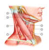
A
- Omohyoid muscle (superior and inferior belly)
- Sternohyoid muscle
- Thyrohyoid muscle
- Stylohyoid muscle
- Posterior belly of digastric muscle
- Sternocleidomastoid muscle
- Scalene muscles (anterior, middle and posterior)
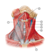
21
Q
Identify the following structures in this image:

A
- Temporalis muscle
- Masseter muscle insertion (cut away)
- Buccinator muscle
- Orbicularis oris muscle
22
Q
- What is this muscle?
- What group of muscles does it belong to?
- What is its origin?
- What is its insertion?
- What is its function?
- What is its innervation?

A
- Stylohyoid
- Suprahyoid muscles
- Styloid process of temporal bone
- Body of hyoid bone
- Elevates and draws hyoid bone posteriorly
- Stylohyoid branch of facial nerve (CNVII)
23
Q
Identify the following structures in this image:

A
- Orbicularis oculi muscle
- Nasalis muscle (transverse and alar parts)
- Buccinator muscle
24
Q
- What is this muscle?
- What group of muscles is it part of?
- What is its origin?
- What is its insertion?
- What is its function?
- What is its innervation?

A
- Levator anguli oris muscle
- Oral group
- Canine fossa of maxilla (inferior to infraorbital foramen)
- Angle of mouth
Some fibres belnd and provide origin for orbicularis oris m. - Elevates angle of the mouth - e.g. smiling
Makes nasolabial furrow more pronounced - Facial nerve - zygomatic and buccal branches
25
1. What is this muscle?
2. What is its origin?
3. What is its insertion?
4. What is its function?
5. What is its innervation?

1. Frontalis muscle (part of occipitofrontalis muscle)
2. Skin and superficial fascia along eyebrows
Adjacent facial muscles - corrugator supercili, orbicularis oculi and procerus
3. Epicranial aponeurosis
4. Elevates eyebrows
Wrinkles forehead
5. Facial nerve - temporal branch
26
Identify the following structures in this image:

1. Thyrohyoid muscle
2. Common carotid artery
3. Sternohyoid muscle
4. Ansa cervicalis (C1-C3 of cervical plexus)
5. Internal jugular vein
6. External jugular vein
7. Anterior jugular vein
27
Identify the following structures in this image:

1. Digastric muscle
2. Mastoid process
3. Styloid process
4. Hyoglossus muscle
5. Mylohyoid muscle
28
1. What is this muscle?
2. What is its origin?
3. What is its insertion?
4. What is its function?
5. What is its innervation?

1. Posterior scalene
2. Posterior tubercles of transverse processes of vertebrae C5-C7
3. External surface of rib 2
4. Bilateral - neck flexion
Bilateral - elevation of rib 2
Unilateral - neck ipsilateral flexion
Unilateral - neck contralateral rotation
5. Anterior rami of spinal nerves C6-C8
29
1. What is this muscle?
2. What group of muscles is it part of?
3. What is its origin?
4. What is its insertion?
5. What is its function?
6. What is its innervation?

1. Depressor labii inferioris muscle
2. Oral group
3. Mandible (inferior to mental foramen)
4. Lower lip
Fibres blend and provide origin for orbicularis oris muscle
5. Depresses the lower lip (e.g. when pouting)
6. Facial nerve - mandibular branch
30
1. What is this muscle?
2. What is its origin?
3. What is its insertion?
4. What is its function?
5. What is its innervation?

1. Occipitalis muscle (part of occipitofrontalis muscle)
2. Superior nuchal line
Mastoid process
3. Epicranial aponeurosis
4. Wrinkles the back of the head
5. Facial nerve - posterior auricular branch
31
1. What is this muscle?
2. What is its origin?
3. What is its insertion?
4. What is its function?
5. What is its innervation?

1. Anterior scalene
2. Anterior tubercle of transverse processes of vertebrae C3-C6
3. Anterior scalene tubercle
Superior border of rib 1 (anterior to subclavian groove)
4. Bilateral - neck flexion
Bilateral - elevation of rib 1
Unilateral - neck ipsilateral flexion
Unilateral - neck contralateral rotation
5. Anterior rami of spinal nerves C4-C6
32
Identify the following structures in this image:
1. Buccinator muscle
2. Zygomaticus minor muscle
3. Zygomaticus major muscle
4. Depressor anguli oris muscle
5. Depressor labii inferioris muscle
6. Mentalis muscle
33
1. What is this muscle?
2. What group of muscles is it part of?
3. What is its origin?
4. What is its insertion?
5. What is its function?
6. What is its innervation?

1. Risorius muscle
2. Oral group
3. Fascia overlying the parotid gland
4. Angle of the mouth
5. Moves the angle of the mouth laterally (e.g. grimicing or grinning)
6. Facial nerve - buccal branch
34
1. What is this muscle?
2. What is its origin?
3. What is its insertion?
4. What is its function?
5. What is its innervation?

1. Longus Capitis
2. Anterior tubercles of transverse processes C3-C6
3. Basilar part of occipital bone
4. Bilateral contraction - head flexion
Ipsilateral contraction - head rotation
5. Anterior rami of spinal nerves C1-C3
35
1. What is this muscle?
2. What group of muscles is it part of?
3. What is its origin?
4. What is its insertion?
5. What is its function?
6. What is its innervation?

1. Medial pterygoid muscle
2. Muscle of mastication
3. Medial surface of lateral pterygoid plate
Maxillary tuberosity
Pyramidal process of the palatine bone
4. Angle of the mandible
Medial surface of ramus of mandible
5. Elevates and protrudes the mandible
Lateral excursion of mandible
6. Medial pterygoid nerve
branches of mandibular division of trigeminal nerve (CNV3)
36
1. What is this muscle?
2. What group of muscles is it part of?
3. What is its origin?
4. What is its insertion?
5. What is its function?
6. What is its innervation?

1. Nasalis muscle
2. Nasal group
3. Maxilla
4. 2 parts: compressor naris (transverse part) + dilator naris (alar part)
1. Compressor naris - compressor naris muscle of opposite side
2. Dilator naris - nasal cartilage
5. Compressor naris - compresses the nostril
Dilator naris - dilates the nostril
6. Facial nerve - buccal branch
37
1. What is this muscle?
2. What group of muscles is it part of?
3. What is its origin?
4. What is its insertion?
5. What is its function?
6. What is its innervation?

1. Levator labii superioris muscle
2. Oral group
3. Maxilla (superior to the infraorbital foramen along the inferior margin of the orbit)
4. Lateral upper lip
Some fibres blend and provide origin for orbicularis oris muscle
5. Elevates the upper lip
6. Facial nerve - zygomatic and buccal branches
38
1. What is this muscle?
2. What group of muscles does it belong to?
3. What is its origin?
4. What is its insertion?
5. What is its function?
6. What is its innervation?

1. Mylohyoid
2. Suprahyoid muscles
3. Mylohyoid line of mandible
4. Mylohyoid raphe
Body of hyoid bone
5. Form the floor of the oral cavity
Elevates hyoid bone and floor of mouth
Depresses mandible
6. Nerve to mylohyoid (of inferior alveolar nerve - CNV3)
39
Identify the following structures in this image:
1. Orbicularis oris muscle
2. Risorius muscle
3. Platysma muscle
40
1. What is this muscle?
2. What group of muscles is it part of?
3. What is its origin?
4. What is its insertion?
5. What is its function?
6. What is its innervation?

1. Zygomaticus minor muscle
2. Oral group
3. Zygomatic bone (anterior to zygomaticus major muscle)
4. Lateral upper lip (medial to zygomaticus major insertion)
5. Helps elevation of the upper lip
6. Facial nerve - zygomatic and buccal branches
41
1. What is this muscle?
2. What group of muscles does it belong to?
3. What is its origin?
4. What is its insertion?
5. What is its function?
6. What is its innervation?

1. Geniohyoid
2. Suprahyoid muscles
3. Inferior mental spine (inferior genial tubercle)
4. Body of hyoid
5. Elevates and draws hyoid bone anteriorly
6. Anterior ramus of spinal nerve C1 (via hypoglossal nerve)
42
1. What is this muscle?
2. What group of muscles is it part of?
3. What is its origin?
4. What is its insertion?
5. What is its function?
6. What is its innervation?

1. Zygomaticus major muscle
2. Oral group
3. Zygomatic bone (anterior to zygomaticotemporal suture)
4. Angle of the mouth
Some fibres blend and provide origin for orbicularis oris muscle
5. Moves the angle of the mouth superiorly and laterally (e.g. borad smile and laughing)
6. Facial nerve - zygomatic and buccal branches
43
1. What is this muscle?
2. What is its origin?
3. What is its insertion?
4. What is its function?
5. What is its innervation?
Hint - 3 parts: superior, intermediate and inferior

1. Longus Coli
2. 3 parts:
1. Superior part - anterior tubercles of transverse processes of vertebrae C3-C5
2. Intermediate part - anterior surface of vertebral bodies C5-T3
3. Inferior part - anterior surface vertebral bodies T1-T3
3. 3 parts:
1. Superior part - anterior tubercle of vertebra C1
2. Intermediate part - anterior surface of vertebral bodies C2-C4
3. Inferior part - anterior tubercles of transverse processes of vertebrae C5-C6
4. Bilateral - neck flexion
Unilateral - neck contralateral rotation
Unilateral - neck ipsilateral flexion
5. Anterior rami of spinal nerves C2-C6
44
1. What is this muscle?
2. What group of muscles is it part of?
3. What is its origin?
4. What is its insertion?
5. What is its function?
6. What is its innervation?

1. Depressor anguli oris muscle
2. Oral group
3. Mandible - the area near the external oblique line
4. Angle of the mouth
Some fibres blend and provide origin for the orbicular oris muscle
Fibres overlap those of the depressor labii inferioris muscle
5. Depresses the corners of the mouth in an inferior and lateral direction
Antagonises levator anguli oris muscle
6. Facial - buccal and mandibular branches
45
1. What is this muscle?
2. What group of muscles is it part of?
3. What is its origin?
4. What is its insertion?
5. What is its function?
6. What is its innervation?

1. Lateral pterygoid muscle
2. Muscle of mastication
3. Infratemporal crest of greater wing of sphenoid bone
Lateral surface of lateral pterygoid plate
4. Articular disc
Capsule of TMJ
Pterygoid fovea
5. Protrudes and depresses mandible
Lateral excursion of mandible
6. Lateral pterygoid nerve
branches of anterior division of mandibular division of trigeminal nerve (CNV3)
46
Identify the following structures in this image:

1. Geniohyoid muscle
2. Submandubular gland
3. Submandubular duct
4. Inferior alveolar artery and nerve (branch of CNV3)
5. Lingual nerve (branch of CNV3)
6. Sublingular gland
7. Mylohyoid muscle
47
Identify the following structures in this image:
1. Sternohyoid muscle
2. Omohyoid muscle - superior belly
3. Omohyoid muscle - inferior belly
4. Thyrohyoid muscle
48
1. What is this muscle?
2. What group of muscles does it belong to?
3. What is its origin?
4. What is its insertion?
5. What is its function?
6. What is its innervation?

1. Sternothyroid
2. Infrahyoid muscles
3. Posterior surface of manubrium of sternum
Costal cartilage of rib 1
4. Oblique line of thyroid cartilage
5. Depresses larynx
6. Anterior rami of spinal nerves C1-C3 (via ansa cervicalis)
49
1. What is this muscle?
2. What group of muscles is it part of?
3. What is its origin?
4. What is its insertion?
5. What is its function?
6. What is its innervation?

1. Mentalis muscle
2. Oral group
3. Incisve fossa of mandible
4. Skin of chin
5. Elevates and protrudes lower lip - e.g. pouting
Wrinkles skin of chin
6. Facial nerve - mandibular branch
50
1. What is this muscle?
2. What is its origin?
3. What is its insertion?
4. What is its function?
5. What is its innervation?

1. Sternocleidomastoid
2. 2 heads:
1. Sternal head - superoanterior surface of manubrium of sternum
2. Clavicular head - superior surface of medial third of clavicle
3. Lateral surface of mastoid process
Lateral half of superior nuchal line
4. Bilateral - Atlanto-occipital joint/superior cervical spine - head and neck extension
Bilateral - Inferior cervical spine - neck flexion
Bilateral - Sternoclavicular joint - elevation of clavicle and manubrium of sternum
Unilateral - Cervical spine - neck ipsilateral flexion and contralateral rotation
5. Accessory nerve (CNXI)
Anterior rami of spinal nerves C2-C3
51
1. What is this muscle?
2. What group of muscles is it part of?
3. What is its origin?
4. What is its insertion?
5. What is its function?
6. What is its innervation?

1. Oribicularis oris muscle
2. Oral group
3. Bone - anterior midline of maxilla and mandible
Muscular - angle of the mouth - fibres blend from levator anguli oris, depressor anguli oris, zygomaticus major and risorius muscles
4. Skin along the mouth
5. Closes, protrudes and purses lips
6. Facial nerve - buccal and mandibular branches
52
Identify the following structures in this image:
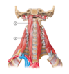
1. Longus capitis muscle
2. Longus coli muscle
53
1. What is this muscle?
2. What is its origin?
3. What is its insertion?
4. What is its function?
5. What is its innervation?

1. Auricularis posterior muscle
2. Mastoid process of temporal bone
3. Posterior part of the auricle
4. Draws auricle posteriorly
5. Posterior auricular nerve (branch of facial nerve - CNVII)
54
Identify the following structures in this image:
1. Lateral pterygoid muscle
2. Articular disc of TMJ
3. Sphenomandibular ligament
4. Medial pterygoid muscle
5. Buccinator muscle
55
1. What is this muscle?
2. What group of muscles does it belong to?
3. What is its origin?
4. What is its insertion?
5. What is its function?
6. What is its innervation?

1. Omohyoid
2. Infrahyoid muscles
3. Superior border of scapula (adjacent to suprascapular notch)
4. Inferior border of body of hyoid bone
5. Depresses and draws hyoid bone posteriorly
6. Anterior rami of spinal nerves C1-C3 (via ansa cervicalis)
56
1. What is this muscle?
2. What group of muscles is it part of?
3. What is its origin?
4. What is its insertion?
5. What is its function?
6. What is its innervation?

1. Buccinator muscle
2. Oral group
3. Pterygomandibular raphe
Alveolar margins of the maxilla and mandible
4. Some fibres belnd and provide origin for the orbicularis oris
Some fibres blend into the upper and lower lips
5. Aid mastication - keeps food bolus between cheek and teeth
Helps forcibly expel air
Helps create a sucking action
6. Facial nerve - buccal branch
57
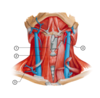
Identify the following structures in this image:
1. Platysma
2. Mylohyoid muscle
3. Thyrohyoid muscle
4. Omohyoid muscle (superior belly)
5. Sternohyoid muscle
6. Sternothyroid muscle
7. Sternocleidomastoid muscle
58
Identify the following structures in this image:

1. Frontalis muscle / frontal belly of occipitofrontalis muscle
2. Mentalis muscle
3. Drepressor labii inferioris muscle
4. Depressor anguli oris muscle
59
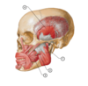
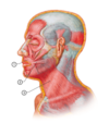
Identify the following structures in this image:

1. Occipitalis muscle (occipital belly of occipitalfrontalis muscle)
2. Auricularis posterior muscle
3. Auricularis superior muscle
4. Auricularis anterior muscle
5. Levator labii superioris alaeque nasi muscle
6. Levator labii superioris msucle
7. Zygomaticus minor muscle
8. Zygomaticus major muscle
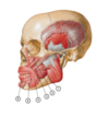
60
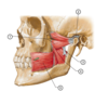
Identify the following structures in this image:
(Ignore labels 3 and 4)
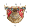
1. Medial pterygoid muscle
2. Sphenomandibular ligament
5. Lateral pterygoid muscle
6. Pterygoid hamulus
61
1. What is this muscle?
2. What is its origin?
3. What is its insertion?
4. What is its function?
5. What is its innervation?

1. Splenius capitis
2. Ligamentum nuchae and spinous processes C7-T3
3. Mastoid process of the temporal bone
Lateral third of the superior nuchal line of occipital bone
4. Bilateral - extension of head and neck
Unilateral - ipsilateral flexion of head and neck
5. Posterior rami of the middle cervical spinal nerves
(Variation C2-C3 or C3-C4)
62
1. What is this muscle?
2. What is its origin?
3. What is its insertion?
4. What is its function?
5. What is its innervation?

1. Levator scapulae
2. Transverse processes of C1-C4
3. Superior portion of the medial (vertebral) border of scapula
4. Elevates the superior angle of the scapula, drawing it medially
Rotates scapula
When the scapula is held in a fixed postion: levator scapulae bends the neck ipsilaterally and rotates the head ipsilaterally
5. C3 and C4 via cervical plexus
Branch of the dorsal scapular nerve (C5) to the lower fibres of the muscle
63
Identify the following structures in this image:

1. Levator Scapulae muscle
2. Splenius Capitis muscle
