Head and Neck Week 2 Flashcards
(11 cards)
Subcutaneous lymph in the head will drain into what collection of lymph nodes?
What are the names of these nodes and where do they drain into?
Superficial Ring (“Collar Chain”) Nodes
Submental, Submandibular, Parotid, Mastoid, Occipital
They converge on the jugular nodes (deep cervical nodes when higher up) and jugular trunk (at the root of the neck)
Lecture: Lymphatics of H&N
Objective 4: Identify the general route of lymph flow from any part of the head and neck to the veins of the neck, and identify lymph nodes that are directly downstream.
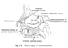
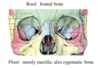
What bones make up each anatomical area of the orbit?
Lateral Wall (2 bones)
Medial Wall (3)
Roof (1)
Floor (2)
Lateral: Zygomatic, Greater wing of the sphenoid
Medial: Maxilla, Lacrimal, Ethmoid
Roof: Frontal
Floor: Maxilla, some zygomatic
Lecture: Orbit Anatomy &Development
Objective 1: Identify the seven bones forming the walls of the orbit, the foramina and fissures, and the structures that traverse them.
Name the branches of the maxillary artery (a terminal branch of the carotid artery) from proximal to distal.
Picture Courtesy of Complete Anatomy

Inferior alveolar artery
Middle meningeal artery
Deep temporal arteries
Posterior superior alveolar arteries
Infraorbital and sphenopalantine arteries
Picture courtesy of Complete Anatomy

Lecture : Infratemporal Fossa Lab tutorial
Objective 3: Identify the branches of the mandibular nerve and maxillary artery and describe their distributions and functions.
Which is the correct pairing of masticatory muscles proper and specific function during mastication?
a. Elevation: Temporalis, masseter, lateral pterygoid
b. Protraction: Masseter, medial pterygoid, lateral pterygoid
c. Retraction: Masseter
d. Lateral excursion (same side): Temporalis
d. Contralateral excursion: Masseter, medial pterygoid, lateral pterygoid
b. Protraction: Masseter, medial pterygoid, lateral pterygoid
Correct parings of muscles involved in…
- *Elevation**: Temporalis, masseter, medial pterygoid,
- *Protraction**: Masseter, medial pterygoid, lateral pterygoid
- *Retraction**: Temporalis
- *Lateral excursion**: Masseter
- *Contralateral excursion**: Medial pterygoid, lateral pterygoid
Lecture: Infratemporal Fossa
Objective 3: Describe the orientation of the muscles of mastication in the temporal and infratemporal fossae, and their role in producing mandibular movement
What are the two parasympathetic neuron pathways emerging from CN VII to their associated peripheral ganglion and target organs?
CN VII (Facial nerve) -> Greater petrosal nerve -> Nerve of pterygoid canal -> Pterygopalatine ganglion -> Maxillary nerve (V2) branches -> Nasal, palatal mucous glands, Lacrimal gland
CN VII (Facial nerve) -> Chorda tympani -> Lingual nerve (V3 branch) -> Submandibular ganglion -> Lingual nerve -> Submandibular and sublingual glands
Lecture: Autonomic Innervation of the H&N
Objective 4: Describe the pathways for CN VII and IX; practice drawing these out and labeling associated components, foramina**and target organs

A pediatric patient presents with a non-tender lump located in their neck midline. Following ultrasound, the lump is surgically excised and is found to be well encapsulated with a thick, yellowish appearance.
What was the most likely diagnosis for this mass and what are its embryologic origins?
Cervical Dermoid Cyst
Results from trapped epithelial elements (ectoderm and endoderm) along embryologic lines of fusion. These are lined by epithelium and can contain skin elements like hair and sebaceous glands
Lecture: Pediatric H&N Masses
Objective 3: Understand the relationship between cervical location and diagnosis of cervical cysts and masses.
Objective 4: Be able to develop a differential diagnosis for a cervical mass based on knowledge of embryology and locoregional anatomy.
A patient has injured CN XI.
How could this nerve have been injured? (Think about its pathway!)
What are some symptoms you would expect to see in this injured patient?
Stretching with a blow to the side of the neck or traumatic lateral bending
Difficulty elevating the shoulder and wasting of the neck counter on the affected side
CN XI arises from the cervical spinal cord, enters the foramen magnum at the base of the skull, and then exits the jugular canal to supply the sternocleidomastoid and trapezius muscles
- Lecture: Overview of the Cranial Nerves, XI – XII*
- Objective 4: Describe unique anatomical aspects of the nerves that have clinical consequences.*
CN IX, the glossopharyngeal nerve has many neuron components. For each category of neuron, state the end destination.
- Branchiomotor
- Visceral sensory
- Special sensory (2)
- Presynaptic parasympathetic
Branchiomotor: Stylopharyngeus
Visceral sensory: Pharynx, posterior 1/3 of tongue, middle ear (pre-trematic tympanic nerve branch)
Special sensory: Taste to posterior 1/3 of tongue and baroreceptors in carotid sinus (at beginning of internal carotid artery)
Presynaptic parasympathetic: Parotid gland (via tympanic nerve and plexus, lesser petrosal nerve, otic ganglion, and auriculotemporal nerve)
Lecture: Overview of the Cranial Nerves, IX – X
Objective 1: Identify the neuron components of the cranial nerves.
What are the names and basic function of the following CNs?
- CN I
- CN II
- CN III
- CN IV
- CN VI
CN I : Olfactory – Smell
CN II: Optic – Vision
CN III: Oculomotor – Motor to the muscles that move the eyeball
CN IV: Trochlear – Innervates one extraocular muscle (superior oblique) which moves the pupil down and out
CN VI: Abducens – Innervates one extraocular muscle (lateral rectus) which abducts the pupil
Lecture: Overview of the Cranial Nerves, I-IV, VI
Objective**3. Describe the functions of the nerves, particularly the functions that form the basis for their clinical testing in the head and neck exam.
What is the diagnostic criteria for acute rhinosinusitis?
How about chronic rhinosinusitis?
Per the American Academy of Otolaryngology-Head and Neck Surger
Acute Rhinosinusitis
A sudden onset of two or more of the following symptoms for LESS THAN 4 weeks
- Nasal blockage/obstruction/congestion
- Nasal discharge (anterior/ posterior nasal drip
- Facial pain/pressure
- Reduction or loss of smell
Chronic Rhinosinusitis
Inflammation of the nose and the paranasal sinuses that is characterized by two or more of the symptoms listed above for 12 WEEKS OR MORE (continuously). This should be supported by some demonstrable objective disease:
- Nasal polyps
- Mucopurulent discharge primarily from middle meatus
- Edema/mucosal obstruction primarily in middle meatus
- CT changes
- Lecture: Paranasal Sinuses & Nose II*
- Objective 5: Describe the diagnostic criteria for the clinical diagnosis of acute and chronic rhinosinusitis*
What arteries are responsible for supplying blood to the nose and which carotid artery did they ultimately originate from?
Anterior and posterior ethmoid arteries: come from the ophthalmic artery, a branch of the internal carotid artery
Sphenopalatine artery: derived from the internal maxillary artery, fed by the external carotid artery
- Lecture 13b: Paranasal Sinuses & Nose I*
- Objective 1: Describe the structural anatomy of the nose and paranasal sinuses*