Health Assessment Test 3 Flashcards
(230 cards)
What are some common CC
- Appetite
- Food intolerance
- Abdominal pain
- Nausea/Vomiting
- Bowel habits/constipations/diarrhea
- Past abdominal history
- S/E of medications
When assessing a pt with abd pain remember….
- History is the most important element in developing and refining your list of diagnostic possibilities
- Localizing the pain to a quadrant helps to refine and narrow your DDX.
- Workup should be targeted based on the history and physical examination.
- Review the list of medicines the patient is taking prior to seeing the patient and validate during the interview. Remember to ask about over-the-counter and herbal medicines.
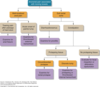
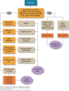
Abdominal Pain
Diagnostic Approach
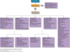
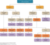
DDX abdmoninal pain

- 24-year-old African American male with no significant past medical history presents with abdominal pain 4 days in duration. Pain started as diffused and pressure sensation, most intensely in the mid abdomen
- Vital signs: 120/70, HR:98 RR: 16 T: 99.2 BMI 23.5
- What are your Differential diagnoses with the available data?
he presents today with worsening of symptoms. Pain is more localized to the Right lower quadrant and increased in intensity, he admits mild chills, nausea, vomiting and anorexia and constipation. Denies any other symptoms.
1.Refine your working diagnosis. Give me your top two?
●
2.What is your leading hypothesis at this point?
DD: Appendicitis
Sickle Cell
Gastroenteritis
Gas
Top Two: Gas, Appendicitis
Leading Hypothesis: Appendicitis
Evaluation of Abdominal Pain in Special Populations

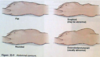
Inspection of the abdomen different shapes
Flat, Rounded, Scaphoid, Protuberant
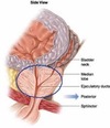
Two ulcers associated with Peptic Ulcer disease
Duodenal Ulcer
Gastric Ulcer
•Nonspecific dyspeptic symptoms: indigestion, nausea, vomiting, loss of appetite, heartburn, and epigastric fullness
Duodenal Ulcer
Midepigastric pain
Gnawing or burning, non-radiating, recurring pain that is often is episodic and relieved by food or antacids because the sphincter closes when food is in stomach stopping the acid from regurgitating
Gastric Ulcer
- Midepigastric pain
- Aggravated by food, relieved by antacids
The pancreas does what and why does pancreatitis happen
The pancreas secretes enzymes to break down protein. This enzyme is not activated until the small bowel. Pancreatitis happens when the enzyme activates prematurely inside the pancreas and it begins to brealk down.
Family history associated with Abdominal Pain
- Colorectal cancer and familial colorectal cancer syndromes
- Gallbladder disease
- Kidney disease
- Malabsorption syndrome
- Hirschsprung disease, aganglionic megacolon
- Familial Mediterranean fever (periodic peritonitis)
Infants at risk for abd pain
- Gestational age and birth weight
- Passage of first meconium stool within 24 hours
- Jaundice
- Vomiting, frequency, projectile
- Diarrhea, colic, failure to gain weight, weight loss, or steatorrhea (malabsorption syndrome)
- Apparent enlargement of abdomen (with or without pain), constipation, or diarrhea
Abdominal disorder causes associated with pregnancy
- Abdominal wall muscles stretch and lose tone
- Organs are displaced and affect functions:
- Heartburn results from alkaline reflux from duodenal contents into stomach
- Gallstones may result from gallbladder changes that produce cholesterol crystals
- Urinary stasis and urgency may occur
- Constipation or flatus is more common
- Hemorrhoids may develop later in pregnancy
- Gastrointestinal concerns common
- Nausea
- Vomiting
- Constipation
- Hemorrhoids
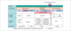
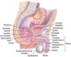
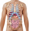
Landmarks of the abdomen
•Nine regions
- Two horizontal lines
- Across the lowest edge of the costal margin
- Across the edge of the iliac crest
Two vertical lines
- Running bilaterally from the midclavicular line to the middle of the Poupart ligament, approximating the lateral borders of the rectus abdominis muscles
Figure. Nine regions of the abdomen. 1, epigastric; 2, umbilical; 3, hypogastric; 4 and 5, right and left hypochondriac; 6 and 7, right and left lumbar; 8 and 9, right and left inguinal.
Order of exam of the abdomen
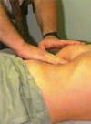
- Inspection
- Auscultation
- Percussion
- Palpation (light and deep)
Inspection of the Abdomen
•Surface characteristics
- •Skin
- •Venous return
- •Lesions and scars
- •Tautness and striae
Contour
- •Contour (abdominal profile from the rib margin to the pubis, viewed on the horizontal plane)
- •Symmetry
- Surface motion
- Inspect abdominal muscles as patient raises head to detect presence of the following:
- •Masses
- •Hernia
- •Separation of muscles
Cullen’s Sign
Ecchymosis around the umbilicus from:
Retroperitoneal hemorrhage
Acute pancreatitis
Pancreatic hemorrhage
Intraperitoneal hemorrhage
Blunt abdominal trauma
Ruptured spleen
Ruptured abdominal aortic aneurysm
Ruptured / hemorrhagic ectopic pregnancy.
Spontaneous bleeding secondary to coagulopathy
Grey Turner Sign
Ecchymosis at the flanks
Retroperitoneal hemorrhage
Acute pancreatitis
Pancreatic hemorrhage
Intraperitoneal hemorrhage
Blunt abdominal trauma
Ruptured spleen
Ruptured abdominal aortic aneurysm
Ruptured / hemorrhagic ectopic pregnancy.
Spontaneous bleeding secondary to coagulopathy
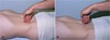
Diastasis recti (abdominal separation)
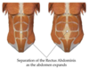
Defined as a separation of the rectus abdominis muscle into right and left halves
Newborns
- increased risk if premature
Postpartum
increased risk if women over 35
after multiple pregnancies
Auscultation of Abdomen

•Auscultate with stethoscope diaphragm for the following:
- Bowel sounds
•Auscultate with bell of stethoscope for the following:
- Bruits over aorta and renal and femoral arteries
Cannot say bowel sounds are absent unless you listen for minutes

Bowel Sounds
- Frequency
- Character
- Usually heard as clicks and gurgles that occur irregularly and range from 5 to 15 per minute
- Generalized so most often they can be assessed adequately by listening in one place
- Loud prolonged gurgles are called borborygmi (stomach growling)
When would you hear increased bowel sounds?
Increased bowel sounds may occur with gastroenteritis, early intestinal obstruction, or hunger
High pitched tinkling sounds suggest:
Intestinal fluid and air under pressure, as in early obstruction
Decreased bowel sounds occur with:
Peritonitis and paralytic ileus
Friction rubs:
- High-pitched sounds that are heard in association with respiration
- Use the diaphragm of the stethoscope
- Rare in the abdomen
- Indicate inflammation of the peritoneal surface of the organ from tumor, infection, or infarct
- Liver and spleen