Heart Sounds Flashcards
(44 cards)
Four Heart Sounds
- hese sounds originate due to the rapid acceleration and deceleration of blood giving rise to vibrations of an audible frequency in the heart and neighbouring structures associated with certain event in the cardiac cycle.
- The sounds are numbered from the onset of systole and are referred to as S1, S2, S3, and S4
S1
- corresponds to the onset of systole and is heard at the time of AV valve closure and the onset of ventricular contraction
S2
- corresponds to the end of systole and is heard at the time of outflow valve closure
S3
corresponds to the rapid passive filling phase of the ventricles
S4
corresponds to the phase of active ventricular filling.
Sounds in Horses v. Dogs/Cats
- In normal horses typically three or four of the normal heart sounds are audible at lower heart rates.
- In dogs and cats normally only the first two heart sounds S1 and S2 are audible.
- When either S3 or S4 are audible in dogs and cats a total of three sounds will be heard this is described as a gallop rhythm and is always abnormal
Heart Mumurs
- An additional noise heard in the cardiac cycle due to the turbulent flow of blood within the heart or great vessels is described as a murmur.
- Murmurs are described according to where they are most clearly heard and also when in the cardiac cycle
- Murmurs are graded in intensity according to how loud they are with relation to the other heart sounds.
Systole v. Diastole
- The period of time between S1 and S2 is systole.
- The period of time between S2and the following S1 is diastole.
Decresendo Mumur
- As is typical of diastolic murmurs this murmur diminishes in intensity during it’s course.
- This would be described as a decrescendo murmur.
Pulmonary Stenosis
- is a condition characterized by obstruction to blood flow from the right ventricle to the pulmonary artery.
- This obstruction is caused by narrowing(stenosis) at one or more points from the right ventricle to the pulmonary artery.
- Pulmonic stenosis is one of the 3 most common congenital heart diseases in dogs (the other 2 are patent ductus arteriosus and aortic stenosis)
- Narrowing of the pulmonic valve results in high velocity blood flow the other side of the valve (in the main pulmonary artery), which results in blood flow turbulence and a heart murmur.
- The point of maximal intensity of the murmur is over the left heart base. The murmur intensity directly correlates with the severity of the disease, so that the more severe the obstruction, the higher the velocity of blood flow, and the louder the murmur.
- The localization and characteristics of a pulmonic stenosis murmur are very similar to that of aortic stenosis and their differentiation on clinical examination is challenging
Cardiac Rythm in Dogs
- Dogs do not normally have a regular cardiac rhythm. The rhythm is often regularly irregular
- beats come in regular cycles with slowing of the rate between. This is quite normal.
Mitral Insufficiency or Mitral Regurgitation
- a disorder of the heart in which the mitral valve does not close properly when the heart pumps out blood.
Which murmur would be most likely to be associated with a patent ductus arteriosus?
PDAs typically have continuous murmurs rather than just systolic murmurs.
What would distinguish a grade IV from a grade V murmur?
A palpable thrill on the skin surface
Holosystolic Murmur
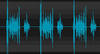
- A holosystolic murmur begins at the first heart sound (S1) and continue to the second heart sound (S2), as illustrated in the phonocardiogram.
- Typically high-pitched, these murmurs are usually caused by ventricular septal defect, mitral regurgitation or tricuspid regurgitation, as discussed below
- A heart murmur between S1 and S2 is a systolic murmur. There is turbulent blood flow during ventricular contraction, that is, the time elapsing between atrioventricular and semilunar valvular closure.
- If the murmur is louder than S1 and S2, and extends throughout the whole of systole, which defines it as loud and holosystolic, respectively. The most common cause of a loud holosystolic murmur with point of maximal intensity over the left cardiac apex is mitral regurgitation.
Differential diagnoses for mitral regurgitation
- Degenerative mitral valve disease (DMVD)
- mitral annulus dilatation secondary to dilated cardiomyopathy
- mitral valve dysplasia and endocarditis
- The first two are by far the most frequent causes of mitral regurgitation.
Diagnosing a Pulmonary Stenosis Murmur
- With pulmonic stenosis the murmur tends to radiate dorsally and the femoral pulses are generally normal, while in aortic stenosis the murmur radiates to the thoracic inlet (carotid arteries) and in severe cases the pulses are weak.
- Echocardiography is necessary to diagnose and stage congenital heart diseases, and this should ideally be performed by a specialist cardiologist.
Balloon Valvuloplasty
- A valvuloplasty, also known as balloon valvuloplasty orballoon valvotomy, is a procedure to repair a heart valve that has a narrowed opening
- A doctor uses a thin flexible tube (catheter) that is inserted through an artery in the groin or arm and threaded into the heart.
- When the tube reaches the narrowed mitral valve, a balloon device located on the tip of the catheter is quickly inflated.
- The narrowed or fused mitral valve leaflets are separated and stretched open as the balloon presses against them.
- This process increases the size of the mitral valve opening and allows more blood to flow from the left atrium into the left ventricle
A loud heart murmur with point of maximal intensity on the right hemithorax
suggestive of a ventricular septal defect or severe tricuspid regurgitation
Ventricular septal defects
(VSDs)
- are the most common congenital heart defect in cats, and the 4th most common congenital heart disease in dogs.
- Most VSDs in dogs are located in the interventricular septum just ventral to the aortic valve, and they are usually small so that relatively little blood flow crosses the defect.
- The systolic pressure in the left ventricle is normally much higher than in the right ventricle, so that a jet of high velocity blood flow crosses from the left ventricle to the right ventricle (left-to-right shunting), creating a very loud murmur heard best on the right hemithorax, cranially and close to the sternum.
- The murmur intensity is inversely related to the severity of the disease, so that the louder the murmur, the smaller the VSD.
Small VSD v. loud VSD
- With a small VSD, the prognosis is usually excellent and no medical or surgical therapy is required.
- A loud systolic heart murmur over the right hemithorax in a young animal is suggestive of a ventricular septal defect, but echocardiography performed by a cardiology specialist is always required for a definitive diagnosis.
A continuous heart murmur at the left heart base
- highly suggestive of a patent ductus arteriosus
- PDA is one of the most common congenital heart diseases in dogs. It is caused by a failure of the ductus arteriosus to close after birth, so that there is a persistent communication between the aorta and pulmonary artery.
- Because aortic pressures are higher than pulmonary artery pressures, there is a constant flow of blood from left (aorta) to right (pulmonary), and the turbulence continues throughout the cardiac cycle.
- Continued aortic blood flow into the pulmonary artery in diastole results in abnormally low diastolic aortic pressure, and a greater than normal difference between systolic and diastolic aortic pressures (hence the hyperdynamic pulses)
Patent Ductus Arteriosus
- A PDA typically causes a very loud continuous murmur with a precordial thrill* at the left heart base (in the dorsal axillary region, under the triceps muscle).
- to avoid missing important heart murmurs always palpate the thorax over the heart (the ‘precordium’) before auscultating a dog, especially a puppy. This will help you to detect any cardiac thrill and indicate the point of maximal intensity of the murmur.
- The murmur is generally louder in systole (higher pressure gradient) than in diastole, because the difference in pressures between the aorta and pulmonary artery is higher in systole.
- Although the characteristics of the murmur (timing, location), breed and age are generally pathognomonic for a PDA, specialist echocardiography should be always performed to confirm the diagnosis, evaluate severity of the disease, and exclude other concurrent congenital heart diseases.
- Treatment is directed at closing the patent ductus.
- This can be done with a catheter interventional procedure (positioning a device in the ductus to close it) or by surgical ligation
Echocardiography

- An echocardiogram, often referred to as a cardiac echo or simply an echo, is a sonogram of the heart. (It is not abbreviated as ECG, because that is an abbreviation for an electrocardiogram.)
- Echocardiography uses standard two-dimensional, three-dimensional, and Doppler ultrasound to create images of the heart