Histo: Cerebrovascular disease and Trauma Flashcards
(39 cards)
What are the two types of cerebral oedema?
- Vasogenic - due to disruption of blood-brain barrier
- Cytotoxic - secondary to cellular injury (e.g. hypoxia, ischaemia)
Result is raised ICP
Which water transporting molecule is found in the brain?
Aquaporin 4
What radiological appearance is characteristic of cerebral oedema?
Loss of gyri
Describe the passage of CSF through the brain.
- The choroid plexus (mainly found in the lateral ventricles) pumps out CSF
- It passes from the lateral ventricles, through the interventricular foramina and into the 3rd ventricle
- It then goes down the cerebral aqueduct into the 4th ventricle
- It then flows down into the medulla and down the spinal cord in the central canal of the spinal cord
- Most of the CSF will leave the 4th ventricle and enter the subarachnoid space throught the lateral and median apertures
- CSF will circulate around the subarachnoid space and will drain via arachnoid granulations into the superior sagittal sinus (and hence back into the systemic circulation)

What constitutes the floor and the roof of the 4th ventricle?
Floor = pons
Roof = cerebellum
Name and describe the two types of hydrocephalus.
Non-communicating - caused by obstruction of CSF flow (usually in the cerebral aqueduct)
Communicating - no obstruction, instead caused by increased production or reduced reabsorption of CSF into the venous sinuses (this could be caused by infection (e.g. meningitis))
What is the normal range for ICP?
7 - 15 mmHg
What are 2 common causes of raised ICP
Cerebral oedema
Space occupying lesion
Name and describe the three sites of brain herniation.
- Subfalcine - the cortex (cingulate gyrus) is pushed across the midline under the falx cerebri
- Transtentorial (uncal) - the posterior cranial fossa is covered by the tentorium cerebelli which has a rigid opening for the brainstem. Supratentorial pressure can result in herniation of the medial temporal love over the rigid end of the opening of the tentorium cerebelli
- Tonsillar - herniation of the cerebellar tonsils through the foramen magnum (this can put pressure on the medulla and cause brain death)

Define stroke.
A clinical syndrome characterised by rapidly developing clinical symptoms and/or signs of focal or global loss of cerebral function with symptoms lasting > 24 hours or leading to death with no apparent cause other than that of vascular origin
Key points
- rapid onset
- localised/ focal symptoms
- requires quick intervention
Which diseases are encompassed by the term ‘stroke’?
- Cerebral infarction
- Primary intracerebral haemorrhage
- Intraventricular haemorrhage
- Subarachnoid haemorrhage (most of the time)
Which diseases are excluded by this definition of ‘stroke’?
- Subdural and epidural haemorrhage
- Infarction or haemorrhage secondary to infection or tumour
What is a TIA?
Same definition as stroke but resolving within 24 hours (typically lasts less than 5 minutes)
Result of clot causing temporary blockage
NOTE: TIA is an important predictor of future infarct (1/3 people with TIA will have a significant infarct within 5 years)
What are the investigations for a TIA?
TIA is a clinical diagnosis that can only be made retrospectively, ie, after neurological symptoms have resolved
What is non-traumatic intraparenchymal haemorrhage?
Haemorrhage into the substance of the brain (parenchyma) due to rupture of small intraparenchymal vessels
What is a big risk factor for non-traumatic haemorrhagic stroke
Hypertension (implicated in over 50% of bleeds)
Where do non-traumatic intraparenchymal haermorrhages tend to occur most frequently?
Basal ganglia
What are some common presenting clinical features of haemorrhagic stroke
- Severe headache
- Vomiting
- Rapid loss of consciousness
- Focal neurological signs
What is an arteriovenous malformation?
- A malformation where blood bypasses quickly from artery to vein without going through a normal capillary network
- They can occur anywhere in the CNS and they can rupture
- As they occur under high pressure, they tend to cause massive bleeds

How are AVMs diagnosed
Cerebral angiography
How are arteriovenous malformations treated?
- Surgery
- Embolisation
- Radiosurgery
Define cavernous angioma.
Well-defined malformative lesion composed of closely-packed vessels with no parenchyma interposed between vascular spaces
NOTE: it is similar to an arteriovenous malformation but there is no brain substance wrapped up amongst the vessels
NOTE: these tend to bleed at lower pressure causing recurrent small bleeds
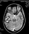
Describe the appearance of cavernous angiomas on MRI.
Shows target sign
What causes subarachnoid haemorrhages?
Rupture of a berry aneurysm (present in 1% of population)
NOTE: berry aneurysms are congenital
