Histo - GI 3 Flashcards
(38 cards)
What are the 5 characteristics of the liver?
- Largest gland in the human body
- 4 lobes
- Capsule
- thickens at porta hepatis (hilum)
- Blood supplied by portal v & hepatic a
- enter at hilum
- Bile, blood, lymph
- exit at hilum
How does liver receive blood supply?
-
Dual blood supply
- hepatic a
- oxygenated blood
- hepatic portal v
- deoxygenated blood
- hepatic a
-
Blood to hepatocytes = mixture of both blood types
- hepatic sinusoids have both
- drains to central v
What is this?

Liver (pig)
Metabolic Functions:
- synthesis of cholesterol & bile salts
- gluconeogenesis
- deamination of AA
- storage of iron, glucose, lipis, vit A
- breakdown of glycogen
- detox
- RBC removal
Exocrine Fxn:
- Bile secretion
Endocrine Fxn:
- Albumin
- Fibrinogen
- Coagulation factors
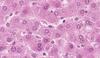
What is this?

Liver (Human)
- Hepatocytes
- polygonal cells w/ large, central nuclei (or 2)
- Hepatic lobules
- cords of liver cells (linear arrangement)
- w/ bile canaliculi & sinusoid in btw
- central v.
- reticular fibers & CT
- cords of liver cells (linear arrangement)
- Portal Triad in space of Kiernan
- (1) portal v (2) hepatic a (3) bile duct
- Sinusoids
-
Space of Disse
- btw endothelial cells & hepatocytes
- aka: perisinusoidal space
- drains 2 types of blood toward central v.
-
Space of Disse
Explain the Organization of the Liver


What is this?

Liver Structure
Blood flow in liver:
- Branches of portal v & hepatic a in Portal Triad –>
- Sinusoids –>
- mixing of oxygenated & deoxygenated blood
- Central v. –>
- Leaves liver via hepatic v.
Note:
- Blood flow is opposite bile flow

What is this?

Portal v & Hepatic a

What is this?

Space of Moll
- btw last hepatocyte & start of CT of space of kiernan
- start of lymph drainage
Note:
- lymph drains towards hilum

What is this?

Liver Microvasculature

What is this?

Liver Sinusoids

What is this?
Liver Sinusoid

What is this?
Sinusoids in the liver

What is this?

SEM of Liver Sinusoids
- You can see the large holes in the sinusoid
How does the Bile Flow in the Liver?

Bile Flow in the Liver
- Hepatocytes –>
- Bile canaliculis –>
- has tight jxns
- Canal of Hering or cholangiole
- has small, low epithelium layer (but still w/in the lobule of hepatocyte, before duct)
- Bile ductule in portal triad –>
- Bile ducts –>
- Leave liver thru R & L hepatic ducts –>
- merge w/ common hepatic duct
- Connects to cystic duct –> gallbladder OR
- Connect to common bile duct –> duodenum
Notes:
- Opposite of blood flow

What is this?

Bile Canaliculis

What is this?

Canal of Hering aka Cholangiole
- bile canaliculi are too small to see at LM

What is this?

Bile Ductule in Portal Triad

What is the relationship btw Blood & Bile flow in the Liver?
Flow in the Liver
- Blood
- from hepatic a & portal v in portal triad –>
- towards central v
- leaves liver via hepatic v
- Bile
- Hepatocytes –>
- Bile canaliculus –>
- canal of Hering (AKA cholangiole) –>
- bile ductule in portal triad –>
- bile duct
What are the 4 structures in this image?

- Portal v
- Hepatic a
- Bile duct
- Space of Kiernan

What is the structure of hepatocytes?
Hepatocyte Structure
- Lots of RER & SER
- Mitochondria w/ both:
- shelflike & tubular cristae
- Lipid droplets
- Microvilli
- Bile canaliculus sealed by tight jxns
- Space of Disse
- space between microvilli & endothelium
- Lots of peroxisomes
What are the Membrane Domains of Hepatocytes?
Btw 2 hepatocytes
- bile canaliculus
- apical domain
- tight jxns seal canaliculus
Btw hepatocyte & endothelial cells of sinusoid
- in basolateral domain
- Space of Disse
- microvilli from hepatocyte
- collagen
- Ito cells (vit A storage)
- Pit cells (NK cells)
- Processes of Kupffer cells
What is this?

Space of Disse (aka Perisinusoidal Space)
- Found in Space of Disse
- Microvilli from hepatocyte
- Collagen
- Ito cells or hepatic stellate cells
- vit A storage
- become myofibroblasts w/ liver injury
- Kupffer cells

What are the 3 Concepts of Liver Organization?
Classic lobule (Hexagon)
- Central vein as the center & portal triads at corners
- Emphasizes flow of blood (endocrine fxn)
- portal triads –> central vein
Portal lobule (Triange
- Bile duct as the center & 3 central veins
- Emphasizes flow of bile (exocrine fxn)
- hepatoctypes –> bile duct
Hepatic acinus (Diamond)
- Diamond shape w/ 2 central veins & 2 portal triads
- Emphasizes metabolic fxn
- oxygenated blood –> hepatocytes
- Zone 1 = most O2 (most toxins!)
- Zone 3 = least O2 (most damage!)

What is this?

Hepatic Acinus
Basics:
- Central axis follows a branch of hepatic a
- in portal space of Kiernan
- 3 concentric arcs = zones
Zones:
- 1 = closest to hepatic a
- most O2
- 2 = intermediate
- 3 = clostest to central v
- least O2 & least nutrients
- most glycolysis, lipid formation, drug biotransformation happens
- 1st area to…
- have fatty accumulation
- undergo ischemic necrosis (least able to fight toxins)


