Histo: Skin Pathology Flashcards
(42 cards)
How thick is a normal epidermis, dermis and subcutaneous fat put together?
6 mm
What types of fibres are found in the layer underneath the epidermis?
Collagen
Elastic fibres
What structures are found within the dermis?
- Blood vessels
- Sweat glands
- Hair follicles
- Sebaceous glands
- Nerve fibres
these are embedded in collagen matrix
How is palmar-plantar skin different from skin in other parts of the body?
There are no sebaceous glands
There is a very thick corneal layer
Describe the effects of ageing on the skin.
Skin becomes fragile with very little epidermis
Collagen and elastic fibres are of poor quality
List some different types of inflammatory reaction patterns in the skin.
- Vesiculobullous - forms bullae
- Spongiotic - becomes oedematous
- Psoriasiform - becomes thickened
- Lichenoid - forms a sheeny plaque
- Vasculitic - associated with vasculitis
- Granulomatous - associated with granulomas
first 4 occur in epidermis
What is bullous pemphigoid? Describe the macroscopic appearance.
- Vesiculobullous condition
- Occurs in elderly patients on their flexor surfaces
- Characterised by the formation of large tense bullae
NOTE: it has a 10-20% mortality

Outline the pathophysiology of bullous pemphigoid.
- Autoimmune disorder driven by IgG and C3 which attack the hemidesmosomes of the basement membrane (specifically BPAg1 and 2)
Hemidesmosomes = specialised structures in epithelial cells that anchor the cells to the underlying basement membrane
How can bullous pemphigoid be definitively diagnosed?
Skin biopsy
Immunofluoresence shows IgG and C3 deposition along the dermo-epidermal junction

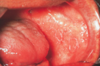
Describe the macroscopic appearance of pemphigus vulgaris.
Blisters are smaller and flaccid meaning that they rupture easily exposing a red raw surface underneath

Outline the pathophysiology of pemphigus vulgaris.
IgG-mediated autoimmune disease against desmosomes within the epidermis
(specifically desmoglein 3 and sometimes desmoglein 1)
What is acantholysis?
- Loss of intercellular connections leading to loss of cohesion between keratinocytes
NOTE: this can occur due to a lot of dermatological conditions so immunofluorescence is needed to identify where the immune-mediated attack is taking place

Describe the macroscopic appearance of pemphigus foliaceus.
- You rarely see intact bullae because they are so thin and fragile
- You are likely to see some flaky remnants of old bullae

Outline the pathophysiology of pemphigus foliaceus.
IgG-mediated attack against desmoglein 1 on the outer layer of keratinocytes
(where the stratum corneum is found)
Describe the appearance of discoid eczema.
- Very itchy
- flexural surfaces
- discoid plaques

Describe the clinical presentation of contact dermatitis.
- Itchy erythematous rash usually on the hands or feet (areas most commonly exposed to irritants)
What is hyperkeratosis? What is parakeratosis?
- In hyperkeratosis, stratum corneum of the epidermis thickens
- In parakeratosis, the superficial cells of the epidermis retain their nuclei

What type of inflammatory skin reaction is eczema?
Spongiotic because there is oedema between the keratinocytes
What are the main immune mediators in eczema?
- T cell mediated
- Eosinophils are also recruited
NOTE: this pattern is also seen in drug reactions
Describe the typical presentation of plaque psoriasis.
- This is a psoriasiform reaction pattern
- Tends to present as silvery plaques on the extensor surfaces
How is the keratinocyte turnover time different in psoriasis compared to normal skin?
- Normal skin turnover = 50 days (time for keratinocyte to go from the bottom of the epidermis to the top)
- Psoriasis = 7 days
- This leads to thickening of the epidermis and you get a layer of parakeratosis at the top
Which layer of the epidermis disappears in plaque psoriasis and why?
Statum granulosum - there is not enough time to form it
What can neutrophil recruitment to the epidermis in plaque psoriasis cause?
Formation of Munro’s microabscesses - cardinal of sign of psorasis (seen within the stratum corneum)
What is lichen planus and what are its main features?
Lichenoid reaction pattern
- T-cell mediated
- Presents with purple papules and plaques on the wrists and arms
- In the mouth it presents as white lines (Wickham striae)