Histology Flashcards

(58 cards)

1
Q

L5.1 Identify and describe the structure of lingual papillae

A

Location: anterior 2/3 of tongue

Types:

1. Filiform:“flame-like”

  • most numerous
  • highly keratinized stratified squamous
  • NO taste buds

2. Fungiform:“mushroom-like”

  • prominent on tip of tongue
  • stratified squamous
  • taste buds (pale staining) on dorsal surface

3. Foliate:

  • lateral edges of tongue
  • deep clefts
  • taste buds on lateral edges of clefts

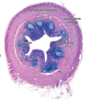
4. Circumvallate:“moat-like/dome-like”

  • invagination of epithlieum
  • Von Ebner’s glands
  • serous secretion for washing away taste to substances
  • taste buds on lateral surface of invagination
2
Q

L5.2 Identify and describe the structure and function of taste buds

A

3 cell types:

1. Neuroepithelial cells:

  • sensory cells; closely associated with nerve
  • microvilli; 1 class of receptor protein
  • 10 day turnover

2. Supporting cells:

  • microvilli
  • 10 day turnover

3. Basal cells

3
Q

L5.3 Identify and describe the distinct 4 layers characteristic of alimentary canal

A

Mucosa:

- epithelium

- lamina propria: loose CT, blood, lymph, GALT

- muscularis mucosa: usually 2 layers: inner circular and outer longitudinal; contraction - movement of mucosa

Function:

  1. protection
  2. absoprtion
  3. secretion

Submucosa:

  • dense irregular CT
  • large BV and lymphatic vessels
  • Submucosal Plexus (Meissner’s)
  • postganglionic parasympathetic neurons
  • neural crest derived

Muscularis Externa:

- Inner circular SM:

  • contracts, compresses, and mixes
  • forms sphincters

- Myenteric Plexus (Auerbach’s):b

  • between inner and outer SM
  • neural crest derived
  • post ganglionic parasympathetic neurons

- Outer Longitudinal SM:

  • contraction propels contents
  • Tenia coli in large intestines

Serosa/Adventitia:

Serosa:

  • CT lined by simple squamous
  • mesothelium: loose CT
  • continuous with mesentery and abdominal cavity

Adventitia:

  • attaches structures to abdominal wall
  • incomplete serosal covering
4
Q

L5.4 Identify and describe the structure, function, localization, and origin of Meissner’s and Auerbach’s plexi

A

Meissner’s Plexus:

  • submucosal plexus
  • postganglionic parasympathetic neurons
  • innervates muscularis mucosa
  • neural crest derived

Auerbach’s Plexus:

  • myenteric plexus; betwene inner circular and out longitudinal of muscularis externa
  • postganglionic parasympathetic neurons
  • innervates muscularis externa
  • persistaltic movement
  • neural crest derived
5
Q

L5.5 Compare serosa and adventitia

A

Serosa:

  • CT lined by simple squamous
  • mesothelium: loose CT
  • continuous with mesentery and abdominal cavity

Adventitia:

  • attaches structures to abdominal wall
  • incomplete serosal covering
  • thoracic esophagus, 2nd-4th parts of duodenum, ascending and descending colon, rectum, and anal canal
6
Q

L5.6 Identify and describe structure and function of esophagus

A

Mucosa:

  • epithlium: stratified sqamous non-keratinized
  • lamina propria: Esophageal Cardia Glands (secretes neutral mucus to protect from regurgitation)
  • muscularis mucosa: single layer of longitudinal muscle that begins at cricoid cartiliage

Submucosa:

  • Meissner’s Plexus
  • Esophageal Glands Proper (secretes slightly acidic mucous to lubricate lumen)

Muscularis Externa:

  • inner circular and outer longitudinal
  • 1st 1/3: skeletal; 2nd 1/3: mixed; 3rd 1/3: smooth
  • myenteric/Auerbach’s plexus

Adventitia: above diaphragm

Serosa: below diaphragm

7
Q

L5.7 Identify and describe structure and function of mucus glands in esophagus.

A

Esophageal Cardiac Glands: neutral mucus to protect from regurgitation

Esophageal Glands Proper: slightly acidic mucus to lubricate lumen

  • excretory duct: stratified squamous
8
Q

L5.8 Identify the structure of the muscularis externa throughout the length of the esophagus

A

1st 1/3: skeletal

2nd 1/3: mixed

3rd 1/3: smooth

9
Q

L5.9 Identify and describe the structure of the 3 regions of the stomach

A

Caridac region:

  • near esophageal orifice
  • cardiac glands

Fundic Region:

  • between cardia and pylorus
  • fundic (gastric) glands

Pyloric Region:

  • distal, funnel-shaped region, proximal to pyloric sphincter
  • pyloric glands
10
Q

L5.10 Describe the change in epithelium of the lower esophagus resulting from chronic acid reflux (Barrett’s Esophagus)

A

Barret’s Esophagus:

  • metaplastic change from stratified squamous to simple columnar with mucus cells or intestinal goblet cells
  • if not treated become dysplasia and can progress to adenocarcinoma
11
Q

L5.11 Identify and describe the structure and function of gastric mucosa

A

Mucosa:

  • gastric pits or foveolae
  • gastric glands
  • extension of muscularis mucosa
  • empties into gastric pits
  • epithelium: simple columnar with surface mucus cells: secrete viscous mucus
  • lamina propria: loose CT surrounding gastric glands
  • muscularis mucosa: inner circular and outer longitudinal
12
Q

L5.12 Compare cardia, fundic, and pyloric glands

A

Cardiac region:

  • short pits & short glands
  • tubular
  • mucus-secreting and enteroendocrine cells

Pyloric region:

  • long pits and short glands
  • branched, coiled, tubular; wide lumen
  • viscous mucus secreting and enteroendocrine cells

Fundic region:

  • short pits with surface mucus cells: thick, bicrabonate rich mucus secretions; elongated nucleus, mucinogen granules
  • long glands:
  • simple, tubular glands
  • 3 regions: isthmus, neck, fundus
  • cells:
  • mucus neck cells: neutral to alkaline soluble mucus, spherical nucleus
  • parietal cells: HCl and intrinsic factor
  • chief cells: pepsinogen –> pepsin and weak lipase
  • enteroendocrine: gastrin, CCK, secrein, VIP, GIP, motilin, somatostatin
  • stem cells
13
Q

L5.13 Identify and describe the structure and function of rugae

A
  • temporary folds of mucosa and submucosa
  • accommodate expansion and filling of stomach
14
Q

L5.14 Identify and describe the structure and function of the muscularis externa of the stomach.

A

3 layers:

1. Innermost Oblique

2. Middle Circular:

  • thickens to form pyloric sphincter

3. Outer Longitudinal

Function: mix chyme and force partial digested food into small intestines

15
Q

L6.1 Identify and desribe the structure of the gastroduodenal junction

A

Mucosa:

  • finger like shaped villi

Submucosa:

  • Brunner’s glands

Muscularis:

  • 2 layers of muscle
16
Q

L6.2 Describe the changes to the wall of the stomach in the development of ulcers

A
  • bacterial infection causes exposure of suface to effects of pepsin and acid
  • irritated and inflammed mucouse membrane become necrotic –> hole forms
  • healing occurs, but continuous irritation makes healing ineffective
  • ulcers can extend deeper, penetrating submucosa, muscularis and serosa is untreated
17
Q

L6.3 Describe the main complication of chronic peptic ulceration

A

Chronic ulcers: bleeding, perforation and peritonitis

18
Q

L6.4 Identify and describe the structure and function of the 3 anatomical regions of the small intestines.

A

1. Duodenum:

  • shortest and widest
  • submucosal glands: Brunner’s glands
  • secretes highly alkaline solution; neutralizrs acidic chyme

2. Jejunum:“Christmas tree”

  • main site of absorption
  • numerous plicae circularis
  • long, prominent villi
  • no submucosal galnds

3. Ileum:

  • submucosa/mucosa: peyer’s patches
  • lymphoid tissue, enteds deom mucosa, into submucosa, and into the lumen
19
Q

L6.5 Identify and describe the structure and function of microvilli, villi, and plicae circulares

A

Plicae Circularis: semi-circular folds

  • Valves of Kerckring
  • permanent transverse folds
  • msot numerous in distal duodenum & jejenum

Villi:

  • finger-like projections; leaf-like mucosal projections
  • central lacteals within lamina propria
  • 1st site of absorption of lipids

Microvilli:

  • feature of enterocytes
  • increase surface area
  • brush border
  • glycocalyx
  • terminal web
20
Q

L6.6 Identify and describe the structure an function of the small intestinal mucosa

A
  • simple columnar
  • GALT
  • Peyer’s patches in Ileum
  • intestinal glands: Crypts of Lieberkuhn
21
Q

L6.7 Identify and describe the structure, function, and localization of the cells of the small intestinal mucosa

A

Enterocytes:

  • simple columnar, primary function: absorptive cells
  • secretory function: digestive enzymes, water, and electrolytes
  • microvilli: contain terminal digestive enzymes
  • tight junctions: selective absorption
  • lateral plications: increase later SA

Goblet cells:

  • unicellular, mucus-secreting
  • mucinogen granules in apical cytoplasm

Panenth cells:

  • intensely acidophilic
  • lyzozymes: anti-bacterical enzyme; digests cell walls of some bacteria
  • alpha-defensin: microbicidal peptides
  • regulation of normal bacteria flora

Enteroendocrin cells:

  • secretion of hormones: CCK, secretin, GIP, and Motilin

M cells:

  • cover Peyer’s pathces and lymphatic nodules
  • modified enterocytes
  • microfolds
  • Ag-transporting cells
22
Q

L6.8 Identify and describe the structure and function of muscularis externa of the small intestines

A
  • inner circular
  • Auerbach’s plexus
  • outer longitudinal

Function: peristaltic movement

23
Q

L6.9 Identify and describe the structure and function of Peyer’s patcher of the ileum

A
  • aggregate of lymphoid tissue
  • immunological function: monitoring intestinal bacteria
24
Q

L6.10 Describe the changes in villi in mal-absorption syndrome

25
L6.11 Identify and describe the structure and function of the anatomical regions of the large intestines
Parts: **- Colon:** ascending, descending, transvers, sigmoid **- Cecum & Vermiform appendix** **- Rectum** **- Anal Canal** **Characteristics:** - Tenia coli: 3 thickened bands of outer longitudinal muscularis externa - Haustra coli: saccualtions b/w TC - Omental appendices: small fatty projections of serosa
26
L6.12 Identify and describe the structure and function of large intestinal mucosa
- "smooth" surface - **Crypts of Lieberkuhn** - Function: - reabsorption of water and electrolytes - elimination of wastes epithelium: simple columnar, no panenth, abundant goblet lamina propria: GALT muscularis mucosa: inner circular and outer longitudinal
27
L6.13 Identify and describe the structure and function of the muscularis externa and large intestine.
- inner circular - outer longitudinal - Tenia coli: prominent longitudinal bands of longitudinal muscle - Myenteric/Auerbach's plexus
28
L6.14 Identify and describe the function of the tenia coli and haustra.
tenia coli: 3 thickened bands that contract lengthwise to produce haustra haustra coli: sacculations with very slow, uncoordinated contractions that increase SA; function to retain, transport, and eliminate waste
29
L6.15 Describe the hsitological appearance of an adenomatous polyp of the large intestine
- slow growing neoplasm - dysplastic epithelium that may form glands or villous processes - 3 types: **1. Tubular:** - most common - branched tubular glands with narroe stalks **2. Tubulovillous:** - combination of tubular glands and villi **3. Villous:** - rare - broad base and papillary appearance
30
L6.16 Identify and describe the structure and function of the appendix
- thin, finger-like extensions of cecum - tenia coli ends at base - **numerous lymphatic nodules**: extends into submucosa
31
L6.17 Identify and describe the structure and function of the rectum and anal canal
**Rectum:** - distal dilated portion of alimental canal - anatomical transverse folds - Mucosa: **crypts of Lieberkuhn & abundant goblet cells** - Muscularis externa: no tenia coli - Adventitia **Anal Canal:** **- Colorectal Zone**: - upper 1/3; simple columnar **- Anal transitional zone:** - middle 1/3, transition b/w simple columnar & stratified squamous **- Squamous Zone:** - lower 1/3; stratified squamous keratinized
32
L6.18 Describe the autonomic nerve plexus in the wall of the GIT that may lead to Hischsprung disease (congenital megacolon)
- faulty migration of neural crest cells --\> lack of myenteric plexus - decreased peristaltic movements of gut - dialted colonic segment
33
L6.19 Explain the consequences of the varicosities of esophagus and the rectum
**Esophageal Varicosity:** varicose in submucosal veins in lower 1/3 of esophagus **Rectum Varicosity (Hemmorhoids):** progressive dilation and bulge of submucosal veins towards lumen - **internal**: above pectinate line - **painless** - **external:** below pectinate line - **very painful** - Tenia coli: 3 thickened bands of outer longitudinal muscularis externa - Haustra coli: saccualtions b/w TC - Omental appendices: small fatty projections of serosa
34
L6.20 Decribe the changes that occur in the wall of the gastro intestinal tract after surgical appendectomy, which may lead to adhesions
- exposure of collagen rich CT - inflammation and scarring - adhesion - obstruction - entrapment of organ - pai
35
L9.1 Identify and describe the structure, function, and localization of the liver.
**_Structure:_** - 4 lobes: right, left, caudate, and quadrate - portal hepatis: main artery (25%) and portal vein (75%) enter and hepatic ducts leave - portal canal: branches of vessels of portal hepatis found between lobules - central vein: union of sublobular veins that drains blood from the liver **_Function:_** 1. production and secretion of plasma proteins (albumins, lipoproteins, glycoproteins, prothrombin, non-immune alph and beta globulin) 2. storage and conversion of vitamins (A,D,K) and iron 3. degradation of drugs and toxins 4. homeostatic pathways
36
L9.2 Identify and describe three structures and functions of Glisson's capsule.
- fibrous CT that encloses and subdivides the liver into lobes and lobules - covered by mesothelium lining, making the liver intraperitoneal - triangular (left & right) ligament, falcilform ligament, and coronary (anterior & posterior) ligament - left triagular ligament: connects left lobe of liver to the diaphragm - right triangular ligament: smalle fold that passes to diaphragm; formed by apposition of anterior and posterior coronary ligament - falciform ligament: attaches the liver to the anterior body wall - coronary ligament: attaches the liver to the inferior surface of the diaphragm
37
L9.3 Describe the flow of blood in the liver.
**Blood supply:** - hepatic artyer: brings 25% fully oxygenated blood to liver - portal vein: brings 75% blood from the intestines, pancreas, and spleen that are rich in nutrients, endocrine secretion and blood cell breakdown products. - hepatic vein: drains most of the blood from the liver via combination of sublobular veins - sublobular veins: collect blood from central veins of liver. **Flow of blood:** portal vein --\> interlobular vein; hepatic artery joins --\> portal canal/triad --\> sinusoids --\> central vein --\> sublobular veins --\> hepatic vein
38
L9.4 Compare the structure and function of the classic lobule, portal lobule, and liver acinus
**_Classic lobule_**: (blood flow) - hexagonal shape with portal area (hepatic artery, portal vein, bile duct, and lymphatic vessel) at each point and central vein at the center. - plates of hepatocytes arranges in radial fashion, radiating from central vein - on cell thick: adults; 2 cells thick: children - sinusoidal capillaries run parallel to radial cords - receives mixed blood from vessels in portal area **_Portal Lobule_:** (exocrine function) - triangular shaped - central vein at each point and portal area at center **_Liver Acinus of Rppaport:_** (blood flow) - 2 central veins at 2 points and 2 portal triad areas at 2 points - 3 zones: 1. zone 1: first to be exposed to entering blood; first to be effected by O2 or toxins 2. sone 2: center zone 3. zone 3: last to be exposed, first to lack blood; easiest to be effected by hypoxemis --\> necrosis
39
L9.5 Identify the component of the portal triad
- hepatic artery - portal vein - bile duct - lymphatic vessel
40
L9.6 Identify and describe the structure, function, and localization of hepatocytes, Ito cells, and Kupffer cells
**Hepatocytes:** - run as radial cords from central vein - microvilli on more than one surface - one or two centrally places nuclei; 4x normal DNA - abundant mitochondria and rER - foamy appearance - glycogen; lipid droplets - lipofuscin granules, lysosomes, peroxisomes function: forms bile canaliculi **Ito cells:** - stellate-shaped fat storing cells; store vit A - located within perisinusoidal space of Disse - reticular fibers, unmyelinated nerve fibers, and blunted microvilli **Kupffer cells:** - stellate sinusoidal macrophages from monocyte lineage - found within discontinuous endothelium
41
L9.7 Compare exocrine and endocrine functions of the liver
exocrine function: - bile synthesis endocrine function: - secretion of IGF-1, angiotensinogen, and thrombopoietin - synthesis of plasma proteins - gluconeogenesis & glycogenesis/lysis - lipid metabolism - Vit A, D, & K
42
L9.8 Describe the flow of bile in the liver.
bile canaliculus (heptocytes) --\> canal of Herring (cholangiocytes & hepatocytes) --\> bile ductules (cholangiocytes) --\> interlobular ducts --\> L & R hepatic ducts --\> common hepatic duct --\> cystic duct to gallbladder or common bile duct to hepatopancretic ampulla of Vater)
43
L9.9 Identify and describe the structure and function of the perisinusoidal space (of Disse).
- space between hepatocytes and endothelial cells of sinusoids; NO basal lamina - functions as blood-liver barrier, allowing for exchange between bloodstreama nd hepatocytes without direct contact - composed of: - Ito cells: storage of Vit A - reticular fibers: maintain sinusoidal architecture - un-myelinated nerve fibers - blunt microvilli of hepatocytes - Kupffer cells
44
L9.10 Describe the changes in the ultrastructure of hepatocytes and stroma due to cirrhosis of the liver
- parenchymal injury and fibrosis, which is irreversible occurs as consequence of injury - collagen deposition in all portions f lobule - alters sinusoidal endothelial cells - disruption of blood flow and decrease in hepatocyte function - nodular appearance due to extensive fibrosis surrounding nodular clusters of healing hepatic tissue
45
L9.11 Describe the changes seen in non-alcoholic fatty liver.
- fat deposition within cytoplasmic vacuoles - associated with obesity, insulin resistance, and metabolic diseases - may progress to cirrhosis or hepatocellular cancer
46
L9.12 Identify and describe the structure, function, and localization of the gall bladder.
**_Structure:_** - key features: highly convoluted, false lumen, Rokitansky-Aschoff sinuses, Ducts of Luschka **Mucosa:** - simple columnar epithelium with microvilli, lateral plications and junctional complexes, and mitochondria and base - abundant fenestrated capillaries - mucus glands priximal to cystic duct - **NO** muscularis mucosae **Muscle Layer:** - thin, oblique layer of smooth muscle **Serosa/Adventitia:** - anterior: Serosa - posterior: Adventitia **_Function_****:** - stores and concentrates bile - contraction under the influence of CCK
47
L9.13 Describe the effects of cholecystokinin on the gall bladder
facilitates contraction
48
L9.14 Identify and describe the structure and function of the biliary tree.
49
L9.15 Describe the changes seen in the lumen of the gall bladder due to excess stasis of cholesterol.
Cholelithiasis aka gallstones - 80% dues to cholesterol excess - can lead to cholecystitis: acute or chronic (usually associated with gallstones) inflammation of gallbladder - thickened walla and increased RA sinuses
50
L10.1 List the extrinsic glands of the digestive system
Salivary glands: - minor: palatine, buccal, molar, labial, and lingual - major: parotid, submandibular, and sublingual
51
L10.2 Identify and describe the structure, function, and localization of the three major salivary glands.
**Parotid Gland**: - purely serous acini, surrounded by myoepithelial cells; compound, tubuloalveolar gland - capsule formed by superficial cervical fascia; trabeculae that form septa, dividing the gland into lobes and lobules - trabecule: carry BV, LV, ducts, and nerves - intralobular (intercalates and striated) ducts - secretion of serous amylase and secretory granules - Stensen's Duct: largest excretory duct the opens into oral vestibule **Sublingual Gland:** - mixed, mainly purely mucous with some demilune serous; compound and tubuloalveolar - striated ducts and short intercalated **Submandibular Gland:** - mixed, mainly serous with groups of mucus - short intercalated ducts & long striated -
52
L10.3 Identify and desribe the structure and function of sequential sequence of salivary gland ducts
**_Sequential segments:_** **1. intercalated ducts:** - low cuboidal - secrete HCO3- - reabsorb Cl- from primary secretion **2. striated ducts:** - high cuboidal to columnar cells - striations from mitochondria - secrete K+ & HCO3- - reabsorb Na+ **3. excretory ducts:** - serous glands contain well developed striated and intercalated ducts - simple epithelium to psuedostratified to cuboidal or columnar
53
L10.4 Describe the composition and function of saliva
**Composition:** - hypotonic secretion - bicarbonate and potassium added - chlorine and sodium removed **Function:** - protection & cleaning - control of bacterial flora - taste sensation - inital digestion of carbs via amylase
54
L10.5 Identify and describe the structure, function, and localization of the pancreas
**Localization:** - retroperitoneal with exocrine and endocrine functions **Structure:** - head, uncinate process, neck, body, and tail - capsule: delicate CT that also forms septa carrying BV, LV, ducts, and nerves - Islets of Langerhan (highest concentration at tail) **Function:** - **Exocrine:** - centroacinar cells at beginning of ductal system with acinar cells surrounding, packed with secretory granules - no striated ducts - secrettion of proenzymes under influence of CCK - secretion of alkaline fluid to neutralize chyme under influence of secretin - **Endocrine****:** - islets of langerhans - alpha: glucagon, beta: insulin, delta: somatostatin
55
L10.6 Identify and describe the structure of pancreatic ducts
- acinus cells with centroacinar cells at center --\> intercalated duct --\> excretory duct - main pancreatic duct of Wirsung delivers pancreatic secretions into the duodenum at the ampulla of vater \*\* NO striated ducts
56
L10.7 Decribe the effect of cholecystokinin and secretin on the exocrine pancreas
CCK: secretion of pro-enzymes (trypsinogen, chymotrypsinogen, peptidase, pancreatic amylase and lipas, and ribo-/deoxyribonucleases) secretin: secretion of intercalated ducts; alkaline fluid to neutralize acidic chyme
57
L10.8 Describe the main clinical and histological feature of acute pancreatitis
- fatty necrosis with micro-hemorrhages - sever inflammation due to blockage and accumulation of pancreatic enzymes which begin to digest the cells of the pancreas
58
L10.9 Describe the outcome in exocrine glands due to mutation in the gene that codes for CFTR protein, which controls the chloride transport in exocrine gland cell membrane.
- abnormal transport of Cl- causes more viscous mucus that obstructs the glands and excretory ducts - alters mucus, digestive secretions, sweat, and tears