Histology Lab 1 Flashcards
(27 cards)
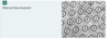
this cell contains steroid secreting cells
false; these mitochondris contain plate-like cristae and are found in cells that need lots of energy (ATP) ; mitochondria that contain tubular reflections (little white circles, as seen in electron microscopy)
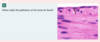
what is the function of the structure in this figure
a. synthesis of membran-packaged proteins
b. synthesis of steroid hormones
c. detoxification of drugs
d. synthesis of ATP

A. the rough ER is where membrane-packaged proteins are synthesized, including secretory, plasma membrane, and lysosomal proteins. In addition, the rER monitors the assembly, retention, and even degradation of certain proteins.
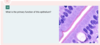
what is the most likely identification of the black circles in this image?
a. lysosomes
b. peroxisomes
c. mitochondria
d. zymogen granules
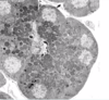
d. zymogen granules congaining enzyme precursors
note the basophilic staining at the tip of the yellow arrow. what is located here?
a. rough ER
b. smooth ER
c. secretory granules
d. golgi apparatus

rough ER
In this hepatocyte, what is the function of the structure enlcosed within the blue circle?
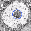
transcription of genes into mRNA; the nucleolus is invloved in the synthesis of rRNA and its assembly into ribosome precursors
In this hepatocyte, what is the structure between 2 yeallow arrows?

heterchromatin
what is the black arrow pointing to?

secretroy granules
what letter represents the trans golgi?
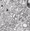
C
what are the small structures collected within the yellow circles in these hepatocytes?
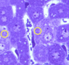
mitochondira
What is the organelle shown within the 4 yellow arrows?
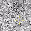
peroxisome
what is the region contained between the two yellow arrows?

golgi apparatus

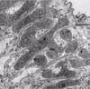
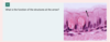
microvilli

absorption (microvilli seen by EM)

microvilli
propel substances along the surfaces (cilia)
trachea; cilia
cilia

this is simple squamous epithelium becuase it is the inside lining of a blood vessel it is also the endothelium
lining of the peritoneum and pleura; simple squamous epithelium; found in the gut

simple cuboidal
absorption

pseudostratified columnar epithelium with cilia

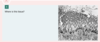
stratified squamous epithelium (nonkeratinized)

stratified squamous epithelium (keratinized)

