Histopathology and Cytopathology Flashcards
(1 cards)
1
Q
A
- Histopathologists (interested in tissue structure): trying to stage disease
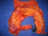
- Biopsies
- Resection specimens
- Frozen sections (real time)
- Post-mortems (hospital or Coroner’s)
-
Cytopathologists (interested in cells):
- Smears
- Fine needle aspirates
Biopsies: (NIC)
- Is it normal?
- Is it inflamed? If so, what’s cause?
- Is it cancer? If so, what type?
Resection specimens:
- How far has cancer spread?
- Is it all out?
Frozen section: 24 hours
- Rapid diagnosis; Is it cancer? Is it all out?
- How sections are obtained:
- Specimen must be properly labelled
- Fix in formalin – crosslinks proteins + slows decomposition
- Embed in paraffin wax
- Cut sections (very thin) and stain
-
Use of sections:
- Stain e.g. Haematoxylin + eosin (H&E), gram, ZN stain (for TB)
- Use antibodies to identify specific antigens = immunohistochemistry
- Carry out molecular tests e.g. oestrogen receptors in breast cancer
Time taken for histopathology result to reach clinician: (probably don’t need to know)
- Frozen section: 30 minutes
- For biopsies: 2-3 days
- For resection specimens: 5-7 days
Cytopathology:
- Looking at individual cells not tissues – pretty much real time, rapid
- Used for fine needle aspirations
- Used for cervical screening
- Most cytology done in real time, VERY quick
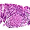
- Billy’s (slides) skin biopsy shows Kaposi’s sarcoma = HIV/AIDS-defining disease
- Immunocytochemistry for CD31 (CD31 marks vascular endothelium) to show vascular tumour infiltrating collagen bundles à endothelial cell tumour
- A fine needle aspiration of one of enlarged nodes revealed mixed cell population
- The diagnosis is of reactive lymphadenopathy