HTC (COMBINED) Flashcards

I combine all topics here, but I also have them seperately. Study all here or sperately by topic names (623 cards)

1
Q

Odontogenic Cysts can be two types

A

Inflammatory
or
Developmental

2
Q

Inflammatory cysts

List ( 4 cysts)

A
  • Periapical (radicular)
  • Residual periapical
  • Buccal bifurcation
  • Paradental
3
Q

Developmental Cysts

List ( 9 cysts)

A

‐ Dentigerous
‐ Eruption
‐ Gingival cyst of newborn
‐ Gingival cyst of adult
‐ Lateral periodontal
‐ Glandular odontogenic
‐ Odontogenic keratocyst
‐ Orthokeratinized odontogenic
‐ Calcifying Odontogenic

4
Q

The following cysts are histologically the same in which way

-Periapical (radicular)
‐ Residual periapical
‐ Buccal bifurcation
‐ Paradental

‐ Dentigerous
‐ Eruption
‐ Gingival cyst of newborn
‐ Gingival cyst of adult

A

all lined by squamous epithelial

5
Q

What are the

Sources of epithelium
within the jaw bone

A

▪ Epithelial rests of Malessez
▪ Reduced enamel epithelium
▪ Fissural cysts – when 2 pieces of bone come together
▪ Odontogenic cysts
▪ Epithelial component of odontogenic tumors
▪ Salivary gland inclusions – rare, incorporated in development

6
Q

radicular cyst, inflammatory cyst are other names for ?

A

Periapical Cysts

7
Q

The most common cyst of the jaws ?

A

Periapical Cysts

8
Q

Periapical Cysts

Demographic and location

A

▪ Any age (peak in 3rd ‐ 6th decades, rare in 1st decade)
▪ No sex predilection
MX > MD (anterior MX most common)

9
Q

Tooth vitality and Periapical Cysts

A
  • Involved tooth usually non‐vital/non‐responsive with thermal and electric pulp testing
  • Should test vitality of tooth if see radiolucency in apex\
  • If tooth vital, and still see radiolucency ► should do biopsy
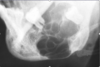
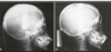
10
Q

Periapical Cyst

(Radiographic)

A
  • Usually appears as well‐circumscribed periapical radiolucency with widening of the PDL space and/or loss of lamina dura
  • Typically small (< 1 cm) but can grow to large dimensions if left untreated
  • Radiographic findings can NOT be used for definitive diagnosis
11
Q

Why the Radiographic findings of Periapical Cyst can NOT be used for definitive diagnosis?

A

‐ similar appearance with:

  • periapical granuloma
  • odontogenic tumors
  • early COD {Cemento Osseous Dysplasia}
12
Q

Lateral radicular cyst appears on the lateral surface of the root of a non‐vital/non‐responsive tooth
‐ A differential for which cyst?

A

lateral periodontal cyst

13
Q
A

Periapical Cysts

►Would need to test both teeth for vitality.
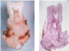
14
Q
A

Periapical Cyst

15
Q
A

Periapical cyst

shows inflammation at site
abscess developed fistula tract thru
soft tissue. Pt will have pain until
pressure is released

16
Q

Periapical Cyst

treatment

A
  • endodontic therapy or extraction of involved teeth
  • larger lesions may require biopsy along with endodontic therapy
  • lesions which fail to resolve should be biopsied
  • follow-up at 1-2 years
17
Q

Residual Cyst

Etiology

A
  • After tooth extracted, not properly cleaned ► the residual cells of the cyst lining and inflammatory cells continue to proliferate
  • Has to be at site where tooth was previously removed
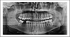
18
Q

Residual Cyst

Radigraphically

A
  • well defined round to oval radiolucency in the site of a previous extraction
19
Q

Residual Cyst

Histologically is identical to which cyst?

A
  • identical to the radicular cyst (periapical cyst)
  • Should biopsy to rule out other causes
20
Q

Residual Cyst

Treatment

A

-Removal

  • Enucleation if small
  • Marsupialization if large
  • Note:*
  • Enucleation* means: removal of an organ or other mass intact from its supporting tissues

Marsupialization means: surgical technique of cutting a slit into an abscess or cyst to empty its contents and suturing the edges of the slit to form a continuous surface from the exterior surface to the interior surface of the cyst or abscess.
Promotes Decompressing and shrinkage.

21
Q
A

Residual Cysts

22
Q
A

Residual Cyst

23
Q

Paradental Cyst

Etiology

A

Some controversy over this designation
‐ some think they are inflammatory cyst
‐ some think they are developmental cysts
▪ Etiology: remains unclear
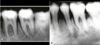
24
Q

Paradental Cyst

Radiographically

A
  • Radiolucent area noted
  • most frequently, along the distal aspect of an impacted or partially erupted third molar
25
Which cyst has been associated w/ enamel extensions into furcation areas of the involved teeth?
**Paradental Cyst**
26
**Paradental Cyst** _Treatment_
**Extraction** of the _tooth along with the lesion_
27
Paradental Cyst
28
Paradental Cyst
29
**Buccal Bifurcation Cyst** is similar to what Cyst ?
_Similar to **a paradental cyst**_ ‐ **EXCEPT**: location is _central on the buccal of mandibular first molars_
30
**Buccal Bifurcation Cyst** _Etiology_
unclear
31
**Buccal Bifurcation Cyst** is most commonly seen with eruption of what tooth?
The eruption of ***the permanent first molar***
32
**Buccal Bifurcation Cyst** _Clinically_
seen as * swelling * tenderness of soft tissue over involved area
33
Which Radiograph type is best to see **Buccal Bifurcation Cyst?**
▪ Radiolucency best seen with **an occlusal radiograph**
34
**Buccal Bifurcation Cyst** *as seen in occlusal radiographs*
35
**Buccal Bifurcation Cyst** *as seen in occlusal radiographs*
36
Buccal Bifurcation Cyst _Treatment_
▪ **Enucleation** **of cyst**; tooth extraction unnecessary ▪ Some cases _resolve w/o surgery_ ▪ Some resolve _w/ daily irrigation_ of buccal pocket with saline/hydrogen peroxide
37
**Dentigerous Cyst** also known as ?
**Follicular Cyst**
38
What is **most common type of developmental odontogenic** **cysts?** 20% of all epithelial lined cysts of the jaw
**Dentigerous Cyst**
39
**Dentigerous Cyst** _Origin_ & _Etiology_
*_Originates_*: by **the separation** of the _follicle_ from the _crown of an unerupted tooth_ *_Pathogenesis_*: **accumulation of fluid** between the tooth and the _reduced enamel epithelium_
40
Dentigerous Cyst _Clinically_
▪ Small cysts typically asymptomatic and picked up on routine radiographic exam ▪ Large lesions may show expansion of bone ▪ Cysts may become infected, especially if partially erupted tooth
41
Dentigerous Cyst _Demographics_ & _Location_
* Mostly **mandibular 3rd molars** (rarely unerupted deciduous teeth) * Most commonly present in **2nd and 3rd decades**
42
What is a key characteristic of **Dentigerous Cyst** _location_?
* **Attached to the tooth at the CEJ**
43
Small Dentigerous Cyst are hard to differentiate radiographically from ---?
**enlarged/hyperplastic follicle** Rule of thumb: * If 4‐5mm or more of radiolucency ► _dentigerous cyst_ * If \<4mm of radiolucency► can be _hyperplastic follicle_
44
dentigerous cyst or follicle ?
_dentigerous cys_t b/c **\*attachment at CEJ**
45
**Dentigerous Cyst**
46
**dentigerous cyst**
47
**dentigerous cyst**
48
**dentigerous cyst**
49
Grossly image of Dentigerous Cyst
50
Dentigerous Cyst Treatment
* **Decompression**: Try to open window in the jawand put tube into cyst lumen and have pt irrigate a few times a day for a few weeks ► release pressure and allows bone to grow back ► **cyst will shrink** * If get rid of whole area surgically► c_an risk_ _fracturing the jaw_
51
What is the Soft tissue counterpart of a dentigerous cyst?
Eruption Cyst
52
**Eruption Cyst** also known as
_eruption hematoma_
53
**Eruption Cyst** Etiology
* Results from accumulation of fluid in the follicular space when the tooth has erupted over the alveolar bone **\*NOT in bone\***
54
**Eruption Cyst** _Demographic_ & _Location_
▪ Usually seen in **1st decade** *(children)* ▪ Most often involves **1st permanent molar** and **maxillary incisors**
55
**Eruption Cyst** Clinically
Frequently _normal mucosal color_, BUT surface trauma (ex. chewing) may result in bleeding into the cystic space► may look **purple or blue** ▪ Usually **soft** or **fluctuant**(like a balloon) _upon palpation_
56
**Eruption Cyst** _Treatment_
* Unless symptomatic, no treatment required, cysts resolve upon eruption of teeth
57
**Eruption Cyst**
58
**Eruption Cyst**
59
**Cysts of the Newborn** can either be --- or ---
**Palatal cysts** or **Gingival cyst**
60
**Palatal cysts** _Types_ _&_ _Location_
**‐ Bohn’s nodules:** scattered over HP (hard palate), often junction of HP and SP (soft palate) **‐ Epstein’s pearls:** along median palatal raphe
61
Cysts of the Newborn: **Palatal cysts** _Demographics_
* Seen in 60‐80% of neonates
62
Cysts of the Newborn: **Palatal cysts** _Clinically_
* 1‐3 mm cream to white papules (keratin filled cysts) ## Footnote **\*NOT in bone\***
63
Cysts of the Newborn: Palatal cysts Treatment
**No treatment is required** ‐ Resolve (degenerate or rupture) on their own in a few months ‐ Once baby eats solid foods, will go away
64
Cysts of the Newborn: Palatal cysts
65
Cysts of the Newborn: **Gingival cyst of the Newborn** Also known as
**Dental lamina cysts**
66
Gingival cyst of the newborn _demographics & Location_
* Found superficially on the **alveolar ridge mucosa** * **MX** \> MD * Rarely seen after 3 mos. of age
67
**Gingival cyst of the newborn** _Treatment_
▪ No treatment is necessary ▪ Spontaneously resolve (degenerate or rupture)
68
**Gingival cyst of the newborn** _Clinically_
* 1‐3 mm creamy white papule (keratin filled cysts) * \*NOT in bone\*
69
Gingival cyst of the newborn/ Dental lamina cysts/Cysts of the Newborn-gingival
70
What is the soft tissue counterpart of the **lateral periodontal cyst ?**
**Gingival Cyst of the Adult**
71
Gingival Cyst of the Adult _Origin_
Derived from dental lamina rests **‐ Rests of Serres**
72
**Gingival Cyst of the Adult** _Demogrophic & Location_
**▪Uncommon lesion** ▪ 60‐75% **mandibular canine/premolar area** ‐ most common location on the _facial or buccal aspect_ ▪ **5th and 6th decade** most common
73
**Gingival Cyst of the Adult** _Clinically_
* Painless, dome‐like swellings up to 5 mm in diamete * Often with a bluish or grayish hue
74
**Gingival Cyst of the Adult** has _similar histology_ to which cyst?
**lateral periodontal cyst**
75
**Gingival Cyst of the Adult** _Treatment_
* **simple surgical excision** * _Unlikely_ to recur/come back
76
**Gingival Cyst of the Adult**
77
Gingival Cyst of the Adult
78
Gingival Cyst of the Adult notice the _bluish hue_
79
**Lateral Periodontal Cyst** represents the intrabony counterpart of which cyst?
**gingival cyst of the adult?**
80
Lateral Periodontal Cyst _Origins_
* Developmental cyst believed to arise from **dental lamina rests**
81
**Lateral Periodontal cyst** is diagonsed when cysts occur in the _lateral periodontal region_ and after what have been excluded?
* **an inflammatory origin cysts** or t**he diagnosis of odontogenickeratocyst** _have been excluded_
82
**Lateral Periodontal Cyst** _Charcterstics_ and _tooth vitality_
▪ Commonly **asymptomatic** and _found on routine radiographic exam_ ▪ Associated teeth tests **vital/responsive with electric pulp test**
83
Radilucency Lateral to a teeth how would you know if it's Lateral Periodontal Cyst or Lateral Radicular Cyst or Lateral OKc
‐ **If pulp alive**► **lateral periodontal cyst** or ***Lateral Okc ( if huge lesion)*** ‐ If pulp dead► **lateral radicular cyst**
84
**Lateral Periodontal Cyst** _Demographic_ and _Location_
▪ Most likely found after age 30 ▪ Males\>Females **▪ ~65% mandibular canine/premolar area** ‐ Can also be seen _between canine and lateral incisor_
85
**Lateral Periodontal Cyst** _Radiographically_
Present as **well circumscribed**, **unilocular** _radiolucencies between 2 teeth,_ located **lateral to tooth root** ▪ Most often **0.5‐1.0 cm** in diameter ▪ Radiographic features are **NOT** diagnostic
86
Which is here is **Lateral Periodontal Cyst** **Lateral Radicular Cyst** **Lateral Odontogenic Kertocyst**
* * Could be differential for lateral Odontogenic keratocyst, except this does not grow in size * Lateral radicular cysts from an accessory canal if tooth is non vital * or it could be Lateral Periodontal Cyst if tooth is vital!
87
**Lateral Periodontal Cyst**
88
**Lateral Periodontal Cyst**
89
Lateral Periodontal Cyst _Treatment_
* consists of **conservative enucleation**
90
What cyst is a **variant of lateral periodontal cyst?**
**Botryoid Odontogenic Cyst**
91
**Botryoid Odontogenic Cyst** _Grossly_ and _Microscopically_
shows **a grape‐like cluster** of small individual cysts
92
**Botryoid Odontogenic Cyst** _Radiographically_
▪ Either unilocular or multilocular on radiographs, depending on size of the lesion ▪ Cyst lining similar to lateral periodontal cyst
93
**Botryoid Odontogenic Cyst** well circumscribed, between 2 teeth (similar to lateral odontogenic cyst), multilocular
94
**Botryoid Odontogenic Cyst**
95
**Glandular Odontogenic Cyst** _Charcterstics_
* **A rare odontogenic cyst** which exhibits features of _glandular differentiation within the epithelium_ * Presumably **represents the pluripotentiality of odontogenic epithelium**
96
Glandular Odontogenic Cyst _Demographics_
▪ Wide age range from 2nd to 9th decades **‐** *_mean age 49_* **▪ ~ 80% of cases in mandible ▪ Anterior lesions** ‐ More common ‐ _May cross the midline_
97
**Glandular Odontogenic Cyst** _Radiographically_
▪ Uni‐ or (more often) multilocular radiolucency ▪ **Well‐defined** with **a sclerotic border**
98
**Glandular Odontogenic Cyst** reccurance rate
(~ 25% recurrence rate) Can be locally aggressive
99
Glandular Odontogenic Cyst _Clinically_
▪ Usually asymptomatic unless inflamed
100
_Histologic differential diagnosis_ of Glandular Odontogenic Cyst includes --?
**mucoepidermoid carcinoma** (salivary gland tumor)
101
**“Primordial” Cyst** Assuming histologically it is *different from OKC*
102
“Primordial” Cyst is **not** a true -------
* lesion, *was actually some other type of cyst* * * it is now thought that most of the reported Primordial cysts were actually **OKCs**
103
**Odontogenic Keratocyst** **OKC** _Also known as_
**keratocystic odontogenic tumor (KOT)** -2005 WHO _but now it's back to **OKC**_
104
**Odontogenic Keratocyst (OKC)** _Etiology_
* **Growth and expansion** of this lesion due not only to osmotic effects/pressure, but to _unusual gene expressions_
105
Which **unusal gene expression** causes _growth and expansion of OKC_ ?
* _Expresses **Ki‐67**_ (high rate of cell proliferation) * O_verexpression of **Bcl‐2**_ (antipoptotic protein) * _Overexpression of **MMP’s 2 and 9**_ (thought to allow growth into connective tissue) * _Mutation of **PTCH**_, a tumor suppressor gene * when PTCH is non‐functional → cell proliferation
106
**Odontogenic Keratocyst (OKC)** _Demographic & Location_
* ~ **60%** present in **2nd and 3rd decade**, but can occur at any age * **Mandible** affected in 60‐80% of cases * tendency to occur in **posterior mandible and ramus** * **25‐40% of cases involve an unerupted tooth** * ‐ **5%** of patients have **_multiple cysts_**
107
**Odontogenic Keratocyst (OKC)** differes from **Meloblastoma** **in its growth pattern**
**Odontogenic Keratocyst (OKC) :**grows in anterior to posterior manner before causing cortical expansion *while* **Meloblastoma:** causes **cortical expansion _early_**
108
Which cyst make up **~10‐15%** of all **odontogenic cysts?**
**Odontogenic Keratocyst (OKC)**
109
5% of **Odontogenic Keratocyst (OKC)** are associated with _which syndrome?_
**nevoid basal cell carcinoma syndrome**(*_**Gorlin syndrome**)_*
110
What are the site distribution of OKC?
Most of OKC in posterior region
111
Odontogenic Keratocyst _Reccurance Rate_
* **HIGH Recurrence Rate** * **Benign**, but _locally aggressive biologic behavior_ * _Solitary OKCs have_ ~**10% recurrence rate with appropriate treatment** * _Multiple OKCs hav_e ~ **30% recurrence rate**
112
**Odontogenic Keratocyst** **(OKC)** _Reccurance Rate Order_ from **highest** to **lowest** reccurance rate
**Syndrome OKC \> Multiple OKC \> Solitary OKC \> Conventional odontogenic cysts**
113
**Odontogenic Keratocyst** **OKC** _Radiographically_
* Usually **a well‐circumscribed** *_radiolucency_* with **smooth, often** **corticated margins** ▪ Cysts may be ‐ **Unilocular** (most common) ‐ **Multilocular** (larger lesions)
114
**Odontogenic Keratocyst** **OKC** _clinically_
▪ **Small cysts** are typically **asymptomatic** and picked up on routine radiographic exam ▪ **Larger cysts** may or may not be asymptomatic ▪ \*Cysts tend to grow in an antero‐posterior direction prior to lateral growth ►_therefore cysts are usually quite large when they start to expand the cortical plate_
115
**Odontogenic Keratocyst** **OKC** Has similar Radiographic findings with ?
* dentigerous cyst * ameloblastoma * and others
116
**Odontogenic Keratocyst OKC** _Treatment_
▪ **Marsupialization** (decompression) ▪ **Peripheral ostectomy** ‐ Carnoy’s solution ▪ **Resection** ▪ **Medications targeted to PTCH** ▪ ***Long term follow‐up***
117
**Odontogenic Keratocyst OKC**
118
**Odontogenic Keratocyst OKC**
119
**Odontogenic Keratocyst OKC**
120
**Odontogenic Keratocyst OKC**
121
similar to *lateral periodontal cyst* but is actually **OKC**
122
**Nevoid Basal Cell Carcinoma Syndrome** is also known as ----- ?
**Basal Cell Nevus or Bifid Rib Syndrome** or **Gorlin syndrome**
123
Which cyst is assoicated with Nevoid Basal Cell Carcinoma Syndrome ?
**Odontogenic Keratocyst “OKC”**
124
**Nevoid Basal Cell Carcinoma Syndrome** **(Gorlin syndrome)** _modes of inheritanc_e
**_Autosomal dominant_ inheritance**
125
**Nevoid Basal Cell Carcinoma Syndrome**
126
Which **_Gene mutation and pathway_** _associated with_ **Nevoid Basal Cell Carcinoma Syndrome** **(Gorlin syndrome)**
* _Mutation_ of **PTCH** (tumor suppressor gene) * in the **Sonic Hedge Hog pathway**
127
**Nevoid Basal Cell Carcinoma Syndrome** _Prognosis_
■ Prognosis _depends on progression of skin tumors_
128
**Nevoid Basal Cell Carcinoma Syndrome** _Treatment_
✎Surgery (typically MOHS) ✎Sometimes curette them ✎ Radiation therapy (RT) is typically not the first line of therapy with small lesions RT ✎Cryotherapy which means they just use a little liquid nitrogen and freeze them ✎Photodynamic therapy with photosensitizer and topical medications ■ New medication: **Vismodegib _inhibits sonic hedgehog pathway by binding smoothened (SMO)_** * *suppressive rather than curative cause it seems to work for short time and after ~7-8 months ..may also helps _suppress growth of OKC_*
129
✎A patient who has **Nevoid Basal Cell Carcinoma Syndrome** ✎We can see **multiple cystics** areas and lesions in _the jaws, maxillary and mandible_ ✎**Both 3rd molar displaced** in the maxilla because of the cyst
130
✎**Multiple lesions**, impacted 3rd molar in mandible and displaced 3rd molar up into the sinus, ✎These too many lesions hard to manage the issue with a surgery ✎This large area on the left mandible – good example of why we do decompression because if you just remove this lesion and the entire area is left open, this would be an area risk for fracture
131
What is this called which can be seen with Nevoid Basal Cell Carcinoma Syndrome
✎An example of the **pitting** that can be seen **palmar and plantar** ~ This is a side of a hand ✎This is an early stage of basal carcinoma which never goes on (like it is aborted)
132
What are these **findings** that is associated with ## Footnote **Nevoid Basal Cell Carcinoma Syndrome?**
* **thousands of basal cell carcinoma** is developing on the skin -very difficult to manage with surgery, ~ That’s why they remove the larger ones, the deeper ones ~ They leave the one that’s less as an issue until they get to a larger size to be removed
133
Why **Basal Cell Carcinoma** is very problematic ?
It’s not the lesion themselves causing metastasis that’s the issue, it’s the lesion growing deeply and in affecting adjacent structures that really is the issue with basal carcinoma
134
What is the **Most common type of skin cancer?**
**Basal Cell Carcinoma (BCC)**
135
**Basal Cell Carcinoma (BCC**) _Demographics_
* 2-3 million cases a year * About 3 out of 4 skin cancers are basal cell carcinomas
136
Basal Cell Carcinoma _Growth_ and _location_ | (BCC)
- Develop in the **lowest layer of the epidermis**, called the **basal Laye**r - Develops on sun-exposed areas: _cumulative DNA Damage_ **- Slow-growing** • If not treated, basal cell c**ancer can grow into nearby areas and invade the bone or other tissues beneath the skin**
137
Basal Cell Carcinoma _Progrssion_ | (BCC)
within 5 years of being diagnosed with BCC►**35%-50%** of people _develop a new skin cancer_
138
**Calcifying Odontogenic Cyst** **​COC** also known as ?
* **Calcifying Cystic Odontogenic Tumor** * **_Gorlin Cyst_ ( don't confuse it with Gorlin syndrome)** * **Ghost Cell Tumor** *
139
**Calcifying Odontogenic Cyst (COC)** can present in **3 types**
1. **■ Cystic Unilocular COC** * COC with odontoma (~ 20%) * Extraosseous/peripheral – present in _older patients_ 2. **■ Solid COC (odontogenic ghost cell tumor)** * Often demonstrate _a more aggressive behavior_ * WHO once considered them all CCOT now back to COC 3. ■ **Odontogenic ghost cell carcinoma** * _very rare lesion_
140
**Collision Tumors** is a term used to describe lesions involving Calcifying Odontogenic Cyst (COC), what does that mean?
* where you see **_both features_ of ameloblastoma with COC** or **adenomatoid odontogenic tumor with COC**
141
**Calcifying Odontogenic Cyst (COC)** _may occur in association_ _with_ **which tumors or cysts?**
* **Odontomas** (a benign tumour linked to tooth development) * **Ameloblastomas** (rare, noncancerous (benign) tumor) * **Adenomatoid odontogenic tumor** (rare tumor of epithelial origin that is benign, painless, noninvasive, and slow-growing)
142
**Calcifying Odontogenic Cyst (COC)** _Demographics & Location_
■ Peak in _second decade_, most **before age of 40** ■ Frequently presents **anterior to molars** ■ ~ **20% extraosseous (peripheral)**, found in **older age group** (~ 50 years of age) ■ Female \> Male ■ **~ 70% occur in MX** ■ One third are associated with **unerupted teeth**, usually a **canine**
143
Calcifying Odontogenic Cyst _Radiographically_ | (COC)
■ Usually **a well-circumscribed unilocular radiolucency,** _infrequent multilocular cases_ ■ **One third to one half** show _radiopaque structures within the radiolucency_ ■ When you see **calcifications** within a lesion, you don't use the term uni or multi locular anymore, but they are called **_mixed radiolucent/radiopaque lesions_** ■ May cause **resorption or displacement of roots** **■** **_One third_** are associated with **unerupted** **Canine**
144
``` **Calcifying Odontogenic Cyst (COC)** ``` * _in the mandible_ and you can see it **well circumscribed radiolucency** * a little bit of blunt root resorption in this area * _No calcifications_ in this one yet ►so this is still **unilocular radiolucency**
145
What are the _clinical_ and _radiographic_ findings here? What is this lesion?
* Clinical finding for this patient was **Obliteration of the vestibule space**, because the mandible is showing expansion * radiographically:we see radiolucency going as far as the first molar * This is a **mixed radiolucent radiopaque lesion** in developing calcifications. * This is an example of **Calcifying** **Odontogenic Cyst (COC)**
146
The hallmark of Calcifying Odontogenic Cyst COC Histology is
**_Ghost cells_** They have that sort of polygonal shape or roundish shape with the pink that looks like the cytoplasm, but in the location where the nucleus would have then, there's an empty spot
147
Histologically speaking, Calcifying Odontogenic Cyst COC, basically looks similar to what epithelium?
**_ameloblastic epithelium_**
148
**Calcifying Odontogenic Cyst COC** _Treatment_
■**Enucleation with peripheral ostectomy** ~ Very similar to odontogenic keratosis ■ **Follow up is long term** because s_ome of the solid tumors have a more aggressive behavior_ **■ Peripheral lesions are treated with excision**
149
When COC is associated with another tumor, ameloblastoma, how would you treat?
■ the treatment is based on **the more aggressive tumor** **~** So you would treat the _ameloblastoma_. ~You wouldn't treat conservatively the COC though
150
151
**Fissural Cysts** | (6)
❑ Nasolabial cyst ❑ Globulomaxillary cyst (historic) ❑ Nasopalatine (incisive canal) cyst ❑ Incisive papilla cyst ❑ Median palatal cyst ❑ Median mandibular cyst (historic)
152
**Nasolabial Cyst** also known as
aka Nasoalveolar cyst
153
where a number of the visual cysts would develop
(1) That's the nasopalatine, which is sort of up in the labial nasal fold and it's in the soft tissue. (2) Sort of where the nasal alveolar cyst would occur. (3) Where the globular maxillary cyst would occur between the canine and the lateral sometimes between the lateral and the first premolar (4) The nasopalatine in the cyst of the nasopalatine papilla (5) Is the median palatal
155
**Nasolabial Cyst** _Etiology_
■ Thought to be caused by: * either **epithelial remnants of the nasolacrimal duct** * or **cells left after fusion of the maxillary, medial and lateral nasal processe**s during _development of the midface_
156
**Nasolabial Cyst** _Location_
_Rare_ **soft tissue cyst of the upper lip**, lateral to the midline (right under the ala of the nose) \*NOT in bone\* ■ Clinically see a swelling which can cause elevation of the ala of the nose ■ Intraorally see a swelling in the maxillary vestibule lateral to the midline (usually sort of in the canine area or just a little bit distal to the canine area) ■ Pain is uncommon, unless cyst becomes infected
157
**Nasolabial Cyst** _Clinically & Intraoray_
■Clinically we see a **swelling** which _can cause elevation of the ala of the nose_ ■ Intraorally see **a swelling in the maxillary vestibule lateral to the midline** (usually sort of in the canine area or just a little bit distal to the canine area) ■ **Pain is uncommon,** _unless cyst becomes infected_
158
**Nasolabial Cyst** _Demographics_
■ Peak in **4th and 5th decades** ■ **3 to 4 times** _more common in **females**_ ■ ~ **10%** of cases are **bilateral**
159
**Nasolabial Cyst** _Treatment_
* **Surgical Excision** via i**ntraoral approach**, * usually do not recur ~ **very low risk of occurrence**
160
Nasolabial Cyst The lesion here just below the nose and you can tell that it's sort of raising the edge of the nose slightly
161
**Nasolabial Cyst** the lesion raising the edge of the nose slightly
162
_Nasolabial Cys_t has a a **respiratory type epithelium** and so it's very similar to what you would see in ?
either in the **sinus** or in the **nasopalatine ducts**
165
Is this ## Footnote **Globulomaxillary Cyst** **lateral granulomas** **OKCs** **COCs**
* we can see the **displacement of the root** * A **teardrop or pear shaped** radiolucency between the lateral and the canine * **Well circumscribed** maybe leaving **a little sclerotic edge** up here * ended up being in **a odontogenic keratocyst (OKC)**
166
Is this Globulomaxillary Cyst , lateral granuloma or OKC?
~ it is kind of **a teardrop or pear shaped size** ~Little less well differentiated in this particular instance but again **unilocular radiolucency between the roots of two teeth** This one ended up being an **OKC**
169
"Globulomaxillary Cyst" _Origin_ _controvesy_ why the name in quotations?
* it's in quotations, because really there is no such thing as a globulomaxillary cyst * because it was thought that this was remnants _after fusion of the globular portion of the nasal process with the maxillary process,_ and now we know that **these two processes are always united from the start and that there is no fusion** * When biopsied these cysts are **odontogenic in origin**
170
what does it mean for **Globulomaxillary Cyst** to be **odontogenic in origin**?
✎This is term used to **describe a cyst in a particular anatomic location** _it is not a diagnosis_ ✎An odontogenic cyst (inflammatory cyst, lateral periodontal or even sometimes OKC) that forms in the area **between the maxillary lateral incisor and the canine roots** ~ It's really associated with a_n anatomic location **not** with any particular cyst._ ✎So it can be any of the odontogenic lesions such as **lateral granulomas or cysts, OKCs, COCs, etc.**
171
**Globulomaxillary Cyst** _Radiographically_
## Footnote ✎Presents as a **“inverted pear”** shaped **well-circumscribed radiolucency** ✎Frequently causes **displacement of the roots**
172
What are two different ways nasopalatine duct cyst arise?
* *A**. It can either be _the cyst totally within bone_ * *B**. It can actually cause _widening of the orifice and causing the soft tissue expansion in this way_
175
Most common non-odontogenic cyst of the oral cavity
**Nasopalatine Duct Cyst**
176
**Nasopalatine Duct Cyst** also known as
**incisive canal cyst** **nasopalatine canal cyst**
177
**Nasopalatine Duct Cyst** ## Footnote ✎This person is edentulous ✎ **an inverted pear shape** ✎The nasal spine is superimposed on your radiolucency ► **a heart shape**
178
**Nasopalatine Duct Cyst** ✎Between the roots of the two teeth, a well circumscribed radiolucency, not showing any changes to the adjacent structures ✎could be an enlargement of the incisive canal due to variation in size ~ **early lesions can be hard to diagnose** ✎**the treatment in such cases**: a follow up with another radiograph in six months to see if there's been any change in size **✎ No surgical intervention until you see the cyst expanding**
179
This is showing you the how the papilla can be enlarged if it's only in soft tissue or if there's a partial soft tissue partial bone expansion *Nasopalatine Duct Cyst*
180
**Nasopalatine Duct Cyst** _Origin_
* arise **from epithelial remnants of the nasopalatine duct** which, _embryologically, connects the oral and the nasal cavities_
181
**Median Palatine Cyst**
182
**Nasopalatine Duct Cyst** _Demographic_ and _Location_
* Peak presentation in the **4th to 6th decades**, _but can occur at any age_ ~ *because it takes a little bit of time for the cyst to grow within the bone* * commonly found on the **anterior palate** ~ typically in the _nasal area of the papilla._
183
Is this Median Mandibular Cyst Or something else
Remember ## Footnote _Median Mandibular Cyst_ is a term used to describe a cyst in a **anterior mandible** not a definitive diagnosis So, this turned out to be an early ameloblastoma. It wasn’t a cyst The lesion radiolucency in the anterior mandible and again
184
Nasopalatine Duct Cyst _Clinically_
■ present with **swelling** o_f the anterior palate_ (in the nasal area of the papilla) ■ Most are **asymptomatic**, but _they may have pain or drainage_
186
**Nasopalatine Duct Cyst** _Radiographically_
■ a **well-circumscribed unilocular** **radiolucency** on _the midline of the anterior hard palate_ _between and apical to the central incisors_ ■ The radiolucency often have an oval or inverted pear shape with a sclerotic border ■ Superimposition with the nasal septum can create an appearance of the classic **“heart” shape**
187
**Cysts of the incisive papilla** **Incisive papilla cyst**
Is a **soft tissue cyst** (no bone involvement) located in the same area as the **Nasopalatine Duct Cyst** _on the midline of the anterior hard palate between and apical to the central incisors_ . They may be symptomatic or asymptomatic and usually are not seen radiographically. some consider them to be uncommon variants of the nasopalatine duct cysts
188
**Surgical Ciliated Cyst of the Maxilla** In this premolar shot (middle image) you can see a **well-circumscribed lesion** ✎Because the maxillary sinus is **radiolucent**, it almost looks like this is **radiopaque** but it's not ✎ If you did a CBCT you would see that **it's an empty space within the bone of the maxilla.** It's not actually radiopaque
189
**Nasopalatine Duct Cyst** _Treatment_
* **surgical excision** * **recurrence is rare**
195
**Median Palatine Cyst** **is** **_a variant of which cyst?_**
**nasopalatine duct cyst** * it represents a more **posteriorly** placed nasopalatine duct cyst * ~ It's probably due to some sort of anatomic variation in the patients; that their palatine duct is just placed more posteriorly * So **instead of being between the roots of these two teeth, it's placed more posteriorly**
197
**Median Mandibular Cyst**
* **A controversial cysts** whose existence is questioned ~ similar to **the globulomaxillary cyst** ■ Originally *thought to arise from the fusion of the “halves” of the mandible, but current embryology finds that the mandible forms from a single bilobed process, therefore, no epithelial remnants would be found* ■ Now, it is thought that cysts in this area represent **odontogenic cysts or tumors** * **Median Mandibular Cyst is a** term used **to describe a cyst in a particular anatomic location not a definitive diagnosis** * ~ It is other lesions that occur in that particular location * **The Anterior Mandible**
199
**Surgical Ciliated Cyst of the Maxilla** _Etiology_
■ Occurs after trauma or sinus surgery (iatrogenic - reactive not neoplastic)
200
**Surgical Ciliated Cyst of the Maxilla** _Formation_
■a portion of the sinus lining is **separated** from the sinus and forms an epithelial lined cavity in bone ■ Cavity f**ills with mucin** produced by the _mucous cells of the cyst lining_ ■ These cysts **enlarge** as the _intraluminal pressure increases_, causing **destruction of bone**
201
**Surgical Ciliated Cyst of the Maxilla** _occurs frequently_ **after** **which procedures?**
* after **a Caldwell-Luc procedure** * *sometimes with **difficult maxillary extractions***
202
In which country **Surgical Ciliated Cyst of the Maxilla** are reported with higher frequency ?
**Japan**
203
**Surgical Ciliated Cyst of the Maxilla** In this premolar shot (middle image) you can see a **well-circumscribed lesion** ✎Because the maxillary sinus is **radiolucent**, it almost looks like this is **radiopaque** but it's not ✎ If you did a CBCT you would see that **it's an empty space within the bone of the maxilla.** It's not actually radiopaque
204
What are **pesudocysts?**
* **They have no epithelial lining.** * They’re called cysts by convention just because that's what everybody is used to * **They're not true cysts.**
205
pesudocysts List (5)
* Aneurysmal Bone Cyst * Antral Pseudocyst * Simple Bone Cyst * Osteoporotic Bone Marrow Defect * Stafne Bone Cyst
206
**Aneurysmal Bone Cyst** Demographics
■ Most common site in the body is **long bones *or* vertebrae** _■ In the jaw_s, most frequently seen in the **1st and 2nd decade** **■ MD \> MX** ## Footnote *it's a pesudocyst*
207
**Aneurysmal Bone Cyst** _Clinically_
* swelling, frequently a rather rapid swelling * often with **pain** and/or **paresthesia** (signs which can be suggestive of the presence of _a malignant or aggressive lesion_)
208
**Aneurysmal Bone Cyst** Etiology
* Etiology is **unclear**, may result from **trauma** or a **vascular malformation** * most agree that it is a _**reactive** and ***not*** a neoplastic lesion_
209
**Aneurysmal Bone Cyst** _Radiographically_
■ **a radiolucency** which can be either **unilocular or multilocular** in appearance ■ **Borders are variable**, *often irregular in shape* and may be **ill-defined** (again, giving the suggestion of malignancy) ■ **Teeth may be displaced** ■ we may see **cortical expansion and thinning** ~ the cortex itself can become quite thin
210
What does this person have?
- you might think that he has an odontogenic infection but he didn't. You can see that there's a pretty significant swelling on the left side of his face This is a **Aneurysmal Bone Cysts**
211
Aneurysmal Bone Cyst you can see that there is kind of a **multilocular radiolucency** in this particular area
212
**Aneurysmal Bone Cyst** ## Footnote ✎ There's a **radiolucency** involving _the second molar_ that's going as far anterior as the first molar and back to the third molar ✎ There is a little bit of **spiking root resorption** and _that's one of the signs that we associate with malignancy_ ✎ It's a little bit **ill-defined** *~ hard to say exactly where it begins and ends*
214
**Aneurysmal Bone Cyst** ✎ It looks like **a blood soaked sponge** ✎ There’s these **open sinusoidal spaces** and then fibrous connective tissue surrounding them. *✎The sinusoidal spaces can vary in size; some of them are fairly small and others are large*
218
**Wall of the aneurysmal bone cyst** can have a **histology** _similar to the following_
**✎ Central giant cell granuloma** **✎ Cherubism** **✎ Brown tumor of hyperparathyroidism**
220
**Aneurysmal Bone Cyst** _Treatment_
■ Treatment is **surgical enucleation and curettage** ■ **lesions can recur** ~ Usually the recurrence is because you didn't get the entire thing out the first time around ■ Some surgeons follow **enucleation** with **cryotherapy** ■ **Irradiation** is _contraindicated_
221
Is **bleeding a concern** during surgical removal of **Aneurysmal Bone Cyst?**
■ No, **vascularity is predominantly “low flow”**, therefore not as much concern for bleeding upon surgical removal ■ As compared with **central hemangioma** where there is a concen for bleeding
222
**Antral Pseudocyst** * a **Dome-shape** **swelling** on the floor of the sinus. * _They can sometimes_ be **fairly subtle** **Antral Pseduocyst** are **NOT Mucoceles** **Mucoceles** would have more of **meniscus-like appearance**where it would come up to**the edge of the sinus**
223
**Antral Pseudocyst**
They are different than **surgical ciliated cyst** in their lining, etiology, location and appearance!
224
**Aneurysmal Bone Cyst** ## Footnote ✎ A **dome shape swelling on the floor of the sinus** that’s associated with some _sort of inflammation of tooth of t_he **premolar** caused inflammation underneath the apex of the bone (right) and then that leads to accumulation of fluid which causes the sinus lining to elevate off the bone and fill with fluid ✎ After root canal therapy and once the infection gets under control, these will typically resolve on their own
225
As opposed of surgical ciliated cysts, **Antral psuedocysts are not ----** *( in term of their lining)*
_Not epithelial lined spaces_
226
As opposed of surgical ciliated cysts, **Antral psuedocysts are not ----** *( in term of location)*
Not within the bone **but are in the sinus**
227
As opposed of surgical ciliated cysts, **Antral psuedocysts Develop as ----** ## Footnote ***( in term of etiology)***
develop a**s an accumulation of an inflammatory exudate** (often serum) _between the sinus epithelial lining and the bone_ -It develops because of an **inflammatory event in the jaw,** usually the **maxilla**, often from the roots of the maxillary teeth that cause inflammation
228
As opposed of surgical ciliated cysts, **Antral psuedocysts appear as ----** ## Footnote *( in term of Radiology)*
Appears as **a dome shaped elevation** _of the floor of the sinus_
230
**Simple Bone Cyst** also known as
aka **traumatic bone cyst**
231
**Simple Bone Cyst** ## Footnote ✎**A well-circumscribed with cortication** in the **body of the mandible**, affecting _the posterior aspect (premolars and the molars )_ ✎Note the **scalloping** that happens up **between the roots**. It doesn't cause root resorption and actually the lesion will grow up between the roots of the teeth
232
**Simple Bone Cyst** * **A well-circumscribed**showing **the scalloping up between the roots of the teeth radiolucency**
233
**Simple Bone Cyst** ## Footnote ✎Big lesion example: It’s going back to the molar area here. ✎You can see that the lesion extends _over to the canine on the other side_ ✎Most lesions are usually **in the anterior mandible**
235
**Simple Bone Cyst** _Charcterstics_
* **A benign**, empty or fluid filled, cavity in bone which is devoid of an epithelial lining – **a pseudocyst** * Thought to be **reactive**, NOT *neoplastic*
236
**Simple Bone Cyst** _Etiology_
_Etiology_ **ununcertain**, theories include: * **trauma** * **ischemic necrosis of medullary space** * **cystic degeneration of a primary bone lesion**
237
**Simple Bone Cyst** _Demographics_
* In jaws, most likely in the **2nd decade** * Almost **exclusively the mandible** * **Twice** as common in **males**
238
**Simple Bone Cyst** _Radiographically_
* a **well-circumscribed radiolucency** with **an irregular outline** * Tendency to **“scallop” around and between roots** *_(highly suggestive_*, but *_not diagnostic of this lesion_*)
239
**Simple Bone Cyst** _Treatment_
* **exploration** and **curettage** of space to create bleeding. Clot will organize and allow bone repair * **Recurrence** is **_rare_**
241
* You can see there's a little bit of radiolucency. * There happened to actually still be teeth in the area, but * when it was _biopsied_ it showed that it was a * **hematopoietic** or **osteopoietic bone marrow defect**
243
**Stafne Bone Cyst** _Charcterstics_
■ An **asymptomatic** focal _concavity_ of the cortical bone on the _lingual aspect of the MD_ ■ A *pseudocyst*, not a true cyst
246
**Osteoporotic Bone Marrow Defect** _Demographic_
■ _Uncommon finding_ ■ \> 75% of cases are in **females** ■ ~ 70% occur in the **posterior MD**, often in _an edentulous area_ it's a *pesydocyst*
247
**Osteoporotic Bone Marrow Defect** _Etiology_
* **Etiology unclear** * may be **hyperplasia of marrow** due to need for RBCs or * **abnormal regeneration of bone** after **an extraction or persistence of fetal marrow**
248
**Stafne Bone Cyst** _This is the classic look._ - a **well-circumscribed corticated radiolucency** - **below the inferior alveolar nerve**, away from the teeth. - They can be either **oval**, like this, or **round** in appearance
249
**Stafne Bone Cyst** Less common location Check if the teeth were vital with vitality test Get a CBCT in that area to see what was going on first and then once you saw the CBCT you'd be able to make the diagnosis.
250
**Osteoporotic Bone Marrow Defect** _Charcteristics_
* it's a pesydocyst * a **radiolucency** in an area typically where tooth has been removed. Instead of filling in with bone, it fills in with marrow. * When we biopsy it, you’re seeing the hematopoietic elements. (fat, early stages of (the -blasts of) red cells, white cells. * We see basic **bone forming marrow** **content**
251
_Stafne Bone Cyst_ What we see on biopsy: ✎It's just **salivary gland tissue** b_ecause the salivary glands grow into that space_ ✎ **It’s an empty space** that they can grow into and that's what they do; they just expand into that location**. It's not that the salivary gland is causing it**
252
Osteoporotic Bone Marrow Defect
**Hematopoietic bone marrow defect**
253
**Osteoporotic Bone Marrow Defect** _Clincalally_
■ Typically **asymptomatic** and found on _routine_ radiographic exam
254
**Osteoporotic Bone Marrow Defect** _Radiographically_
* **Irregularly shaped radiolucency** with _either_ **a well-defined or ill-defined border** (It can be in the _differential diagnosis with malignancy_)
255
**Osteoporotic Bone Marrow Defect** _Treatment_
■ **Must biopsy** to make a definitive diagnosis ■ **No further treatment is then necessary** ~ You don't have to remove it; you can just leave it as it is
258
**Stafne Bone Cyst** also known as
**static bone cyst, Stafne defect**
260
**Stafne Bone Cyst** _Demographics and Location_
* Most commonly found near **the angle of the mandible** **below the inferior alveolar nerve** (but also _seen in the anterior MD)_ * **\> 80% in Males** * usually noted **only in adults**
261
**Stafne Bone Cyst** _Radiographically_
Oval round well-circumcribed radilucency Below the Inferior Alveolar Nerve
262
**Stafne Bone Cyst** _Etiology_
* Believed to be **developmental** in origin, *but usually noted only in **_adults_***
263
**Stafne Bone Cyst** _Treatment_
* lesions in the posterior MD are usually pathognomonic * **no further treatment is necessary**
266
**Dermoid Cyst** _Charcterstics_
* **Benign** developmental cystic lesion * Considered a form of teratoma Remember: _Teratomas_ have **all four embryologic** **layers** and so you can see these cysts that have **teeth, bone, hair, muscle, and nerves.** **Dermoid cyst** is sort of a lesser version of a teratoma in that **it just has dermis, rather than all the other layers**
267
**Dermoid Cyst** _Clinically_
* Depending on whether the cyst is above or below the mylohyoid muscle►the lesion will cause **swelling into the oral cavity elevating the tongue** or **under the chin in the submandibular area**, _respectively_ * Usually **found on the midline** * **Painless** and **slow growing,** _if not infected_ * _Upon palpation, cyst feels **doughy or rubbery**_ * _Usually **roundish to oval-ish swelling**_
268
**Dermoid Cyst** _demographic and locations_
* Most common in the 1st and 2nd decade ( young pts) * Can be found anywhere, but in the oral cavity they are ususally located i**n the anterior floor of the mouth (FOM) -** *usually* ***on the midline***
269
**Dermoid Cyst** _Treatment_
* **surgical excision** * _recurrence_ is ***rare***
270
_Dermoid Cyst_ a **dome shaped swelling**in _the floor of the mouth._ If these were left long enough, they could _cause issues with swallowing_
271
**Dermoid Cyst** ## Footnote ✎This is **a larger lesion on the floor of the mouth**, causing elevation of the tongue ✎If you let this go/grow, it would be similar to **Ludwig’s angina** where you would basically **eventually obstruct the airway** ✎**The difference** is this is **very slow growing** while **Ludwig’s happens** rather quickly. with **fever and other symptoms.**
272
**Dermoid Cyst** * This is showing you when they occur below the **mylohyoid muscle.** * You get **an elevation under the chin.** * This is **a fairly small on**e but they can get much larger
277
**Epidermoid Cyst** also known as
**infundibular cyst** **epidermal inclusion cyst** **“sebaceous” cyst** (laymen’s term, not really sebaceous) ~
278
**Epidermoid Cyst** _Charcterstics_
* A very common skin cyst
279
**The epidermoid cyst** is similar to which cyst?
similar to the dermoid cyst, except we don't see those **adnexal structures**
280
**Epidermoid Cyst** _Etiology_
* Often occur after _***inflammation*** of a hair follicl_e
281
**Epidermoid Cyst** _Demographics and Location_
■ **_Males_** \> *Females* ■ **Young adults** _more likely to have cysts of the_ **fa**ce ■ **Older adults** _have cysts of the_ **back**
282
**Epidermoid Cyst** Associated with which **syndrome**?
Associated with **Gardner’s syndrome** **Gardner syndrome** is associated with **polyps in the intestine**. **Gardner syndrome** is associated with **epidermoid cysts.**
283
Epidermoid Cyst **A dome-shaped swelling.** There’s **no change** in the overlying skin color, no redness, no pain
284
**Epidermoid Cyst** _Clinically_
■ **Subcutaneous** ***nodular***, ***firm to fluctuant,* _papule_** ~ It tends to be **a subcutaneous, dome-shaped nodule that can be either firm to fluctuant**, _depending on how much stuff is within the lumen_
285
What is the key difference between _a dermoid and epidermoid cyst?_
* The key difference between a dermoid and * epidermoid cyst, **is that there's no adnexal structures in an epidermoid cyst. _There are adnexal structures in a_** **_dermoid cyst._** * The adnexal structures are: **sebaceous glands, sweat glands, hair follicles, etc.**
286
**Epidermoid Cyst** _Treatment_
■ Treatment is **_excision_** ■ _Recurrence_ is ***rare***
288
Thyroglossal Duct Cyst This is **NOT** a goiter. It looks like an enlargement of the thyroid, but this ended up being just a cyst, so they had a thyroglossal duct cyst
290
Thyroglossal Duct Cyst _Etiology/Origin_
* _A developmental cyst_ that develops **from epithelial remnants of a tract which forms when the thyroid anlage descends into the neck** _from an area that later forms **the foramen caecum**_ * Follows a path that goes **anterior to the hyoid bone and ends below the thyroid cartilage**
291
Thyroglossal Duct Cyst Clinically
■ Cysts are typically **painless fluctuant swellings,** _unless infected_ ■ **If the cyst remains attached to the hyoid bone or the tongue ►** i_t will move up and down when swallowing or protruding the tongue_ ■ **~ 1/3 will present with a fistulous tract** ~ _so they'll be draining._
292
Thyroglossal Duct Cyst Treatment
■ **surgical excision** ■ _recurrence_ are ***not uncommon*** ■ *Rare cases* of **thyroid carcinoma** developing in these cysts have been reported
293
Thyroglossal Duct Cyst Demographics and locations
■ **60-80% of cysts** are _below the hyoid bone_ ■ **Most commonly present in the first 2 decades** (~ 50% prior to 20 years of age) ■ Cyst classically **forms at the midline** ■ _The most common developmental cyst of the neck_
295
What is **the most common** ## Footnote **developmental cyst of the neck?**
Thyroglossal Duct Cyst
296
Branchial Cleft Cyst **a small one in a child.** You can see that there's a small cystic lesion here on the neck
297
**Branchial Cleft Cyst** Then you can see it in an older person; this is getting to be maybe 4-5 centimeters at least in size. He left his for a little bit longer
299
Branchial Cleft Cyst Also known as
**cervical lymphoepithelial cysts**
300
**Branchial Cleft Cyst** _Demographic and location_
■ Most commonly presents in the **3rd to 5th decades** ■Located on the **lateral aspect of the neck**, usually **anterior to the sternocleidomastoid muscle** ■**2/3** of the reported lesions have been **on the left side** ■Although cyst are uncommon in the parotid gland, can see multiple lymphoepithelial cysts bilaterally in **HIV positive patients** ■These cases present as painless uni- or bilateral swellings of the parotid glands
301
Branchial Cleft Cyst _Clinically_
* presents as a **soft fluctuant swelling** ranging from **1 to 10 cm** _in diameter_
302
**Oral Lymphoepithelial Cyst** **A pale dome-shape swelling in the floor of the mouth. ​**because the lesion is so close to the surface; you're seeing _little capillaries of the mucosa lining the lesion_
303
Branchial Cleft Cyst _Etiology_
Etiology is _disputed_ * Some think it is _from_ **remnants of the branchial cleft** * Others think it is _cystic change of parotid gland epithelium_ which **became entrapped in a cervical lymph node during development**
304
Branchial Cleft Cyst & HPV patients
We can see _multiple Branchial Ceft cysts_ bilaterally on the parotid gland Painless swelling bilaterally or unilaterally on the parotid gland
305
**Branchial Cleft Cyst** _Treatment_
surgical excision, recurrence is rare
309
Oral Lymphoepithelial Cyst _Demographics and Location_
■ **Uncommon lesion** ■ **The Most frequent location is _the floor of the mouth (FOM)_** (\> 50%)
310
**Oral Lymphoepithelial Cyst** _Clinically_
■ Usually **less than 1 cm** _in diameter_ ■ May feel **firm or soft** _on palpation_ ■ Typically **creamy to yellow** _in color_ ■ **Painless** _unless infected_
311
**Oral Lymphoepithelial Cyst** _Treatment_
* **Surgical Excision** * _Reccurance_ is ***Rare***
312
**Oral Lymphoepithelial Cyst** **A pale dome-shape swelling in the floor of the mouth. ​**because the lesion is so close to the surface; you're seeing _little capillaries of the mucosa lining the lesion_
313
What is the relationship between lesion's **agrressivness**, **rate of reccurance** and **follow up duration ?**
the more aggressive the biologic behavior, the higher risk of recurrence, and the longer the follow up needed for the patient
314
What is the spectrum of benign and malignant lesions
316
Which lesions are considered benigns
Things that have a very low rate of recurrence when you do a conservative excision or a nucleation ►they're going to be very benign and they're not going to be likely to be recurrent: ▪ **Odontoma and radicular cysts** are way down here near the benign side ▪ **AOT** (Adenomatoid Odontogenic Tumor) is benign. ▪ **COCs** (Calcifying odontogenic cyst) are benign. ▪ **OKCs** (Odontogenic keratocyst) ‐ they’re benign. ▪ Even **Ameloblastomas** are benign
317
which lesions are on the malignant side?
But eventually you get over to the side over here where you can have something like an ***Ameloblastic carcinoma*** ‐ truly malignant --\> W*e know that it can metastasize and it can lead to death* ▪ Lesions like **Ameloblastomas and CEOTs** will need to be _managed more aggressively._ (**Not just curettage, aka surgical scraping or cleaning**) o You have **a resection ‐ either a portion of the mandible is removed or a segment of the mandible is removed.**
318
What are the **3 Classification of benign tumors?**
1. **Epithelial** 2. **Mesenchymal** 3. **Mixed**
319
What are the list of Epithelial Benign Tumors? (5)
**▪ Ameloblastoma ▪ Adenomatoid odontogenic tumor ▪ Calcifying epithelial odontogenic tumor ▪ Squamous odontogenic tumor** ▪ **Odontogenic keratocyst** (aka Keratocystic odontogenic tumor)
320
What are the list of ***Mesenchymal*** Benign Tumors? (5)
**▪ Odontogenic myxoma ▪ Central Odontogenic fibroma ▪ Cementifying fibroma ▪ Cementoblastoma ▪ Granular cell odontogenic tumor**
321
What are the list of ***Mixed*** Benign tumors? 5
**▪ Odontoma** (complex and compound) **▪ Ameloblastic fibroma/odontoma ▪ Primordial odontogenic tumor ▪ Dentinogenic ghost cell tumor ▪ Calcifying cystic odontogenic tumor**(aka COC, ghost cell tumor)
322
**Ameloblastoma** _Charcterstics_
* **An epithelial odontogenic neoplasm (**Tumor of Epithelial Origin) * with a close histologic **resemblance to the enamel organ**
323
**Ameloblastoma** _Origin_
**_Potential sources of epithelium include:_** **o Enamel organ** – look like they’re about to deposit a substance but never do **o Odontogenic rests (Malassez, Serres)** **o Reduced enamel epithelium** **o Epithelial lining of odontogenic cysts** ‐ can actually have an ameloblastoma arise within a dentigerous cyst
324
Ameloblastoma _Radiographically_
**-Osteolytic tumor** **(radiolucent – no hard tissue formed)** - **Well-circumscribed uni**- or **multilocular radiolucency** - Often with **sclerotic** or **corticated borders** - May see **blunt root** **resorption** and **displacement of teeth** - Frequently seen **in association with unerupted teeth**
325
**Ameloblastoma** Clinically
* Rather **slow growing tumor** * Larger lesions present as ***painless*** ***expansion*** or ***swelling*** of bone * Smaller ones are ***asymptomatic***, can be seen on routine imaging * **Buccal and lingual cortical expansion is common** * May perforate cortical plate and invade surrounding soft tissue * Can arise in a dentigerous cyst (see transition from stratified * squamous to ameloblastic epithelium)
326
**Ameloblastoma** _Types_
Conventional/multicystic/solid/ (~ 80%) * **Unicystic** (~6-15%) need entire specimen (excision) to know * **Desmoplastic** * **Peripheral** * **Malignant**
327
**Ameloblastoma** _Demographics_
▪ **11‐18% of non‐cystic lesions** of the _maxillofacial bones_ ▪ **4th and 5th decade most common,** but occurs over a broad age range *(rare in first decade*) o Usually starts 2nd decade, can go up to 80‐90s. **Late 30s/early 40s are usually the peak** **▪ \> 80% occur in the mandibl**e _(most in molar/ramus area)_
328
What is **_the second_** **most common** **odontogenic neoplasm**?
**Ameloblastoma** _(after odontoma)_ o although ***odontomas*** are more _like hamartomas_
329
Which tumor can arise in **a dentigerous cyst?**
**Ameloblastoma** (we see transition from stratified squamous to ameloblastic epithelium)
330
**ameloblastoma** _location_
▪ Almost 80% or a little over 80% (of ameloblasts) are down in the mandible. ▪ And the vast majority are in the posterior mandible ▪ Do occur in maxilla but at lower rate
331
**Unilocular and unicystic** ‐ An example of **a unilocular ameloblastoma** that is not associated with an impacted tooth ▪ Is between roots of two teeth, may be confused with lateral periodontal cyst. **Well‐circumscribed radiolucency**
332
▪ Typical appearance for **ameloblastoma** **Multilocular**, **very well‐circumscribed,** _associated with impacted tooth_. Can see bowing of _inferior aspect of mandible_ _lateral oblique radiograph.was used here_
333
**Ameloblastoma** **clinically:** Have expansion of the buccal plate, obliterating the vestibule in this area. **Radiographically:** Root resorption of molar, unilocular radiolucency in mandible
334
**Ameloblastoma** * **Small lesion** distal to impacted tooth. * **Unilocular radiolucency** with _elevation of alveolar ridge + some expansion of soft tissue_
335
**Ameloblastoma** ▪ **Well‐circumscribed radiolucency** with **a sclerotic or corticated margin**. ▪ If you had a CBCT, it would probably show you that there was a thin septa in this area of residual bone trabeculae. ▪ **Fracture could be caused by very large cysts.** ▪ **Resolve by decompressing unless with odontogenic tumor** – need to remove the mandible **1cm+** on either side of lesion
336
**Ameloblastoma** _Etiology_
▪ Over expression of **Bcl‐2 (**anti‐apoptotic protein) ▪ **Expression of fibroblast growth factor (FGF)** ▪ **Over expression of matrix metalloproteinases (MMPs 9 and 20**) – like in OKC, allowing tumor to grow into surrounding area ▪ Surprisingly, _no significant increase in Ki‐67 expression_ (cell proliferation marker) **– ameloblastomas do NOT turn over rapidly**
337
**Case** **16yo female** Describe the lesion and what is the diagnosis?
▪Left mandible, **multilocular radiolucency associated with impacted tooth** ▪ It's **well circumscribed,** edge may be a little **bit sclerotic or corticated** ▪ It has _displaced an impacted tooth down_ towards the inferior aspect of the mandible ▪ Appears to be expanding the cortex of the mandible in areas ▪ There's **blunt resorption of the teeth adjacen**t (PMs and molar) **Ameloblastoma**
338
Case ## Footnote ▪ **Well‐circumscribed radiolucency, no impacted tooth** ▪ But notice that **it’s coming up to posterior aspect of first molar** ▪ _Surgery done to remove lesion, left inferior aspect of mandible_ Follow‐up image: **conservative surgery but still removed bone up to mesial aspect**. _Less conservative would be removing entire mandible_ ▪ Concern with that is **paresthesia** (from removing the nerve as well)
**Conventional/Solid Ameloblastoma**
339
**Unicystic ameloblastoma** _types_ Subgroup of ameloblastomas
▪ **Unicystic** (Simple or luminal) * lumina- confined to the surface lining of the cystic space ▪ **Plexiform** (intraluminal) * intralumina-one or more areas of the ameloblastic epithelial lining, proliferate into the lumen of the cystic space ▪ **Mural** – hard to distinguish from conventional, so pathologists think they should NOT be treated the same as unicystic (which would be a more conservative treatment) ▪ **Ameloblastoma arising in a cyst** ‐ can usually be treated in a similar way ***as unicystic ameloblastoma.***
343
**Unicystic Ameloblastoma** but could be **Dentigerous Cyst** based on clinical presentation! So radiograph is not diagonstic
347
Desmoplastic Ameloblastoma * **Spherical growth**. Within it, has both radiodense and radiolucent areas (is * **mixed radiolucent‐radiopaqu**e)– similar appearance to *_benign fibro‐osseous lesions._* * **Well‐circumscribed, corticated**.
348
**Conventional/Solid Ameloblastoma** _Treatment_
* **Resection** (treatment depends on extent of the lesion and anatomy of involved bone) * **Segmental** * **Composite** * **Long term (decades) follow up** is needed for these patients
350
**Peripheral Ameloblastoma**
353
**Unicystic Ameloblastoma** _Demographcics and Locations_
▪ **Younger initial presentation** (~ 50% in ***2nd decade***) ▪ **90% in MD (mandibular)** ▪ Typically _asymptomatic and found on routine radiographic exam_
354
**Unicystic Ameloblastoma** _Radiographically_
* Commonly **a well‐circumscribed radiolucency** that _surrounds the crown of an unerupted tooth_ * Commonly accompanied by **_root resoprtion_**
355
**Unicystic Ameloblastoma** _radiographically_ can be confused **with which cyst?**
* Radiographically can be confused with **dentigerous cyst** * **Presence of root resorption** should _increase your suspicion of ameloblastoma_
357
**Unicystic Ameloblastoma** _Treatment_
* Treatment is typically **enucleation and curettage** * Reports of lower rate of recurrence (10‐20%) than conventional ameloblastoma (50‐90%) with similar treatment * Some recommend decompression prior to surgery * Use of **Carnoy's solution** ***after enucleation- resulted in a recurrence rate _lower_***
358
**Desmoplastic Ameloblastoma** _Location_
* **Anterior jaws** (particularly **maxilla**)
359
**Desmoplastic Ameloblastoma** _Radiographically_
* looks **“fibro‐osseous”** due to **mixed radiolucentradiopaque** **appearance** * Mineralization of **dense collagen** * **Well‐circumscribed**, _corticated_.
360
**case**
Calcifying Epithelial Odontogenic Tumor (CEOT) **Clinically:** we see **a little expansion** on _inferior aspect of mandible + lingual_ too Radiographically: we see **well‐circumscribed**, **a little corticated/sclerotic edge**, **impacted tooth** ▪ we can see bowing of inferior aspect of mandible ▪ Within areas of radiolucency, see areas of opacity **(calcified lesions = classic CEOT)** ▪ _When smaller► could have looked ***unilocular***_
362
**Calcifying Epithelial Odontogenic Tumor(CEOT)** * **flecks of calcifications.** * **Calcifications** all around crown is common
363
**Calcifying Epithelial Odontogenic Tumor(CEOT)** * **Multilocular radiolucency with calcifications**. * **an expansion** *up to PMs and back to 2nd molar* * as well as **bowing of mandible.**
364
**Calcifying Epithelial Odontogenic Tumor(CEOT**) * ***Fewer calcifications here***, **well‐circumscribed and corticated, impacted** tooth. * **periosteal reaction** _causing elevation_ at the bottom of image!
365
**Calcifying Epithelial Odontogenic Tumor(CEOT)** * **well‐circumscribed radiolucency with calcifications** *_in lower anteriors_*
366
**Peripheral Ameloblastoma** _Origin and Charcterstics_
* Thought to arise from **epithelial rests or basal cells in the gingiva** * *Uncommon* * **Does not invade underlying bone** * *Histology is the same as _conventional type_*
367
**Peripheral Ameloblastoma** _location_
* Found on **gingiva or alveolar mucosa (**\*that's why it's named *peripheral* or extraosseous)
369
What are the two types of **“Malignant” Ameloblastomas ?**
1. **Malignant ameloblastoma** 2. **Ameloblastic carcinoma**
370
▪ Malignant ameloblastoma o Primary lesion and metastasis have normal welldifferentiated ameloblastic (benign) histology o Most commonly to lung ▪ Ameloblastic carcinoma o Primary lesion has atypical poorly‐differentiated neoplastic(malignant) histology, may metastasize
371
Adenomatoid odontogenic tumor (AOT) _Origin_
* thought to arise from ***remnants of the dental lamina in the gubernacular cord /canal***
372
What is **Ameloblastic carcinoma?**
* **a primary lesion** has _atypical poorly‐differentiated_ * _neoplastic_(malignant) histology * **may metastasize**
373
What is the DD?
**well‐circumscribed radiolucency at crown of an impacted canine** _Hard to tell if attaches at CEJ._ If further down, *less likely **a dentigerous cyst*** and more likely **AOT, ameloblastoma, or OKC**
374
we see **calcifications** forming, with both radiolucent and radiolucent areas. *▪ Dentingerous cyst, ameloblastoma, and OKC are NO LONGER in the* **differential diagnosis.** **_This is clearly AOT_** **_(_Adenomatoid odontogenic tumor)**
375
Adenomatoid odontogenic tumor (AOT) **Swelling in maxillary vestibule**
376
**Adenomatoid odontogenic tumor** **(AOT)** fibrous capsule of AOT is at least partially encapsulated. Easy to remove; “popped right out”.
377
**Adenomatoid odontogenic tumor** **(AOT)** An expansion _into lingual area_ as well as _into vestibule_
378
_Adenomatoid odontogenic tumor_ _(AOT)_ **Snowflake‐like calcifications** within ***mixed, well‐circumscribed radiolucency***
379
Adenomatoid odontogenic tumor (AOT) * **Teardrop shape / inverted pear** _between roots of teeth_. * **Well-circumscribed, corticated margin** & **snowflake‐like calcifications** *within*
380
What is a **malignant ameloblastoma?**
▪ Malignant ameloblastoma o Primary lesion and metastasis have normal welldifferentiated ameloblastic (benign) histology o Most commonly to lung
381
**CALCIFYING EPITHELIAL ODONTOGENIC TUMOR** **CEOT** also known as ?
**Pindborg Tumor**
382
Calcifying Epithelial Odontogenic Tumor (CEOT)
▪ Uncommon (~1% of odontogenic tumors) ▪ Does not have inductive effect
383
Calcifying Epithelial Odontogenic Tumor (CEOT) _Demographics and Location_
o **2nd to 10th decade**s, peak **~ 4th decade** o **MD (mandibular) 2 : 1 MX (maxillary)** o Usually ***posterior mandible***
384
Calcifying Epithelial Odontogenic Tumor (CEOT) Clinically
* Presents as **painless slowly expanding swellings** * sessile swellings **of the gingiva or alveolus** *( 2 times more on mandible than maxilla* * **Peripheral lesion** may be seen, but are **rare**
385
**Calcifying Epithelial Odontogenic Tumor** **(CEOT)** _Radiographically_
* *May be **unilocular***, but **most commonly as a multilocular lesion** * ***May be entirely radiolucent*** or ***a mixed radiolucent-radiopaque lesion*** * **Often associated with an unerupted tooth** * *MD third molar most common* * **Calcifications** in the lesion, if present, are typically *prominent around the crown of the impacted tooth.*
386
In addition to *fracture*, there is **semilunar loss of bone around the molars ►** (SOT) **Squamous Odontogenic Tumor**
387
**SOT** **Squamous Odontogenic Tumor** * ***Semilunar** loss of bone*. * **Alveolar bone** is gone due to **impacted canine** that is visible
389
**Calcifying Epithelial Odontogenic Tumor** **(CEOT)** have clinical presentation _similar to what lesion?_
* CEOT clinically is similar to **ameloblastoma** * Also**, CEOT** has potential _to be locally invasive_, if in the right anatomic location, *but has **a less aggressive biologic behavior** compared to **ameloblastoma***
394
**Calcifying Epithelial Odontogenic Tumor** **(CEOT)** _Treatment_
▪ **Enucleation** _with peripheral ostectom_y ▪ **Resection** _with rim of normal bone_ ▪ _Recurrence_ rate is ***~12%*** ▪ **~ 2%** demonstrate *malignant transformation*
395
Central odontogenic fibroma (COF) * **well‐circumscribed radiolucency** *posterior to molar*
396
Central odontogenic fibroma (COF) round mass of opacity due to FCT. **Ground glass‐like appearance**
397
**Adenomatoid odontogenic tumor** **(AOT)** _Demographics and Location_
▪ 2/3 **anterior jaws** ▪ 2/3 **females** ▪ 2/3 associated with **an impacted canine** ▪ 2/3 **MX** ▪ 2/3 **2nd decade** – kids and teenagers\ That's why it's known as **the tumor of two thirds** ▪ ~ 3‐7% of all odontogenic tumors *New research showing more in **_ant md_** though*
398
**Adenomatoid odontogenic tumor** **(AOT)** _Clinical charcterstics_
* Frequently ***_asymptomatic_***, discovered upon routine radiographic exam or when lesion becomes large enough to expand bone * Tumor of *Epithelial Origin*
399
**Adenomatoid odontogenic tumor** **(AOT)** _Radiographically_
▪ ***~ 75%*** are **well‐circumscribed unilocular radiolucency** involving the **crown of an unerupted tooth** o ***less often,*** they are **found between the roots of teeth** **▪** ***Mixed radiolucent/radiopaque appearance*** is likened to **“snowflake” calcifications** *▪ May be totally radiopaque in some cases* **▪ Divergence of roots** _is frequently seen_
400
If an **Adenomatoid odontogenic tumor (AOT)** is not showing any calcifications yet, it’s in the differential diagnosis with ------ ?
**a dentigerous cyst.**
402
**Odontogenic Myxoma** * Classic example of **enlargement of the mandible** caused by **multilocular radiolucency.** * Enlarged into oral cavity ‐ **alveolar ridge elevated**
403
**Odontogenic Myxoma**
404
Case
**Clinically**: obliteration of vestibule on patient’s left mandible **Radiographically**:lesion running from posterior by third molar all the way anterior to canine. Well‐circumscribed, multilocular radiolucency is scalloping up between teeth, causing some root divergence **Grossly**: gelatinous appearance of myxoma makes it hard to remove **After treatment:** post‐surgery; had excised all the way to right 2nd PM This is **Odontogenic Myxoma**
405
**Adenomatoid odontogenic tumor** **(AOT)** _Treatment_
* Treatment is usually **_enucleation_** * _recurrence_ is ***rare***
411
**CEMENTOBLASTOMA (True Cementoma)** * First molar has **tumor attached to the root.** * Mostly **radiopaque but has some less radiodense areas within** = *_classic for cementoblastoma._* * **Radiolucent halo** around region.
413
**Cementoblastoma** **Grossly** continuous growth from tumor to the root of the tooth. Brownish areas were more vascular. **Histologically:** tubular dentin in tooth, attached to mass of tissue with calcifications
415
**Squamous Odontogenic Tumor** **(SOT)** _Demographics and Location_
▪ Typically _involves alveolar ridge_ **▪ Anterior \> Posterior jaws** ▪ Seen from 2nd to 7th decade (mean 40 years of age)
416
**Squamous Odontogenic Tumor** **(SOT)** Clinical charcterstics
* Tumor of *Epithelial Origin* * ***Rare*** * Usually ***asymptomatic***, but _may present with tooth mobility and slight pain_ * **Multiquadrant** ~ 20-25% * A couple reported cases *in families*
417
**Squamous Odontogenic Tumor** **(SOT)** _Radiographically_
* **Well‐circumscribed radiolucency** , often ***a semilunar radiolucency of alveolar ridge*** * Can mimic _periodontal disease_
418
**Squamous Odontogenic Tumor** **(SOT**) _Origin_
* Thought to arise ***from epithelial rests (Malassez) in the periodontal ligament space***
419
**Squamous Odontogenic Tumor** **(SOT)** _Treatment_
* Treatment is **conservative local excision** * _Recurrence_ is ***rare***
420
**Squamous Odontogenic Tumor** **SOT** Histologically may be mistaken for what other lesions?
* **Ameloblastoma** * **Squamous cell carcinoma (SCCa)**
421
Compound Odontoma Vs Complex Odontoma
**Compound Odontoma** * Mature normal appearing pulp, enamel and dentin * Organization like teeth, with enamel surrounding dentin which surrounds pulp ( Well developed rudimentary “tooth” forms) * appear as small tooth‐like structures in the Anterior jaws (esp. MX) **Complex Odontoma** * Mature pulp, enamel and dentin * No organization, mass of dentin and enamel matrix and pulp tissue (\*\*Poorly developed mass of calcified deposits) * appear as masses of radiopaque material with * variable densities in the Posterior jaws (esp. MD)
422
Classic appearance of **Odontoma** * **multiple tooth‐like shapes** *aggregated together* * Typically with some sort of **radiolucent halo around them**
423
**Compound Odontoma** _little teeth‐like structures_ blocking canine eruption
424
**Complex Odontoma** * _2‐2.5cm mass overlaying the molar_. * ***radiolucent rim/halo*** that is **mixed**, *mostly* **radiopaque**
427
**Central odontogenic fibroma (COF)** also known as ?
* *Odontogenic Fibroma (central) **
428
**Central odontogenic fibroma** **(COF)** _Origin_
- Tumors of Mesenchymal Origin - Some believe represents **the counterpart to the peripheral ossifying (odontogenic) fibroma** *(in soft tissue)*
429
**Central odontogenic fibroma** **(COF)** _Location_
* **MX ≈ MD** * lesions in **MX** tend to be **anterior to first molar** * those in **MD** **anterior ≈ posterior** * **1/3** associated with an **unerupted tooth**
430
Case
Primordial Odontogenic Tumor (POT) **unilocular radiolucency**
431
**Central odontogenic fibroma (COF)** _Clinically_
*_-Small lesion_* tend to be **asymptomatic** -*_Larger lesions_* can cause **cortical expansion and tooth mobility**
432
**Central odontogenic fibroma** **(COF)** _Radiographically_
* *Small lesions* tend to be **well-circumscribed unilocular radiolucencies** * **often periradicular** * can mimic periapical granulomas and cysts * *Larger lesions* tend to be **well-circumscribed multilocular radiolucencies** * ***Borders*** are usually **sclerotic** * **Root resorption** or **divergence** may be seen * ***~ 10- 15%*** will show **radiopaque flecks** _within the radiolucency_
433
**Central odontogenic fibroma** **(COF)** _Treatment_
* **Enucleation** with **curettage** or **excision** * usually d**on’t recur**
434
**Ameloblastic Fibroma (AF)** 1‐3 potential locules, no impacted tooth associated
435
What is the Microscopic *_Differential Diagnosis_* of **Central odontogenic fibroma (COF)**?
**o Desmoplastic fibroma** ‐ *a more aggressive lesion* o **Fibromyxoma *‐*** *variant of odontogenic myxoma with abundant collagen* **o Hyperplastic tooth follicle** *‐ typically loose immature stroma, but when hyperplastic can have abundant collagen*
436
**Ameloblastic Fibro-odontoma (AFO)** * _well‐circumscribed radiolucency_ * ***corticated edg***e + **calcification**
437
**Ameloblastic Fibro-odontoma (AFO)** has expansion into oral cavity. Flecks of calcification in lesion with impacted tooth = odontoma
440
**Odontogenic Myxoma** _Origin_
* Tumors of *Mesenchymal Origin* * Thought to arise from ***the tooth follicle or dental papilla***
441
**Odontogenic Myxoma** _Demographics and Location_
* **~3-5%** of all odontogenic tumors * Wide age range, but **3rd decade most common** * Found anywhere in the ***MD or MX***
442
**Odontogenic Myxoma** _Radiographically_
* *Small lesions* present as **asymptomatic radiolucencies** found upon routine exam * *Larger lesions* can cause **painless expansion of bone** * All are **radiolucent lesions**, but the appearance can vary from **well‐ circumscribed to irregular and diffuse** * ***Unilocular*** to, **more commonly, multilocular** _(“soap bubble” or “honeycomb”) radiolucency_ * **Borders** are often **scalloped**, can see sc_alloping around the roots of teeth_ * But can cause **displacement of teeth and resorption of the roots of teet**
443
**Odontogenic Myxoma** _Grossly_
* the tumor is described as **loose**, **soft** and **gelatinous**
444
**Ameloblastic Fibrosarcoma** in the mandible developed after two years from AF
445
**Odontogenic Myxoma** _Treatment_
* **Surgical excision** or **resection** * Because the lesion is not encapsulated and has a gelatinous loose consistency► it is difficult to remove completely * this is thought to be why myxoma **has a fairly high recurrence rate** * _**Maxillary posterior lesions** should be treated more aggressively_
449
**CEMENTOBLASTOMA** _Origin_ | (True Cementoma)
* Tumor of *Mesenchymal* origin * **Benign tumor** of *cementoblasts*
450
CEMENTOBLASTOMA _Demographics and Location_ | (True Cementoma)
451
**CEMENTOBLASTOMA (True Cementoma)** _Clinically_
* **2/3** of cases have **pain and swelling** * Can cause **cortical expansion *_if large enough_***
452
**CEMENTOBLASTOMA (True Cementoma)** _Radiographically_
* **Radiopaque mass** _fused to_ *the root of the affected tooth* * Usually has a **thin radiolucent halo or rim** _surrounding the radiopacity_
453
CEMENTOBLASTOMA _Treatment_ | (True Cementoma)
* **surgical extraction** _of the involved tooth with attached tumor_ * **Root amputation (with attached tumor) and endo** is an option *_for smaller lesions_* * _Recurrence_ is ***unlikely***
454
CEMENTOBLASTOMA | (True Cementoma)
456
**Cementoblastoma** has similar histologic presentation to what?
**osteoblastoma** Difference is Osteoblastoma is **NOT** a_ttached to the root of a tooth_ (whereas cementoblastoma must be)
458
What are **Odontogenic Tumors of Mixed Origin** (Epithelial and Mesenchymal)
They are tumors in which the _odontogenic epithelial componen_t causes **induction** of the _mesenchymal component_ **to produce a product** **Examples:** * Odontomas * Ameloblastic fibroma * (and Ameloblastic fibrosarcoma) * Ameloblastic fibro-odontoma
459
**Odontoma** _Origin_
* Odontogenic Tumors of Mixed Origin (Epithelial and Mesenchymal * They are **_hamartomas_** *rather* than _true neoplasms_ * They are **_masses of enamel and dentin_** *with variable amounts* ***of*** **_pulp and cementum_**
460
What is **the most common odontogenic “tumor”**?
**Odontoma**
461
**Odontoma** _Demographics and Locations_
▪ **First 2 decade** *_most common_* (mean age of ***14***) Location is based on *the type of Odontoma* * **Compound Odontoma** * _Anterior jaws (esp. MX)_ * \*\***Well developed rudimentary “tooth” forms** * **Complex Odontoma** * _Posterior jaws (esp. MD_) * **\*\*Poorly developed mass of calcified deposits**
462
Odontoma _Charcterstics_
* Often associated with _an unerupted tooth_ * Lesions **may prevent eruption of teeth** * Usually ***small* in size,** but ***rare cases of \> 6cm reported*** * *large lesions* **can cause bone expansion**
463
**Odontoma** _Radiographically_
▪ Radiographically see **a radiopaque structure(s)** *surrounded by* **a radiolucent rim** ▪ As with any calcified lesion, those found *early in development* may appear _totally or *predominantly radiolucent*_ ▪ **Compound odontomas** *appear as* **small tooth‐like structures** ▪ **Complex odontomas** *look like* **masses of radiopaque material** *with* **variable densities**
464
**Odontoma** _Treatment_
▪ **Simple excision** or **enucleation** ▪ **Unlikely** *to* **recur**
469
**Primordial odontogenic tumor (POT)** **Origin**
* Tumor of mixed origin * Very rare! first reported in 2014 -**less than 30 cases so far**
470
**Primordial odontogenic tumor (POT)** _Demographics and Location_
* Most common in **1st and 2nd decades** * Mean age **12.5 years** * **MD:MX 6:1**
471
**Primordial Odontogenic Tumor (POT)** _Clinical Charcterstics_
- **Asymptomatic** found on routine imaging - Can cause **tooth displacement** and **cortical expansion**
472
**Primordial Odontogenic Tumor (POT)** _Radiographically_
* **Well-defined radiolucency** _associated with_ **an impacted tooth** * Most commonly **a third molar**
473
Primordial Odontogenic Tumor (POT) _Treatment_
* **conservative excision/enucleation** * So far ***no recurrence***
475
Ameloblastic fibroma (AF) and ameloblastic fibro-odontoma (AFO) _Charcterstics_
* _Uncommon_ ***benign* mixed** odontogenic neoplasms. * **Considered together b**ecause it is thought they are _variations of the same process_
476
**Ameloblastic fibroma (AF) and ameloblastic fibro-odontoma (AFO)** _demographics and location_
* Typically presents in **first 2 decades** * mean is 12 years of age * **~ 70%** occur in **the posterior mandible** * **~ 75%** associated with **unerupted teeth**
477
**Ameloblastic Fibroma (AF)** _clinical and radiographic presentations_
* **Small lesions** are **asymptomatic** and found on routine exam * **Large lesions** _can cause bone expansion_ * **Smaller lesions are unilocular Radiolucencies** * **Large ones are multilocular radiolucencies** * **Border** is **well defined and often sclerotic** * ***Untreated***, _can grow to very large size_
479
**Ameloblastic Fibro-odontoma (AFO)** _Clinical and Radiographic features_
- Clinical features similar to **Ameloblastic Fibroma AF** *(Small lesions are asymptomatic and found on routine exam & Large lesions can cause bone expansion)* Radiographically, we see **a mixed radiolucent/radiopaque lesion** _because of the formation of odontomas_
482
**Ameloblastic fibroma (AF) and ameloblastic fibro-odontoma (AFO)** _Treatment_
* **Conservative surgical excision** or **curettage**, _easily_ _removed from surrounding bone_ * _Prognosis_ is **excellent**, _recurrence_ is ***unusual*** * ▪ **Rare cases** reported of **development of ameloblastic fibrosarcoma** _in area of AF or AFO_
483
**Ameloblastic Fibrosarcoma** _Charcterstics & Origin_
* _Malignant counterpar_t of **ameloblastic fibroma** * **Rare lesion** which may arise in the site of a previous AF/AFO or arise de novo
484
**Ameloblastic Fibrosarcoma** _Demographics and location_
* **1.5 times** _more common_ in **males** * ~ **80% MD**
485
**Ameloblastic Fibrosarcoma** _Clinically_
* **Pain**, **swelling** and **rapid growth** are *common presenting signs*
486
**Ameloblastic Fibrosarcoma** _Radiographically_
* presents as an **ill-defined destructive radiolucency** *with* **irregular borders**
487
**Ameloblastic Fibrosarcoma** _Treatment_
- **Radical surgical excision** as the tumor is ***very aggressive*** *and* ***infiltrative*** - _Prognosis_ is *dependent on* **complete removal of tumor**
489
**Odontogenic carcinomas** **List them (5)**
-**Ameloblastic carcinoma** **-Primary intraosseous carcinoma, NOS** **-Sclerosing odontogenic carcinoma** **-Clear cell odontogenic carcinoma** **-Ghost cell odontogenic carcinoma** All fairly rare lesions!
490
What is the Differential Diagnosis D/D of **Multilocular Radiolucency**
**MOCHA** * **M odontogenic **_M_**yxoma** * **O **_O_**dontogenic keratocyst** * **C **_C_**entral giant cell granuloma** * **H Central **_H_**emangioma** * **A _A_meloblastoma** **Others:** * Aneurysmal bone cyst * early CEOT * ameloblastic fibroma AF * central MECa
491
**Key Concepts of Malignant Lesions on Imaging**
**Rapidly growing and infiltrative** –finger-like extensions into surrounding anatomy Remember: in some instances, a benign lesion can mimic a malignant one. Therefore we should be wary of all the information that is available.
492
Key Concept of Malignant Lesions on Imaging
ill-defined invasive borders followed by bone destruction
493
Key Concept of Malignant Lesions on Imaging
Destruction of the cortical boundary (floor of maxillary antrum) with an adjacent soft tissue mass (arrows)
494
Key Concept of Malignant Lesions on Imaging
Tumor invasion along the periodontal membrane space causing irregular thickening of this space
495
Key Concept of Malignant Lesions on Imaging
Multifocal lesions located at root apices and in the papilla of a developing tooth destroying the crypt cortex and displacing the developing tooth in an occlusal direction (arrow)
496
Key Concept of Malignant Lesions on Imaging
Four types of effects on cortical bone and periosteal reaction, from top to the bottom: •**cortical bone destruction** *without* **periosteal reaction** ***•laminated periosteal reaction*** *with* **destruction of the cortical bone and the new periosteal bone** **•destruction of cortical bone** *with* **periosteal reaction at the periphery** *forming* **Codman's triangles** **•a spiculatedor sunray** _type of periosteal reaction_
497
**Key Concept of Malignant Lesions on Imaging**
Bone destruction around existing teeth, producing an appearance of teeth floating in space.
502
**Chondrosarcoma** * its consistent widening as opposed to seen in periodontitis and inflammatory disease
503
Case CC of loose teeth wanted extractions and a denture
**Chondrosarcoma** * Ill defined lesion of anterior maxilla * Areas of radiolucency * Classic area of moth‐eaten look * Circular area of radiolucency with trabecular * pattern * Patient left without surgery, not heeding medical advice Then patient came back ▪ Someone was willing to do the dentures for her ▪ CC‐ denture was not fitting ▪ Expansion of cortical plate ▪ Hyperkeratotic because of denture rubbing ▪ Still has malignancy ▪ Advise for surgery ▪ Refused again Then the lesion kept on Lesion still growing ▪ Metastasize to lungs ▪ Admitted to breathing issues ▪ About 5 ½ years from initial dx to pt passing away
504
**Chondrosarcoma** * Alveolar process and floor of mouth affected * **Limitations of movement of the tongue**
507
Case * 83 year old female with nodular areas under denture on anterior mandibular ridge * ▪ c/c of her denture rocking
Can see in the anterior region there’s an elevation histology shows it's not chondrosarcoma because it contained **_Cutright lesion_** **papule or nodule on alveolar ridge** * Osseous and/or chondromatous metaplasia within the soft tissue of the gingiva o **Lesion is NOT central in bone or connected to bone** *_▪ NOT a malignant lesion_* o Thought to be **reactive metaplasia due to a poorly fitting denture**
509
**Chondroma** & **Chondrosarcoma**
* *Chondroma –** benign * *Chondrosarcoma**‐ malignant * Both are listed here _because_ * _a benign cartilaginous tumor central In the jaw is extremely rare_ (or may not exist) * _Lesions tend to recur many times_ and **eventually metastasize *( Basically chondromas are not really benign)***
510
**Chondrosarcoma** _Charcterstics_
* **Malignant tumor** that _forms cartilage_ * **10%** of all primary bone tumors, but ***rare in the jaws***
511
**Osteosarcoma** ## Footnote ▪ Swelling on left side of face ▪ Difficult opening
512
**Osteosarcoma** * See something in the operculum * Infection in third molar?
513
**Osteosarcoma** * AP Plain Film * Most of jaw was missing * Radiolucency affecting entire ramus and condyle
514
**Osteosarcoma** * Classic _sunburst pattern_ * **Fuzzy appearance** _on outer edges of cortex_
515
**Osteosarcoma** * _cloudy bone formation_ on surface of cortex on facial and lingual aspect
516
**Osteosarcoma** a patient with swelling with side of face
517
**Osteosarcoma** ▪ _Lytic lesion_ ▪ *Slightly* **ill defined** ▪ **Loss of bone** in the _inferior aspect of mandible_
518
**Chondrosarcoma** Clinical presentations
* Patient’s chief complaint is **painless swelling,** *may be associated with tooth mobility* * **Symmetric widening of the PDL space** * Can be _initial presentation with chondrosarcoma and osteosarcoma_ * **Along radicular surface of the tooth** there is the _same rate of widening all the way down the tooth_ * ***In contrast to periodontal disease***, where there is _a triangular shaped loss of space_
519
**Chondrosarcoma** Radiographically
* **Poorly defined radiolucency**, often with ***scattered radiopaque foci*** * Radiopaque foci can be seen since the cartilage in the tumor can ossify
523
**Chondrosarcoma** _Treatment_
* **Radical surgical excision** _on initial treatment_ * _Maxillectomy/Mandibulectomy_ * If anterior region they remove the entire anterior portion of the jaw * These lesions *don’t respond to radiation or chemotherapy* * ***Although used as adjuncts for lesions that can’t be treated surgically*** * *For example **a posterior sinus** **lesion** _since that is the base of the skull_* * *These patients have **poorer prognosis** than those with more accessible sites such as the mandible* * **Prognosis is poorer** *than for osteosarcoma* (which contrasts with the prognosis in extragnathicsites) * **Treatment failure** (and ***mortality***) is usually **due to uncontrolled local disease** *not metastasis*
524
Why any diagnosis of **chondroma** in the jaws **should be viewed with suspicion?**
* **Since 20% of chondrosarcomas of the jaw are initially misdiagnosed as chondromas** ► any diagnosis of chondromain the jaws should be viewed with suspicion * **All cartilaginous tumors arising in the jaws should be excised widely** * (\>60% of cartilaginous tumors of the jaw recur and ~7% metastasize to the lung and/or bone )
526
**Osteosarcoma** _Demographics & Location_MD \> MX, Male \> Female
* **Most common malignant bone tumor in the jaws** is _metastatic disease_ * **Most commonly primary** (meaning started at this location) **malignant bone tumor in patients under 40 years old** * 2nd most common overall after *multiple myeloma* * _Mean age at presentation for jaw lesions_ is **33 years old,** *10‐15 years older than that for long bones* *
527
**Osteosarcoma** _Clinically & Radiographically_
* **Swelling and pain** are the common presenting symptoms **(25% have “toothache”)** * Can also have ***loosening of teeth,** **paresthesia of lip** and **nasal obstruction*** * **Symmetric widening of the PDL** is often _an early radiographic change_ * Lesions vary from ***dense sclerotic, mixed sclerotic*** *and* ***radiolucent*** *to* ***all radiolucent***
528
**Osteosarcoma** _Treatment_
* _Important to distinguish from chondrosarcoma as treatments are different_ * **_Osteosarcoma_** is currently treated with **pre-op multi-agent chemotherapy followed by surgery** * ***Radiation therapy*** alone is ***insufficient*** for cure * **_Favorable jaw site – MD symphysis_** * **_Worst site – MX sinus_** * **5 year survival is ~ 20%** (***_up to 80% if caught early_*** and *_treated with radical resection)_*
531
**Langerhans Cell Disease** Infant with _Acute disseminated type_ ▪ See lesions on head/ear
532
Langerhans Cell Disease we see _lesions on maxilla_
533
**Langerhans Cell Disease** ▪ Older child ▪ Chronic disseminated form ▪ Alveolar ridge involvement ▪ Lot of bone loss and mobility ▪ Painful to brush
534
**Langerhans Cell Disease** Torus and molar involvement
538
**Eosinophilic Granulations** Erythematous area
539
▪ Child with _disseminated form_ ▪ **Punched out radiolucency** in the skull
540
▪ Child with bone loss surround the teeth ▪ Floating teeth _disseminated form_
541
▪ Floating teeth ▪ Only attached by soft tissue due to extensive bone loss _disseminated form_
542
**Eosinophilic granuloma**
545
**Peripheral (juxtacortical) Osteosarcoma** _Location_
* **Arise on the surface of the bone** (vs. medullary site for usual forms of osteosarcoma) * Usually **long bones**
546
What are the two types of **Peripheral (juxtacortical) Osteosarcoma ?**
**parosteal** – *_well differentiated_*, but will **recur** with less than an en bloc or radical surgery **periosteal** – _higher grade_ with prominent **cartilaginous** component
547
**_Parosteal_ Peripheral Osteosarcoma** _Charcterstics_
o **Mushroom like growth** on bone surface o **No** *elevation of periosteum* o **No** *new bone formation* o **Low grade**
548
**Periosteal Peripheral Osteosarcoma** Charcterstics
o Usually **sessile** **growth** on bone surface o **Elevation of periosteum** o **New bone fills in** space under periosteum o **Prognosis is _better_** *than medullary osteosarcoma* ***but worse than**parosteal type*
549
**Langerhans Cell Disease** also known as ?
* **Histiocytosis X (old name**) * **Langerhans cell granuloma** * **Eosinophilic granuloma** * **_Langerhans cell histiocytosis_**
550
**Langerhans cell histiocytosis** _Etiology_
Etiology unclear o Demonstration that LCH cells are **clonal**, along with the recent discovery of **activating BRAF mutations in LCH cells**, ►**strongly suggests that LCH is a neoplastic disease**
551
**Langerhans cell histiocytosis** _Demographics & Location_
* **Males**\>\>\> Females * More than half of cases seen **under the age of 10** * **Jaw** affected in **10 ‐20% of cases**
552
**Langerhans Cell Histiocytosis** _Clinically & Radiographically_
**_Clinically:_** Common sites: **skull, rib, vertebrae, and mandible** ▪ Often have associated **pain or tenderness** **_Radiographically:_** ▪ In jaws we see loss of alveolar bone in molar area o Mimics severe periodontitis
553
* **Punched out** radiolucency * **Lytic** radiolucency *without cortication* MM Multiple Myeloma
554
* more punched out radiolucency in iliac crest in the image * Bone marrow biopsy usually done in this area since it’s frequently involved multiple Myeloma MM
555
Multiple Myeloma ## Footnote ▪ Radiolucency without sclerotic border ▪ Multiple and separated
556
Swelling of gingiva ## Footnote **▪ Plasmacytoma**
557
Types of Langerhans Cell Disease List 3
**▪ Acute disseminated form (Letterer‐Siwe disease) ▪ Chronic disseminated form (Hand‐Schuller‐Christian Disease, ▪ Eosinophilic Granuloma (chronic localized)**
558
**Acute disseminated form** (Letterer‐Siwe disease) charcterstics
* **Multisystem** (bone, skin, liver spleen and lymph nodes) * **Infants** * **high mortality** * **rapidly progressive**
559
**Chronic disseminated form (Hand‐Schuller‐Christian Disease, Multifocal Eosinophilic Granuloma)** _Charcterstics_
* **Unisystem** (frequently *bone*, but _also skin and viscera)_ * **Children** * ***fairly* high mortality** * ***more* chronic progression**
560
**Eosinophilic Granuloma (chronic localized)** _Charcterstics_
* **Solitary or multiple bone lesions** *without visceral involvement* * **Adults** * **very low mortality** * Some reports of **association with smoking marijuana** * In these cases ***the lungs are involved***
561
**Ewing Sarcoma** ## Footnote ▪ Long bone ▪ Large expansion
562
**Ewing Sarcoma** * an _expansion of tissu_e * **Dissolution of bone** in that area
563
The site of invovelement of Langerhans Cell Disease depends on ?
Site of involvement depends on clinical form * Neoplastic proliferation of Langerhans cells * Langerhans cells * Dendritic mononuclear cells normally *found in the epidermis and mucosa* * Antigen presenting cells to T lymphocytes
565
Metastatic Carcinoma to Jaw Bones ▪ A. Metastatic breast carcinoma surrounding the apical half of the second and third molar roots and extending inferiorly. It has destroyed the inferior border of the mandible.
566
Metastatic Carcinoma to Jaw Bones B. Bilateral metastatic lesions from the lung destroying the mandibular rami.
567
Metastatic Carcinoma to Jaw Bones D. Destruction of the left mandibular condyle (arrows) from a thyroid metastatic lesion
568
Metastatic Carcinoma to Jaw Bones A. Partial panoramic image of prostate metastatic lesions involving the body and ramus; note the sclerotic bone reaction (arrows).
569
Metastatic Carcinoma to Jaw Bones B. Occlusal image of prostate lesions causing sclerosis and spiculated periosteal reaction (arrows)
570
Metastatic Carcinoma to Jaw Bones C. Periapical image of a metastatic lesion of breast carcinoma; note the irregular widening of the periodontal membrane spaces and patchy sclerotic bone reaction, especially around the roots of the molars
574
**Chronic disseminated form** (Hand-Schuller-Christian Disease, Multifocal Eosinophilic Granuloma) Has a classic Triad?
* **Exophthalmus** * **Diabetes insipidus** * **Lytic defects of bone**
575
**Chronic disseminated form** *_of Langharan Disease_* Clinically and radiographically
Clinically: * we see _mobility of teeth_ Radiographically: * picture of child with **“teeth floating in air”** * **Sharply punched out radiolucent lesion**s, *ill- defined radiolucency in some cases*
576
**Eosinophilic Granuloma** _Charcterstics & Demographics_
* **Elderly men** often present with **lung lesions** *_(increased incidence with smoking)_* * **Localized lesions,** usually affecting **one bone** * If found to be a*n accessible solitary lesion,* **treatment** is usually **curettage and possibly low dose RT** * in some instances it can be **initial presentation of systemic disease** – but that would _typically be in children or young adults_
582
What is the Differential Diagnosis for
583
Langerhans Cell Histiocytosis Histology It contain rod shape called
**Birbeck granules**
584
What is the differential diagnosis for **Alveolar Bone Loss in Children?**
**o Juvenile periodontitis** **o Langerhans cell histiocytosis** **o Papillon‐Lefevre syndrome** **o Cyclic neutropenia/agranulocytosis** **o Burkitt’s Lymphoma**
585
**Multiple Myeloma** _charcterstics_
**Monoclonal Expansion of malignant plasma cells** * Plasma cells make a lot of immunoglobulin ► So we will see a lot of immunoglobulin
586
**Multiple Myeloma** _Demographics and Location_
* Most common in **40‐70 year old** * Means 63 years old * Bones most commonly involved include **ribs, vertebrae and skull** * **70‐90%** will have ***_jaw involvement at some point_***
587
**Multiple Myeloma** _Clinically_
* Present with **bone pain (\>70%)** and **pathologic fractures** * ***Anemia, thrombocytopenia and neutropenia*** _due to crowding out of normal cells within bone marrow by proliferating malignant cells_ * _50‐60%_ have **Bence‐Jones proteins in urine** (light chains, usually kappa)
588
▪ Solitary plasmacytoma Vs Extramedullary plasmacytoma Vs Multiple myeloma – multifocal disseminated disease
**▪ Solitary plasmacytoma** * *Individual lesion **in bone*** * When affecting oral cavity, ~95% of the cases are ramus and angle of the mandible * In many cases p_rogress to more systemic disease_ **Extramedullary plasmacytoma** * *individual **soft tissue** lesion* (not central In bone) **Multiple myeloma** * ***multifocal*** *disseminated disease*
589
Multiple Myeloma Radiographically
* **“punched out” radiolucencies** (no sclerotic margin) often _with an irregular outline_
590
**Multiple Myeloma** Lab findings
* **Elevated M spike in serum** * *Elevation of immunoglobulin in serum* (**hyperglobulinemia**) most **commonly IgG** * *Deposition of amyloid in tissues* (**macroglossia**)
591
**Multiple Myeloma** _Treatmet_
Treatment can include : * **chemotherapy** _with or without Radtiaion Therapy_ * bone marrow transplant * interferon * antibodies made against tumor cells * thalidomide * Even with treatment, many patients do not survive more than 18‐24 months, however treatment modalities are improving * Older patients are treated less aggressively
596
Ewing Sarcoma Ewing’s family of sarcomas _Charcterstics_
▪ Highly malignant, undifferentiated, small round cell tumor/small blue cell tumor o The cell of origin is in question, may be of neural crest origin
597
**Ewing Sarcoma** _Demographics and Location_
* ​**Primarily a disease of children and adolescents (**90% of patients are between 5 and 30 years old) ▪ **\>60% males** ▪ * _Twice as common_ **in mandible** * **Caucasians\>\>\>\>Blacks and Asians** * Make up **~6% of primary malignant bone tumors** * In the jaws, the ratio of primary tumor to metastasis is 14 to 1 * **50%** of cases i**n the femur and pelvic bones** * **~1%** occur in the **jaw bones**
598
**Ewing Sarcoma** _Clinically_
* Patients often present with **pain, swelling, fever and elevated ESR** (similar to signs of inflammation) * May be misdiagnosed as **an infection or osteomyelitis** * **Paresthesia** and **loosening of teeth** are _common findings with jaw lesions_
599
**Ewing Sarcoma** _Radographically_
* **an irregular lytic lesion** *with* i**ll‐defined margins** * **Root resorption** _may be seen_ * May see **thickening of the periosteum** with a characteristic **“onion skinning” pattern** (like with peripheral periostitis) * seen more in long bones
602
**Ewing Sarcoma** Treatment
* **Combined therapy with multi‐agent chemotherapy, radiation therapy and surgery** has led to 40% ‐ 80% survival rates * **Gnathic Ewing sarcoma** has *_a lower mortality rat_*_e_ than all other primary sites
603
What are the most common carcinomas that metastasize to the jaw
The most common carcinomas to jaw: **B**reast **L**ung **T**hyroid and **C**olon **K**idney **P**rostate ▪ “B. L. T. and Cold Kosher Pickle”
610
Malignant tumors of jaw _Review_
Metastatic tumors to the jaw are more common than primary lesions ## Footnote ● Think osteosarcoma, chondrosarcoma, or osteoblastic metastasis (breast or prostate) if lesion radiographically looks malignant and has radiopaque internal pattern ● Ewing’s sarcoma is clinically accompanied by signs of inflammation ●Multiple myeloma characteristically presents as multiple, well‐ defined, punched‐out radiolucencies ● Bone affected by Non‐Hodgkin’s lymphoma radiographically appears to be dissolving ● Langerhans cell histiocytosis characteristically presents as scooped‐ out radiolucencies at the mid‐root level ● Remember to include metastatic carcinoma to jaw as a radiographic differential diagnosis for malignant lesions
611
612
**Hyperparathyroidism** _What is it?_ _What are its types?_
_What is it?_ * **Excess of circulating parathyroid hormone important in formation of osseous structures** * Increase bone remodeling but tips the balance of osteoblastic and osteoclastic activity in favor or osteoclastic resorption * it's a systemic endocrine disease _What are its types?_ **o Primary hyperparathyroidism Type** 1 ▪ Uncontrolled parathyroid hormone production by parathyroid neoplasm (adenoma \>\> hyperplasia \> adenocarcinoma) **o Secondary hyperparathyroidism Type 2** ▪ Continuous parathyroid hormone production in response to chronic low serum calcium levels ▪ Renal osteodystrophy
613
Which systemic disease is this?
Hyperparathyroidism Here we see we see * _a granular appearance of the max and mand bone everywhere,_ it is **not localized**. * There is a **loss of bone density** and **the loss of definition of cortical bone.** * Here we see a **loss of definition of lamina dura** as well _because it is now granular, and is not as clear._
614
Which systemic disease shows radiographically like this ?
**Hyperparathyroidism**; this is the **brown tumor** which is sometimes **well or ill defined,** **multi or unilocular radiolucency** with **granular septation.** If you have a patient that is younger than 15-20 years old that has **a central giant cell granuloma** ► you have to check that patient for hyperparathyroidism, because it could be a brown tumor.
615
Which systemic disease shows radiographically like this ?
This is another medical CT scan. You see the granular appearance of the maxilla, skull, and well-defined multilocular radiolucency with granulation. We call this a brown tumor because it is associated with **hyperparathyroidism.** **( s**ystemic endocrine diseases)
616
Which systemic disease shows radiographically like this ?
Hyperparathyroidism ## Footnote On our intraoral radiographs, we see **loss of definition of lamina dura** because the bone now has **a granular appearance** which extends to the lamina dura. The teeth are usually normal, but there is a loss of lamina dura around the teeth. These teeth are not mobile.
617
Which systemic disease shows radiographically like this?
a medical CT scan of a patient with **secondary hyperparathyroidism.** We see a **lack of cortical bone** – no normal cortical bone. Inside the skull we have **a granular appearance**, with **radiolucent and radiopaque dots**, we call this **a salt and pepper dots**. This is why we call this a salt and pepper appearance, _there is no normal cortical bone._ *( systemic endocrine diseases)*
620
Which systemic disease shows radiographically like this?
we have 2 Pas of patients **with pseudohypoparathyroidism**. * hypoplasia of enamel, tooth material * hypoplastic tooth bud ( hypoplastic means arrested development) * delayed eruption, * external root resorption.
623
Which systemic disease shows radiographically like this?
Acromegaly (Hyperpituitarism) * enlargement of the mandibular bone with a high degree of enlargement * a class III appearance * enlargement of sella tursica because of the pituitary gland enlargement
625
Which systemic disease manifests radiographically like this?
Dwarfism * hypopituitarism* * We see* multiple dental anomalies: **hypodontia, radicular fusion,** fused roots of left lateral incisor and left canine **and impacted permanent teeth.** * (from google)*
626
**Hyperparathyroidism** Radiographic features
▪ **Stones, bones, moans, and groans** ▪ **Radiolucent appearance** (_generalized osteopenia_) What we see in the bones is one of the earliest radiographic features that we see = sudden erosions in the phalanges in the bones of the hands followed by mineralization of the skeleton including max and mand, skull base. As a result, you have generalized osteopenia. **10% of these patients have brown tumors** ▪ **Brown tumors:** Brown tumors are the same as central giant cell granulomas. When they are associated with hyperparathyroidism, we call them brown tumors. ▪ **Punctate or nodular calcifications** in the joints and kidneys =stones ▪ **Entire calvaria** has _a granular appearance_ classically known as the **“salt and pepper”** skull as a result of the generalized osteopenia, we have this granular appearance in the skull
627
Which systemic disease shows radiographically like this?
_Osteoporosis_ * **reduction in bone density,** * **larger bone marrow spaces.** We **need more tests to confirm osteoporosis** besides dental radiographs.
628
**Hypoparathyroidism** _Radiographic features_
What is Hypoparathyroidism: ## Footnote _Insufficient secretion of PTH o Damage or removal of the parathyroid glands during thyroid surgery_ Principal radiographic change: calcification of the basal ganglia Sometimes we have **pseudohypoparathyroidism.** This happens when we have a normal amount of PTH, but there is something wrong with the response of the target tissue. So, we have normal PTH but abnormal response to PTH. **dental enamel hypoplasia, external root resorption, delayed eruption, or root dilaceration**
630
Which systemic disease shows radiographically like this?
medical CT scan with a patient with **osteopetrosis**- very dense. Not a nice definition of the cortical bone. We see decreased in size of skull foramina.
631
Which systemic disease shows radiographically like this?
pt with Osteopetrosis we see **Hypovascular bones** so they are *more prone to osteomyelitis*. This is a sign of sequestrum which is a sign of osteomyelitis.
632
pt with Osteopetrosis * **Generalized increase in bone density,** increased trabeculation, loss of large bone marrow spaces. * These patients are more prone to **osteomyelitis** _because they are Hypovascular_. * We have to be careful in **extractions** because they don’t have the same vascularity as other healthy patients have. * We see an **onion skin appearance** by the white arrow.
633
Hyperpituitarism What is it? What is its types
_What is it?_ * Hyperfunction of the anterior lobe of the pituitary gland caused by a benign functioning tumor of the anterior lobe * it's a systemic endocrine disease _What is its types ?_ ▪ Types based _on age of onset_ * **Gigantism** happens in ***children***, **generalized overgrowth of most long bones, hard and soft tissue**. These patients are usually very tall. Proportion in these patients may be normal, but very large appearance. * **Acromegaly** Increased hyperfunction in **adult patients**. In these patients, the epiphysis of the long bones already closed, **you can see in the mand or max.** One of the observable features is **enlargement of the whole mandible/skull/sinuses/soft tissue.** Patients usually present with _“My hat is not fitting anymore, my denture is not fitting anymore”_
634
**Hyperpituitarism** _Radiographic Features_
**▪ General features** * _Enlargement_ (ballooning) of the **sella turcica** * _Enlargement_ of the **paranasal sinuses** (especially the **frontal sinus**) * _Thickening_ of the outer table of the **skull** **▪ Jaws** * _Enlargement of the **jaws**,_ most notably the **mandible** * **Class III skeletal** as a result of the mandible enlargement with growth centering in the condylar head **▪ Teeth and associated structures** * _Spacing of the teeth, enlargement of tongue_, it could result in spacing of teeth in the anterior region o **Hypercementosis the forces** are higher now * so you may see H**_ypercementosis_** * Hypercementosis is **excessive deposition of non-neoplastic cementum over normal root cementum,** which alters root morphology. This cementum may be either hypocellular or cellular in natur*
636
Which systemic disease has this radiographic manifestation?
Ricket / Osteomalacia hyperplasia or thinning of mineralization of teeth. We can see hyperplasia of enamel in patients.
637
Hypopituitarism What is it? What is it called? How it appears radiographically?
_What is it?_ Reduced secretion of pituitary hormones it's a systemic endocrine disease _What is it called?_ **Dwarfism** _How it appears radiographically?_ ▪ Finding of the jaws o **Normal eruption of primary dentition** but _delayed exfoliation_ o **Small jaws -**\> crowding and malocclusion because there is not enough space for the teeth to erupt
638
Which systemic disease manifest radiographically like this?
**Renal Osteodystrophy 1** *Presentation is variable*. Sometimes you see **denser or granular appearance of bone.** You see _increase here_ but you sometimes will also see loss of definition of lamina dura, sometimes a sclerotic appearance and trabeculation.
639
Which systemic disease manifest radiographically like this?
**Renal Osteodystrophy 2** sometimes you see: * **increase in bone density** * **loss of definition of lamina** * **dura and cortical bone**
641
**Osteoporosis** What is it? Why it happens? What the bone are like?
_What is it?_ ▪ **_Generalized decrease in bone mass_ i**n which the histologic appearance of bone is normal, it is a metabolic bone diseases (MBD) _Why it happens?_ * **Aging process (postmenopausal women)** bone mass usually increases until 30 years of age, and then there is a gradual decrease- about 8% loss in females and 3% loss in males * **Nutritional deficiencies** * **Hormonal imbalance** * **Inactivity** * **Corticosteroid or heparin therapy** _What the bone are like?_ ▪ **More prone to fracture** (distal radius, proximal femur, ribs, and vertebrae)
642
Which systemic disease manifests radiographically like this?
Hypophosphatasia
643
Which systemic disease manifests radiographically like this?
large root canal structures, large root chambers, premature loss of teeth = **hypophosphatasia.**
645
Osteopetrosis What is it? How is the bone is like? What are the effects on other structures? What are the consquences?
_What is it?_ **Defect in the differentiation and function of osteoclast**s, (osteoclasts that are used to resorb bone in the bone modeling process. They are not functioning properly ►so we have an increase in bone density.) It is a metabolic bone diseases (MBD) _How is the bone like?_ Bone is **dense, fragile, and _susceptible to fracture_** _and **infection ►**_we now have a_n osseous structure but small bone marrow spaces_ _What are the effects on other structures?_ It compromised vascular structures and cranial nerves
646
Which systemic disease manifests radiographically like this?
**Hypophosphatemia** * Periapical lesions with radiolucency but no caries on the crown. * There is loss of definition of cortical bone. * On the teeth, you have: * large pulp chambers * hypoplasia of enamel and dentin * periodontal and periapical lesions.
647
**Osteopetrosis** _What are the consquences?_
* Compression of the cranial nerves as they pass through the narrowed skull foramina -\> **blindness, deafness, vestibular nerve dysfunction, and facial nerve paralysis.** * Compromises hematopoiesis Poor vascularity -\> **osteomyelitis**
650
Which systemic disease manifests radiographically like this?
Progressive Systemic Sclerosis (scleroderma) ## Footnote sharp areas of resorption in the bones near muscles attached to the angle of the mandible= masseter and medial pterygoid. You see resorption at the coronoid process at the attachment of the temporal bone as well.
651
Which Systemic disease manifests radiographically like this?
**Progressive Systemic Sclerosis (scleroderma)** * presence of **widening of the PDL space everywhere** around the root of the tooth.
653
Rickets and Osteomalacia _What are they_ _Differences_ _Effects on Bones_
***_What happens?_*** They are metabolic bone diseases (MBD) Inadequate serum and extracellular levels of ***calcium*** and ***phosphate***, ***failure of normal activity of vitamin D.*** There are different levels in the conversion from I,12- dihydroxy vitamin D that can have errors, not just the consumption of vitamin D. ***_Differences_*** **Rickets**: disease affects the growing skeleton in i**nfants and children** **Osteomalacia**: disease affects the **mature skeleton in adults** **Effects on bones:** A softening and weakening of bones
654
Which systemic disease mainfest radiographically like this?
On a **sickle cell anemia** patient, you see: * **loss of this cortical bone area** * **the hair-on-end appearance on the skull**
655
Which systemic disease mainfest radiographically like this?
Sickel Cell Anemia enlargement of bone marrow spaces, less trabeculation, more osteoporotic bone. You see periapical pathology associated with teeth with no obvious reason. You see the radioluscencies around the apex of the mandibular teeth.
656
In Rickets and Osteomalacia, there is Failure of normal activity of vitamin D, what causes it?
o Lack of vitamin D in the diet o Gastrointestinal malabsorption problems o Lack of exposure to UV light o Liver disease o Kidney disease o Defect in the intestinal target cell response
657
Clinical presentation Ricket vs Osteomalacia
_Rickets_ o Growth retardation o Short stature o **Bowing of long bones** of the legs, waddling gait o **Radiograph manifestations in the teeth** (especially \<3 years of age) **and jaws** _Osteomalacia_ o Weak fragile bone structure _o Diffuse skeletal pain_ o Susceptible to fracture with minor injury o _Radiographic manifestations in the jaws_ **are uncommon** o you may see **hyperplasia** *or* **thinning of mineralization of teeth. You can see hyperplasia of enamel in patients.**
658
Which systemic disease mainfest radiographically like this?
**Thalassemia** * **osteopenic bone (***loss bone mass and bones get weaker*) * **radiolucent appearance of bone** * thinning of cortical bone around the mandible and maxilla. * Usually there is **hypoplasia of the paranasal sinuses.**
660
**Renal Osteodystrophy** What is it? what can it lead to?
_What is it?_ **Chronic renal failure** produces **bone changes** by _interfering with the hydroxylation of vitamin D in the kidneys_ -\> _hypocalcemia_ -\> **inhibit the normal calcification of bone and teeth** *(it is a metabolic bone disease)* _what can it lead to?_ (secondary hyperparathyroidism) **level of calcium is low** leads to having **hyperparathyroidism secondary to osteodystrophy**
663
**Hypophosphatasia** _What is it?_ _What are the types?_
**_What is it?_** * Rare metabolic bone disease due to **lack of tissue-nonspecific alkaline phosphatase** **_What are its types?_** **Four types: prenatal, infantile, childhood, adult** * _The younger the age of onset, the more severe the_ * _manifestations_ * It may have premature loss of these patients because loss of function of the lungs in these patients
664
**Hypophosphatasia** Common factors? Dental manfestations?
Common factors? * _Low levels_ of **tissue-nonspecific alkaline phosphatase** * **High** blood and urinary **phosphoethanolamine** * **Rickets-like skeletal malformations** Dental manfestations? * _Premature shedding_ of **primary incisors** * **Enamel hypoplasia** * _Enlarged_ **pulp chambers** and **root canals**
667
**Hypophosphatemia** _What is it?_ _Clinical Manifestations? Dental Manifestations?_
_What is it?_ * A **rare**, **heterogeneous** group of **inherited** metabolic disorders characterized by **decreased phosphate reabsorption** _in the distal renal tubules_
668
**Hypophosphatemia** Clinical Manifestations? Dental Manifestations?
_Clinical Manifestations_ * **Rickets-like skeletal malformations** _Dental Manifestations_ * **Enlarged pulp chambers** and **root canals** * **Periapical and periodontal abscesses** of no obvious cause * **Enamel hypoplasia**
670
**Progressive Systemic Sclerosis (scleroderma)** What is it? Demographics?
_What is it?_ * **Excessive collagen deposition** resulting in **hardening (sclerosis) of the skin and other tissues** can have involvement of _GI tract, heart, kidney,etc_ _Demographics?_ * Middle aged female
671
Progressive Systemic Sclerosis (scleroderma) _Clinical symptoms_ _Oral Manifestations_
_Clinical Presentation_ * Thickened, leathery quality skin * joint pain * exaggerated response to cold (Raynaud's disease) * heartburn. * more prone for heart problems and respiratory insufficiencies, _Oral Manifestations_ * microstomia (small mouth) * Xerostomia (dry mouth) * telangietasia ("spider veins") * Increased decayed, missing and filled teeth. * higher Gingivitis scores (usually) * Deeper Periodontal Pockets
672
Progressive Systemic Sclerosis (scleroderma) _Treatment_
Treatment for generalized symptoms may involve: * **corticosteroids** * **immunosuppressants, such as methotrexate or Cytoxan** * **nonsteroidal anti-inflammatory drugs** Depending on your symptoms, treatment can also include: * blood pressure medication * medication to aid breathing * physical therapy * light therapy, such as ultraviolet A1 phototherapy * nitroglycerin ointment to treat localized areas of tightening of the skin *(from google)*
675
**Sickle Cell Anemia** _What is it?_ _What causes it?_
_What is it?_ **Chronic hemolytic blood disorder** _What causes it?_ ▪ **Abnormal hemoglobin, resulting in anemia** -\> by increasing the production of red blood cells -\> **requires compensatory hyperplasia of the bone marrow**
676
**Sickel Cell Aniema** Radiographics findings
* loss of this cortical bone area * See the hair-on-end appearance on the skull * enlargement of bone marrow spaces * less trabeculation, more osteoporotic bone * periapical pathology associated with teeth with no obvious reason
679
**Thalassemia** What is it? What causes it?
What is it? * **Defect in hemoglobin synthesis** What causes it? * **RBC with reduced hemoglobin content** and _short life span_
680
**Thalassemia** _Clinical and Radiographic findings_
* **Hyperplasia of the bone marrow** **component** of the bone which results _in fewer trabeculae per unit area and can **change the overall shape of the bone**_ * **Protrusive premaxilla** * **Radiographic appearance** very similar to _Sickle Cell Anemia_
682
Suspect systemic endocrine and metabolic disease if radiographs show what?
generalized decrease in bone density of jaws (thin cortices; granular cancellous bone; loss of lamina dura). * Refer to physician for lab tests to make the diagnosis
683
We can include certain systemic endocrine and metabolic diseases as radiographic differentials in the presence of: o **Localized focus of radiolucency**- think what? o **Mandibular prognathism and incisor flaring**- think what? **o Premature shedding of primary incisors**-think what? o **Enamel hypoplasia, enlarged pulp chambers and periapical abscesses of no obvious cau**se- think what?
o Localized focus of radiolucency► **think brown tumor and hyperparathyroidism** o Mandibular prognathism and incisor flaring► **think acromegaly** o Premature shedding of primary incisors► **think hypophosphatasia** o Enamel hypoplasia, enlarged pulp chambers and periapical abscesses of no obvious cause► **think hypophosphatemia**
684
What are the 3 types of Benign Fibro‐Osseous Lesions?
1. Fibrous Dysplasia 2. Cemento-osseous Oysplasia 3. Ossifying Fibroma
685
Fibrous Dysplasia Cemento‐osseous Dysplasia are both type of
Type of Bone Dysplasia
686
Ossifying Fibroma is a ---------
Benign neoplasm
687
What are the types of Fibrous Dysplasia?
** Monostotic  Polystotic  McCune‐Albright Syndrome  Craniofacial  Mazabraud Syndrome**
688
What is the most common type of Fibrous dysplasia?
Monostotic
689
What is Monostotic Fibrous dysplasia?
a Fibrous dysplasia involving **one bone** o Ex: when only the mandible involved or only the maxillae (Most common type (70%)
690
What is Polystotic Fibrous dysplasia?
a Fibrous dysplasia involving **more than one bone**
691
What is the Second most common type of Fibrous dysplasia?
Polystotic Fibrous dysplasia
692
What are the two types of Polystotic Fibrous dysplasia?
** Jaffe Type  McCune‐Albright Syndrome** (involving multiple bones with endocrine abnormalities)
693
What is **Craniofacial Fibrous dysplasia?**
-a Fibrous dysplasia limited to **Skull and Facial Bones.**..
694
What is Mazabraud Syndrome
-Fibrous dysplasia with **intramuscular myxomas**
695
What happens in Fibrous Dysplasia?
an aberrations in osteoblastic/osteoclastic function ► normal bone turn over affected ► normal bone structure will be affected!
696
What is fibrous dysplasia ?
Developmental lesion characterized by substitution of normal bone by poorly organized woven bone and fibrous tissue.
697
What is the etiology of fibrous dysplasia?
**_GNAS1 gene mutation_** in fibrous dysplasia is a potential diagnostic adjuvant, as it is not found in normal bone tissue (etiolog
698
What gender and age affected by Fibrous Dysplasia?
* No gender predilection * commonly seen in pediatric patients and young adults
699
What are the clinical charcterstics of **fibrous dysplasia?**
* Clinically, it causes **bone expansio**n and **asymmetry** * most typical presentation of monostotic fibrous dysplasia is **a slow growing painless enlargement in the affected area.** * so the patient may notice a slight asymmetry that won’t bother them intially, but over time~months to years ►they’ll notice that this area is slowly and steadily growing more and more ( slow and painless)
700
What are the most common sites of **Fibrous Dysplasia**?
• Most common sites of FD include the **ribs, femur, tibia, maxillae and mandible**  the **Maxillae** is affected more than the mandible
701
What is this disease? Patient CC: painless mass that is growing on one side
**FD** Radiographically: you have altered trabecular pattern Clinically: Painless mass slowly growing over time is is typical presntation of Fibrous Dysplasia
702
What is this disease?
**Fibrous Dysplasia** * ill‐defined radiolucent/radiopaque/mixed radiolucent‐radiopaque entities that blend with normal bone. * The left side is affected. Left body of the mandible and the ramus. * The cortical outlines have been expanded near the inferior border of the mandible.
703
What is the **radiographic features** of Fibrous dysplasia (FD)
**• Maxillae affected more than mandible** • **Ill‐defined borders, blends in with the surrounding bone (not necessary to be corticated)** • **Variable density and orientation of the trabecular pattern** (radiolucent, radiopaque or a combination) * *• Ground‐glass appearance** (common) * *• Peau d'orange (surface of an orange)** (common) * *• Cotton wool appearance** (common) * *• Fingerprint pattern** ( uncommon pattern) • Typically the lesionss in the **maxillae are more homogenous and radiopaque,** whereas they may appear **more heterogenous and mixed in the mandible.** *\*typically you’ll see the ground glass appearnce and Peau d’orange on the maxilla as they are homogenous the cotton wool appearance more commonly found in the mandible since it is heterogenous.*
705
What is this trabecular pattern of the FD?
**Fingerprint pattern** The arrow indicating the inter‐radicular area of this molar. You can see the trabecular bone has been altered into a fingerprint pattern. This is a case of localized fibrous dysplasia. Very uncommon.
706
What is this trabecular pattern of the FD?
**Cotton wool appearance** Irregullary shaped and outlined radiopacities blending in with adjacent bone
707
What is this trabecular pattern of the FD?
**Peau d'orange** surface of an orange – the bone shows a “pitting” appearance.
708
What is this trabecular pattern of the FD?
**Ground Glass Pattern** it appears granular in nature. (Grainy)
709
What is this Radigraphical finding?
**Fibrous dysplasia on the right mandible.** Note the superior displacement of the IAN Canal This is not odontogenic ( as they are usually above the canal) **Anything below th canal ►think of it as originiating from the bone itself**
710
What is this Radigraphical finding?
Fibrous Dysplasia in the left of the maxillae. Always compare both side left and right * We see granular/glass appearance of the bone ( blue arrow) and compare it to the contralateral maxillae. The trabucular pattern has changed signficantly. * Also compare the maxillary sinus space. The left maxillary sinus appears radiopaque. * That is because the maxillae has been enlarged to the point where it is pushing the floor of the maxillary sinus superiorly and reducing the total volume of the sinus. * The purple arrows indcate the displaced floor of the maxillary sinus.
711
what is this radiographic finding?
**FD** Note the normal left maxillary sinus and the obliterated space of the right maxillary sinus ( blue arrows). **A ground glass appearing entity** (humogenous radiopaque lesion) has obliterated the space secondary to expansion of the right maxilla. These findings are consistent and quiet common in **advanced cases Firbous dysplasia**
714
How does **Fibrous dysplasia** affects the Surrounding structures?
* **May have no effect** ( especially if it hasn’t reached the cortical outlines) * **Expansion and thinning of cortical walls** * **Displacement of teeth** ( espically in advanced cases) * One of the few entities that cause **SUPERIOR displacement** of the mandibular canal
720
What is this radiologic finding
Periapical COD green arrow Simple bone cysts may develop in regions of COD ( periapical or florid type) So look for areas void of trabucular bone and has scalopping of the lesion. red arrow
721
What is this radiologic finding
mature Periapical COD Coronal cross section of the posterior mandible in the region of the premolar ~ mixed radiolucent/radiopaque entity, the center is radiopaque and the periphery is radiolucent.
722
What is this radiologic finding
mature Periapical COD You can have these lesions in endentulous areas as well. So this is is an endeulous area, but if there was a tooth here, this would be in the periapicel region or near it with mixed radiolucent/radiopaque entity  the differential diagonsis of this area could include Periapical COD.
723
What is this radiologic finding?
mature Periapical COD You see a nice radiolucent rim and radiopaque center  so mixed radiolucent/radiopaque entity in the periapical region of tooth #31  most likely diagonsis would be Periapical COD.
724
What is this radiologic finding
A very mature Periapical COD ( purple arrow) Well‐defined radiopacity in the periapical region. Sometimes you may or may not be able to differentiate a very thin radiolucent line as in this case. you might include ddx of other lesions which might present with radiopacity in the preapical region There are certain tests you can do clinically to differentiate too. or do clinical tests
725
What is this radiologic finding
Periapical COD Sagital cross section of the anterior Mandible. Mixed radiolucent/radiopaque area (green circle)
726
What is this radiologic finding
Early & mature Periapical COD **Preapical radiographs of the anterior mandible of the same patient at different times.** Note the differnece in density between the two radiolecency: Note the internal structure of the radiopacity is quite radiolucent ( purple arrow) The Preapical radiographs of the same region taken at a later time shows a more radiopaque internal structure (pink arrow). This reflect the maturing of the COD lesion. The lesion with Pink arrow can be described as well‐defined mixed radiolucent/radiopaque entity. ( because you have radioleucent part and radipaque part)
727
What is this radiologic finding
Early Periapical COD well defined radiolucency surrounding the apeces of these two central When looking at such cases, it is also imporant to: 1. look at the crowns to look for carious lesions. If there is no restorations or no evidence of caries, it is likely that these lesions are arising from the bone and not secondary to pulpal involvement. 2. It also important to look at the PDL and Lamina dura, typically in COD lesions they should be visisulized and intact ( but because this is a 2D image and this area may superimpose on this region and obsecure the visulizing of the lamina dura and the PDL) **what’s the difference between COD and Inflammatory lesion?** COD won’t have effect on the PDL space itself, because it orginates from the bone! Vitality testing can help us differntiate between inflmmatory lesion and something that happeing inside of the bone.
730
What is this radiologic finding
Florid COD Axial section of the mandible. * Notice **mixed radiolucent/radiopaque entity on the patient right side**. And **on the left the area is more radiopaque centrally and has a thin radioluceny around it.** * The arrows indicate **well‐defined radiopacities immedietly surrounded by radiolucent rims.** * Note that **the radiolucent rim on the left side is thin when compared to the lesion on the right. This likely means that the lesion on the left is more mature** ( more time has passed for the entity to produce more woven bone).
731
What is this radiologic finding
Florid COD ## Footnote Two periapical radiographs of the left and right posterior mandible of the same patient.  In this case, note **the areas of radiolucency and radiopacity are rather ill defined but widepsread** to affect most of the teeth. Most of the teeth noted here are restored.  So even if the radiographs suggest a cemento‐osseous process, it is important to keep an eye for these teeth in term of vitality and prevent a periapical infections which would otherwise **secondarily infect** the altered bone of COD.
732
What is this radiologic finding
Florid COD Multiple regions of COD. Notice the wide areas of scelortic/radiopaque areas on mandible and also on maxillae
733
What is Differential Diagonsis for Firbous Dysplasia (FB) ?
**Generalized FD** * Metabolic bone diseases (hyperparathyroidism) (any disease that incrase trabacular bone density) * Paget's disease **Localized FD** * Osteomyelitis * Osteosarcoma
734
What is the management of Fibrous Displysia?
* Consultation with an OMFR is advisable. Monitoring of the area is also advised. * Typically treatment is not needed unless there are clinical symptoms or patients present with cosmetic concerns if clinical symptoms are severe * Implants and surgical intervention should be avoided when possible as these areas are void of blood supply
735
**Cemento‐ossifying Fibroma** _Types_ and _Location_
There are two types: **classic and juvenile form** -**juvenile** seen in younger patients and tend to be more aggressive in nature ( both radiographically and clinically aggressive features) • Young adults and females affected mostly • Bony expansion and displacement of teeth can be seen in many cases ( not suprising since it acts like a neoplasm) • _More commonly_ seen i**n the mandible and in posterior regions**
736
what is this radiographic finding?
**Cemento‐ossifying Fibroma** ## Footnote Note **the internal granular appearance of the trabucular bone** ( black arrows) The purple arrows shows the wall of the expanded buccal/facial and lingual cortical plates caused by the neoplastic entity. This was a confirmed cased of **ossifying fibroma\*** mainly because of two things: **1. siginficant cortical expansion on buccal and lignual side 2. altered trabacular pattern.** **ddx of fibrous dysplasia b**ut you should look for a radiolucent rim if you can and if not, they maybe considered under the same differential diagosnsis. Note that may help you in differentiating : **** The **maxilla** is affected more in **_Fibrous dysplasia_** **** The **mandible** is affected more in **_Ossifying fibroma_**
737
What is this radiographic finding?
Cemento‐ossifying Fibroma Axial section of the mandible * The granular radiopacity immediately surrounded by a radiolucent rim ( purple arrow) * **Also note the extent of the expansion of the buccal and lingual cortical plates, a feature which is common of ossifying fibroma.** * This is more clear radiographically where we see a radiolucent rim surrouding a mixed radiolucent/radiopaque center and there is a siginficant expansion of the buccal and lingual plates. ► **very common in ossifying fibroma**
738
**Cemento‐ossifying Fibroma** is classifed as ----------- and why?
Classified as **a benign neoplasm** ## Footnote Because _it acts like a benign neoplasm_. Once you examine it under microscope, you’ll see features _resmeble_ **benign fibrous osseous lesions.** You’ll see: ** Fibrous connective tissues  cementum like material  even altered bone material (woven bone)**
739
Cemento‐ossifying Fibroma Radiographic features
* **Well‐defined, round or oval lesion** * **Periphery of the lesion is corticated** and may **exhibit a radiolucent periphery** (sometimes referred to as **a soft tissue capsule**) * Internally, the lesion is typically **granular or radiopaque** but may show variations (**mixed radiolucent/radiopaque**) * _Strong tendency to_ **displace teeth and cortical outlines**
740
what is this radiographic finding?
Cropped Panaromic showing **a case of** Cemento‐ossifying Fibroma * Appreciate the **radiolucent rim** indicated by the black arrows. * Also note the internal structure of the trabucular bone and compare it to adjacent unaffected areas. It is **more granular and radopaque** compared to adjacent areas. * Another important feature to appreciate is **the displacement of the anterior teeth** **(diverging roots)** * These features are usually seen **in lesions with benign neoplastic characterstics.**
741
What is CEMENTO‐OSSEOUS DYSPLASlA (COD)?
Dysplastic lesions that are confined to the jaws.
742
What is this disease?
**Cleido-Cranial Dysplasia** **Treatment:** For children, facial reconstructive surgery on the bones of the face to reshape the forehead or cheekbones. Spinal fusion procedures to support the spinal column. Lower leg surgery to correct knock knees (knees that bend inward toward the center of the body)
743
What is this disease?
Gardner's Syndrome Synonym: Familial Multiple Polyposis. Remember this:  GarDENse Bone Island.  GARDEN-FOREST:  F- Familial adenomatous polyposis.  O- Osteomas.  RE- Retinal epithelial hypertropy.  ST- Supernumerary teeth. Treatment: Because people with Gardner’s syndrome have a higher risk of developing colon cancer, treatment is usually aimed at preventing this. Medications such as an NSAID (sulindac) or a COX2 inhibitor (celecoxib) may be used to help limit the growth of colon polyps. Treatment also involves close monitoring of the polyps with lower GI tract endoscopy to make sure they do not become malignant (cancerous). Once 20 or more polyps and/or multiple higher risk polyps are found, removal of the colon is recommended in order to prevent colon cancer. If dental abnormalities are present, treatment may be recommended to correct problems.
744
What is this disease?
**Osteopetrosis** **Osteomyelitis** is a complication in patients with **osteopetrosis** as can be seen in pan image! Treatment: Bone marrow transplant (to stimulate osteoclast formation).
745
What is this disease?
Paget's disease stages ## Footnote Early Linear lines: The bone is being resorbed in a very distinct pattern \> linear patterns of trabeculation Middle -MAY see the "cotton wool" appearance (but this is more pronounced in the third stage) -the trabecular pattern may or may not be slightly affected Late - There is MORE bone deposition -the "cotton wool" appearance is very very clear in this stage! Also-hypercementosis and spacing of teeth.
746
What is this disease?
Paget's Disease Also Known As: **Osteitis Deformans.** **Skeletal disorder involving osteoclasts** **_Treatment:_** Osteoporosis drugs (bisphosphonates) are the most common treatment for Paget's disease of bone
747
What is this disease?
Cherubism **Treatment**: Usually not needed as the cyst-like lesions fill in with granular bone during adolescence- conservative surgical procedures may follow for cosmetic reasons.
748
What is this radiographic finding??
Idiopathic Osteosclerosis AKA: Dense Bone Island  Not associated with any dysplastic, neoplastic, inflammatory or systemic disorder.  **Common incidental finding.**  Slow growing, typically stops growing by the time of skeletal maturity.  Peak prevalence in the third decade of life. ** No treatment required; monitoring is suggested.**
749
Idiopathic Osteosclerosis vs -----------
**PCOD** ## Footnote **Differentiating factor:** Radiolucent zone surrounding the radiopacities of COD lesions. No such radiolucent areas for idiopathic osteosclerosis.  Important: The root of the tooth #28 appears to be resorbed but is likely not. Look at the root of #29. These teeth are still undergoing development in a young patient.  Left is showing dense bone island.  Right is showing Periapical COD- this is intermediate stage, not fully mature. You should see a radiolucent rim.  Another more obvious radiographic feature: you see radiolucent areas surrounding in both images, the common feature is: because PCOD and dense bone island do not affect the PDL spaces – you should be able to see the PDL spaces.  Sometimes it’s not the case. Look for the PDL, look for radiolucent area surrounding possible central radiopacity, idiopathic osteosclerosis can cause resorption; PCOD hasn’t been associated with root resorption.
750
Idiopathic Osteosclerosis vs -----------
**_Hypercementosis_** ## Footnote  **Differentiating Factor:** A well-defined radiolucent border that is continuous with the PDL of the tooth, in the case of hypercementosis. This means that whatever is happening is within the confines of the tooth-bearing region. In this case, the cementum.  Hypercementosis- Cementum is overraeacting, so there’s enlargement of cementum.  If cementum is larger, it should be pushing the PDL out (black arrow). So the PDL is enlarged meaning you should be able to see a radiolucent rim around the area. And the radiolucent rim should be continuous with the PDL of the remaining root structure.
751
Idiopathic Osteosclerosis vs -----------
**Cementoblastoma** ## Footnote ** Differentiating Factor:** A well-defined radiolucent border that is continuous with the PDL of the tooth, similarly seen in the previous case of hypercementosis. (sometimes it is difficult to differentiate hypercementosis and cementoblastoma, in this case, the beige arrow indicates the resorbed root surface, which normally occurs in benign neoplastic cases, such as cementoblastoma.  Cementoblastoma is more of a heterogenous radiopacity- meaning you may see gaps, like radiolucent voids, in between the areas.  See radiolucent rim.  But more common feature include root resorption because it’s a neoplastic condition. It should act like a neoplasms in which it’s destroying some of the root structure.
752
Idiopathic Osteosclerosis vs -----------
 For condensing osteitis, look for heavily restored or carious teeth. Condensing osteitis typically surrounds the initial rarefying osteitis lesion. The teeth in these cases are non-vital as they represent a condition that is secondary to pulpal necrosis.  Open necrosis, eventual PDL space widening and then once the infection reaches the bone, you have bone loss.  Now you have an overreaction or inflammatory reaction surrounding the initial inflammation.
753
What is this disease?
**Focal Osteoporotic Marrow Defect** A large marrow defect that may mimic a cystic/neoplastic radiolucency in the jaw. common incidental finding a variation of normal anatomy within trabecular bone.
754
What is this disease?
Simple Bone Cyst Also known as  1.) Solitary Bone Cyst.  2.) Traumatic Bone Cyst.  3.) Idiopathic Bone Cyst.  4.) Hemorhhagic Bone Cyst. Remember radiographic feature tend to Scalop between teeth  Treatment includes surgical curettage- spontaneous healing has been reported.
755
What are these two disease?
Sometimes, simple bone cysts should be differentiated from odontogenic keratocysts (OKCs)
756
 CGCG should be differentiated from -------------
brown tumor.
757
What is this disease?
central giant cell granuloma CGCG
758
What is this disease?
Aneurysmal Bone Cyst
759
What is this radiographical presentation?
**Mucositis** Thickening of the mucous of the sinus - normally we don’t see mucosa because it is very thin membrane 10 to 15 times thicker. Most common incidental findings that we can see on radiographs
760
What is this radiographical presentation?
**Sinusitis** Sinus mucosa becomes inflamed and thickened from infection or allergen, which may lead to ciliary dysfunction, retention of sinus secretions and blockage of sinus drainage ● 10% of maxillary sinusitis are related to dental infections
761
What is this radiographical presentation?
**Sinusitis** In this cone beam CT scan, we can see opacification of the sinus + sometimes we see gaseous bubbles. It is fluid that is forming inside the sinus.
762
What is this radiographical presentation?
**Sinusitis** Sinusitis can be secondary to oral antral communication (communication between maxillary sinus and oral cavity) this can happen due to extraction
763
What is this radiographical presentation?
``` Antral Pseudocyst (Retention Pseudocyst) ``` Localized, submucosal accumulation of fluid forming a sessile, domeshaped swelling along a sinus border ● Common incidental radiographic finding on panoramic ● Well-defined, non-corticated and dome shape radiopacity mostly along the floor of maxillary sinus
764
What is this radiographical presentation?
``` Antral Pseudocyst (Retention Pseudocyst) ```
765
What is this radiographical presentation?
``` Antral Pseudocyst (Retention Pseudocyst) ```
766
What is this radiographical presentation?
**Antrolith** ## Footnote Deposition of mineral salts (calcium phosphate/carbonate/magnesium) around an exogenous or endogenous (due to blood, pus or mucous) nidus ● Pediatric and adult population ‐ **Small antroliths** – incidental radiographic finding ‐ **Large antroliths** – sinus obstruction, sinusitis, nasal discharge, pain ● Mostly adjacent to the floor of the maxillary sinus ● We see combination of radiopacity and radiolucency due to layers of calcification.
767
Radicular Cyst VS Antral Pseudocyst
One of the ways to differentiate antral pseudocyst from radicular cyst is by the presence of cortication. ‐ Reticulosis is a cystic lesion that happens around the apex of the nonvital tooth. ‐ They have a round and domed shaped appearance too, but they are from a different origin 􀀀 happening inside the alveolar processes
768
What is this radiographical presentation?
**Antrolith** ## Footnote Deposition of mineral salts (calcium phosphate/carbonate/magnesium) around an exogenous or endogenous (due to blood, pus or mucous) nidus We see a presence of radiopacity which is antrolith inside the maxillary sinus
769
What is this radiographical finding?
**Foreign Body in Maxillary Sinus**
770
What is this radiographical finding?
Foreign Body in Maxillary Sinus
771
What is this radiographical finding?
**Periostitis** ## Footnote **•**Lamellar, periosteal reaction **from periapical inflammatory disease** •**Exudate** from infected tooth diffuses through the cortical bone, (elevation of periosteum) lifts and stimulates the periosteal lining to produce layer(s) of new bone **•Floor of maxillary sinus** •Inferior, buccal, lingual mandibular cortices
772
COD can be divided in to:
1) Periapical COD 2) Focal COD ( not covered) 3) Florid COD Two types of COD instead of three Perapical COD & Florid COD
773
Cemento‐Osseous Dysplasia is Similar to Fibrous dysplasia where
cancellous bone is replaced with fibrous tissues & cementum‐like material.
774
CEMENTO‐OSSEOUS DYSPLASlA COD is it symptomatic? is there bony expansion?
* Typically **asymptomatic**, almost always captured as incidental finding * Generally, no bony expansion but is not uncommon in the florid type
775
Cemento‐osseous dysplasia COD Gender? Age? Ethincity?
• Commonly seen in middle‐aged patients female predilection * more commonly seen in the **Black population** * also frequently seen in **Asian population**
776
What are the Radiographic features of **Cemento‐osseous dysplasia COD**?
* Usually **well‐defined borders** in perapical type (may be ill‐defined in florid type). * **Early lesions show radiolucent features** in the periapical area of the teeth _As times progresses, the lesion matures =_t**he entity may become radiopaque.** ‐ as a result there is also **an intermideate mixed phase** **• The lesions mature from the center outwards** ‐ as a result, **_a radiolucent rim surrounding the lesion is commonly noted._** • Typically **the periodontal ligament space** of affected areas are intact but my not be _visualized_ because _they are superimposed over the region._
785
What is Florid COD?
Widespread form of periapical COD ## Footnote There is some discreppency on how to consider a lesion a florid COD Some people say the radiopacity entities have to be on at least 2 qudarants And other says if you have the radiopacity entities crossing the midline of the bone. If it is in 3 quadrant ► then you can defentitly say it’s the Florid type, but even if you see itone bone and it has crossed the midline ► you may consider it Florid COD
786
What is differential diagnosis for Periapical COD? PCOD
Florid COD ## Footnote  **Rarefying osteitis** (radiolucent lesions) ( it would differentiate in the more early stages of the COD ( the radiolucent stage)  **Condensing osteitis** (considered in the differential when the lesion is more mature and more radiopaque lesions)  **Cementoblastoma** ( benign neoplasm of the cementum so we should be able to see certain features that reflect benign neoplastic lesions )  **Dense bone islands** ( a common differential when considering COD)
790
What is differential diagnosis for florid COD? FCOD
* **Paget's disease** ( t generalized areas) * **Osteomyelitis** ( localized area because we have mixed radioluecent/radiopaque areas)
791
What is the **Management of COD**?
* Typically, no treatment is required **unless these regions show clinical/radiographic evidence of secondary infection** * Ex if patient complain of some pain of that area –\> we want to follow up that region * **Surgery** within the dense bone has a high risk of causing **osteomyelitis** * **Patients should be seen regularlyfor preventive treatment** Want tooth supported rather than tissue supported RPD and CD
798
Treatment of Cemento‐ossifying Fibroma
* Surgical excision ( need to send to **biopsy** in order to confirm the diagosnsis of that) * Wider resection with bone maybe necessary in soma larger or more clincally aggressive cases.