Hypo & Hypercalcaemia Flashcards

1
Q

What range of calcium suggests hypercalcaemia?

A

Serum > 2.6 mmol/l

2
Q

Commonest causes of hypercalcaemia.

A

Primary hyperparathyroidism

Malignancy

3
Q

How can one distinguish between primary hyperparathyroidism and malignancy?

A

Primary hyperparathyroidism is associated with normal or high PTH

Malignancy will be associated with a low PTH

4
Q

What is hypercalcaemia with suppressed PTH until proven otherwise?

A

Malignancy

5
Q

Which malignancy is usually the cause of hypercalcaemia?

A

Squamous cell epithelial tumours of the lung due to the secretion of PTH-rP

However rarely lymphoma can also cause hypercalcaemia.

6
Q

In what stages of malignancy does hypercalcaemia usually present?

A

In large or advanced tumours.

Bony metastases are not always present.

7
Q

When other than in malignancy might hypercalcaemia with a low PTH present?

A

Benign granulomatous disease such as TB or sarcoidosis

8
Q

What is hypercalcaemia with non-suppressed PTH until proven otherwise?

A

Primary hyperparathyroidism

9
Q

What is primary hyperPTH usually caused by?

A

A single parathyroid adenoma

10
Q

What does parathyroid hyperplasia in more than one gland suggest?

A

Genetic cause such as multiple endocrine neoplasia.

It can also be present in CKD due to secondary hyperPTH, however calcium levels will be low in this case.

11
Q

What might a very high serum calcium > 3.5 mmol/l with a large parathyroid tumour indicate?

A

Parathyroid cancer.

This is very rare

12
Q

What might parathyroid cancer rarely occur in association with?

A

Jaw tumours

This is called Hyperparathyroidism-jaw tumour syndrome.

13
Q

Most common clinical presentation of primary hyperPTH.

A

Often asymptomatic and discovered incidentally during routine blood tests.

14
Q

Give examples of non-specific symptoms of hypercalcaemia.

A

Tiredness

Generalised aches and pains.

15
Q

Specific symptoms of hypercalcaemia.

A

Polyuria

Polydipsia

Nephrogenic DI

16
Q

Other symptoms of hypercalcaemia.

A

Abdominal pain

Constipation

Psychiatric symptoms

Kidney stones

Remember Bones, Groans, Stones and Moans.

17
Q

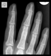
What can severe metabolic parathyroid bone disease cause?

A

Classical cystic appearance on X-ray.

18
Q

What is the classical cystic appearance on X-ray associated with severe metabolic parathyroid bone disease?

A

Brown Tumours

Osteitis fibrosa cystica

Subperiosteal erosions

Cysts

Acro-osteolysis

Pepper-pot skull

19
Q

Investigations of primary hyperPTH.

A

Bloods

Imaging

20
Q

Blood findings in primary hyperPTH.

A

High or non-suppressed PTH

Hypercalcaemia

Low phosphate

High ALP

21
Q

Why might ALP be high in primary hyperPTH?

A

Co-exisiting vitamin D deficiency can lead to increased bone turnover.

22
Q

Imaging findings of primary hyperPTH.

A

X-ray showing reduced bone density, sub-periosteal erosion of the phalanges.

Renal ultrasound can show nephrocalcinosis.

You might get chondrocalcinosis in joints that looks like pseudogout.

23
Q

What is nephrocalcinosis?

A

Calcium depositions in the kidneys.

Associated with nephrolithiasis.

24
Q

What is familial hypocalciuric hypercalcaemia (FHH)?

A

A genetic defect in the calcium sensing receptor.

25
FHH and primary hyperPTH can show similar levels of PTH and calcium. How can the two be distinguished?
FHH shows a low urine calcium/creatinine ratio Usually a family history of mild hypercalcaemia.
26
Why is it important to exclude FHH?
So you don't send the patient to have an unnecessary neck exploration.
27
If it is deemed necessary to do parathyroid surgery, what is important to do?
Localise the adenoma.
28
How is a parathyroid adenoma localised?
Experienced hands might be able to do it by **parathyroid ultrasound**. It is however very operator-dependent. SETAMIBI isotope scanning alongside of ultrasound is commonly performed. SPECT CT / MRI and 4-D CT can also be used.
29
When should surgery be performed in hyperparathyroidism?
Surgery should be considered if serum calcium \> 2.85 mmol/l or if symptoms are severe. Young patients and severe acute hypercalcaemia.
30
What treatment can be considered if the patient is ineligible for surgery?
Drugs such as calcimimetics which are effective at lowering calcium in primary hyperPTH.
31
What are the dangers of acute severe hypercalcaemia?
Medical emergency characterised by profound dehydration and renal impairment.
32
Treatment of acute severe hypercalcaemia.
First off stabilise hydration by giving IV saline. Give IV bisphosponates such as zoledronic acid to counter the calcium levels. When everything is stabilised you can consider glucocorticoids, calcitonin, calcimimetics or parathyroidectomy.
33
When might glucocorticoids be given in hypercalcaemia?
In lymphoma or granulomatous disease.
34
When does symptomatics **hypo**calcaemia occur?
When serum calcium is \< 1.9 mmol/l or at higher values if there is a **rapid** drop in calcium.
35
Most common cause of hypocalcaemia.
Post-surgical hypoPTH following **thyroidectomy**.
36
Prognosis of post-surgical hypoparathyroidism.
Often temporary, usually there is recovery of the parathyroid. However it can be permanent if there is damage or inadvertent removal of the parathyroid. This means that **long-term** **follow-up** is needed to assess if there is recovery of the parathyroid.
37
What else can cause hypocalcaemia?
Vitamin D deficiency Hypomagnesaemia
38
In which populations should vitamin D deficiency be considered?
High risk groups such neonates and elderly. Northern africans Populations which commonly covers themselves in clothes entirely.
39
Presentation of vitamin D deficiency in neonates.
Seizures Tetany This is due to hypocalcaemia.
40
How can you distinguish via bloods between hypoparathyroidism and vitamin D defiency?
Phosphate will be low in vitamin D deficiency due to elevated PTH levels. In hypoparathyroidism phosphate will be high due to low PTH levels.
41
What does hypomagnesaemia cause?
Functional hypoparathyroidism with normal or low PTH levels due to inhibition of the parathyroid.
42
Common causes of hypomagnesaemia.
GI loss Alcohol Drugs such as PPis.
43
Clinical features of acute severe hypocalcaemia.
Laryngospasm Prolonged QT interval Seizures
44
How does hypocalcaemia usually present?
Muscle cramps Carbo-pedal spasms Peri-oral and peripheral paraesthesia Neuro-psychiatric symptoms
45
Tests to assess hypocalcaemia.
Positive Chvosteks sign Positive Trousseaus sign
46
Explain a +ve Cvhosteks sign.
Facial spasm when the cheek is gently tapped with a finger.
47
Explain a +ve Trousseaus sign.
Carpo-pedal spasms induced by the inflation of a sphygmomanometer (blood-pressure cuff)
48
Treatment of acute hypocalcaemia.
IV calcium
49
Treatment of severe vitamin D deficiency.
Loading dose of cholecalciferol 20000 IU per week given for 7 weeks. Then a maintenance dose of 1-2000 IU per week.
50
Treatment of hypoparathyroidism.
1-alfacalcidol or calcitriol Sandocal or Adcal D3 (oral calcium supplements) are given in combination with 1-alfacalcidol.
51
Dose regiment of 1-alfacalcidol in hypoparathyroidism.
Starting dose of 0.25 microgram/day
52
Aim of treatment of hypocalcaemia.
Symptom relief Keep calcium levels at **lower end of reference range**.
53
Why is the treatment aim to keep the calcium levels at the lower reference range?
To reduce the risk of nephrocalcinosis.
54
Treatment of acute magnesium deficiency.
Any drugs that caused it should be stopped. IV magnesium replacement started. Usually given as MgS04 24 mmol/24 hours.
55
Treatment of magnesium deficiency if there is chronic GI loss or alcohol ingestion?
Appropriate specialist input.
56
What is pseudo-hypoparathyroidism?
A rare condition caused by a mutation in a G protein coupled to the PTH receptor leading to **PTH resistance**.
57
Parathyroid hallmarks of pseudo-hypoparathyroidism.
Hypocalcaemia High phosphate **High PTH** **Normal vitamin D**
58
How can you distinguish between pseudo-hypoparathyroidism and hypoparathyroidism?
Low levels of calcium and high levels of phosphate usually suggest hypoparathyroidism. This is because of low levels of PTH. Howevery the **high levels** of PTH alongside of hypocalcaemia and hyperphospataemia suggests PTH resistance rather than a deficiency.
59
Clinical presentation of pseudo-hypoparathyroidism.
Syndromic appearance with short stature, round face and short 4th and 5th metacarpals.
60
Pathway to determine cause of hypercalcaemia.
61
Treatment of acute hypercalcaemia.
If Ca2+ is \>3.5 mmol/L and symptomatic: 1 - Correct dehydration with IV 0.9% saline 2 - Bisphosphonates to prevent bone resorption by inhibiting osteoclastic activity. E.g. Pamidronate, Zoledronic acid. 3 - Chemotherapy can help in malignancy, steroids are used in sarcoidosis.
62
Causes of hypocalcaemia with increased phosphate.
CKD Hypoparathyroidism Pseudohypoparathyroidism Acute rhabdomyolysis Hypomagnesaemia
63
Causes of hypocalcaemia with normal or decreased phosphate.
Vit D def Osteomalacia Acute pancreatitis Over hydration Respiratory alkalosis
64
Features of hypocalcaemia.
SPASMODIC Spasms (Trousseau's sign) Perioral paraesthesiae Anxious, irritable, irrational Seizures Muscle tone increased Orientation impaired and confusion Dermatitis Impetigo herpetiformis Chvostek's sign, choreoathetosis, cataract, cardiomyopathy.
65
Treatment of hypocalcaemia.
Calcium 5 mmol/6h PO if mild In CKD may require alfacalcidol PO Severe =\> 10 ml of 10% calcium gluconate IV over 30 min