Imaging Relevant to Endocrine Disease Flashcards
(37 cards)
Pituitary gland location?
Found in the sella turcica and is closely related to the sphenoid sinus
It is inferior to the optic chiasm and hypothalamus and the carotid arteries are located laterally
Pituitary gland is connected to the brain via the pituitary stalk
Label the MRI of the pituitary gland and the structures surrounding it?

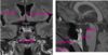
How does the posterior pituitary appear on MRI?
As a bright spot; if this is not present, it indicates posterior pituitary pathology
Features of bleeding (e.g: pituitary apoplexy)?
Sudden onset headache + bright, uniform area on MRI scan
Features of lymphocytic hypophysitis?
Hypopituitarism during pregnancy
+
Thickened pituitary stalk, loss of bright spot and enhancing pituitary gland
Features of pituitary pareidolia?
Imagined perception of a pattern where it does not actually exist, in this case a “big bird” for a pituitary macroadenoma
Location of the thyroid gland?
Deep to the strap muscles of the neck
Anterior to trachea and oesophagus
Medial to common carotid arteries and to internal jugular veins
Inferior relations include sternum, great vessels and the aortic arch
Label the stuctures surrounding the thyroid gland?


Label the inferior relations of the thyroid gland?


Imaging modalities used to visualise the thyroid gland?
CT and USS
Radio-isotope studies
Structures that are at risk during thyroid surgery?
Recurrent laryngeal nerves
Parathyroid glands
Causes of a midline neck mass in adults?
Enlarged thyroid gland
Enlarged lymph nodes are common (usually obvious)
Others include thyroglossal cysts, cystic hygroma (these are rare outwith childhood)
Aim of imaging the thyroid gland?
Differentiate between diffuse causes of goitre (e.g: Grave’s, thyroiditis) and focal causes (e.g: dominant nodule)
Advantages of thyroid USS?
Safe (no ionising radiation) and it is well-tolerated
Can be combined with FNA
When is thyroid USS used?
In euthyroid patients with goitre/palpable nodules
In hyperthyroid patients with focal masses/radio-isotope uptake
What does thyroid scintigraphy involve?
Radio-isotope injected IV and patient is imaged after 20 minutes; image is assessed for pattern and quality of tracer uptake
Features assoc. with Grave’s disease on thyroid scintigraphy?
Homogenously increased tracer uptake (>3% total tracer in gland)
Features assoc. with thyroiditis on thyroid scintigraphy?
Homogenously reduced tracer uptake
Features assoc. with multi-nodular goitre with dominant nodule on thyroid scintigraphy?
Focal uptake at right upper pole
Features of well-differentiated thyroid cancer on imaging?
Heterogeneous microcalcification
Location of adrenal glands?
Right adrenal - posterior to IVC
Left adrenal - lateral to aorta and left diaphragmatic crus
Label the structures surrounding the adrenal glands?


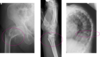
Different bone types on X-ray?
Cortical, trabecular
Medulla can also be seen

Formation of the medulla and cortex of bone?
Osteoblasts replace the cartilage with osteoid, which mineralises to form bony trabeculae
Trabeculae are loosely packed in the medulla (cancellous bone) but condense towards the cortex (compact bone)