Immuno: Primary Immune Deficiencies 1 Flashcards
(34 cards)
In which physiological states might you expect a relative degree of immunodeficiency?
- Neonates
- Elderly
- Pregnancy
List some examples of secondary immunodeficiency.
- Infection - HIV, measles
- Biochemical disorders - malnutrition, zinc/iron deficiency, renal impairment
- Malignancy - myeloma, leukaemia, lymphoma
- Drugs - corticosteroids, cytotoxic
What are some major clinical features of immunodeficiency?
- 2 major OR 1 major + recurrent minor infections in one year
- Unusual organisms
- Unusual sites
- Unresponsive to treatment
- Chronic infections
- Early structural damage
List some features that may suggest primary immunodeficiency.
- Family history
- Young age at presentation
- Failure to thrive
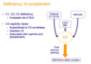
Broadly speaking, what are three mechanisms of phagocyte deficiency?
- Failure to produce neutrophils
- Defect of phagocyte migration
- Failure of oxidative killing
- Cytokine deficiency
Give three examples of failure of neutrophil production and outline their mechanism.
Reticular dysgenesis
- Autosomal recessive severe SCID with no production of lymphoid or myeloid cells
- Caused by failure of stem cells to differentiate along lymphoid or myeloid lineage
Kostmann syndrome
- Autosomal recessive congenital neutropenia
Cyclic neutropaenia
- Autosomal dominant episodic neutropaenia
- Occurs every 4-6 weeks

Name a phagocyte deficiency caused by failure of phagocyte migration.
Leukocyte adhesion deficiency

Describe the pathophysiology of leucocyte adhesion deficiency.
- Caused by deficiency of CD18
- CD18 normally combined with CD11a to produce LFA-1
- LFA-1 normally binds to ICAM-1 on endothelial cells to mediate neutrophil adhesions and transmigration
- A lack of CD18 means a lack of LFA-1, so neutrophils cannot enter tissues
- During an infection, neutrophils will be mobilised from the bone marrow (HIGH neutrophils in the blood) but they will not be able to cross into the site of infection (NO pus formation)
Name a phagocyte deficiency caused by failure of oxidative killing mechanisms.
Chronic granulomatous disease
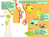
Outline the pathophysiology of chronic granulomatous disease.
- Absent respiratory burst (deficiency of components of NADPH oxidase leads to inability to generate oxygen free radicals)
- Excessive inflammation (persistent neutrophils and macrophage accumulation with failure to degrade antigens)
- Granuloma formation
- Lymphadenopathy and hepatosplenomegaly
Describe the cytokine cycle between macrophages and T cells.
- Macrophages produce IL12 which stimulates T cells, which then produce IFN-gamma
- IFN-gamma acts back on the macrophages and stimulates the production of TNF-alpha and free radicals
- Deficiencies in IL12, IL12R, IFN-gamma or IFN-gamma receptor can cause immunodeficiency

What type of infection do patients with IL12/IL12R or IFN-gamma/IFN-gamma receptor deficiencies tend to present with?
Organisms that infect macrophage (usually atypical mycobacteria)
Name and decribe the colour changes of two tests used to investigate chronic granulomatous disease.
- Nitroblue Tetrazolium (NBT) - yellow to blue
- Dihydrorhodamine (DHR) - fluorescent
NOTE: both of these tests are looking at the ability of neutrophils to produce hydrogen peroxide and oxidative stress
Which types of infection tend to occur in patients with phagocyte deficiency?
- Recurrent skin and mouth infections
- Bacteria - Staphylococcus aureus, enteric bacteria
- Fungi - Candida albicans, Aspergillus fumigatus
- Mycobacterial infections (particularly with IL12 deficiency)
- TB, atypical mycobacteria
For each of the following diseasesm state the expected neutrophil count, leucocyte adhesion markers, NBT/DHR test and presence of pus:
- Kostmann syndrome
- Leucocyte adhesion deficiency
- Chronic granulomatous disease
- IL12/IFN-gamma deficiency
-
Kostmann syndrome
- Absent neutrophil count
- Normal leucocyte adhesion markers
- No neutrophils for NBT/DHR
- No pus
-
Leucocyte adhesion deficiency
- High neutrophil count
- Absent CD18
- Normal NBT/DHR
- No pus
-
Chronic granulomatous disease
- Normal neutrophil count
- Normal leucocyte adhesion markers
- Abnormal NBT/DHR
- Pus present
-
IL12/IFN-gamma deficiency
- Normal neutrophil count
- Normal leucocyte adhesion markers
- Normal NBT/DHR
- Pus present
Outline the treatment of phagocyte deficiencies.
- Aggressive management of infection (infection prophylaxis and oral/IV antibiotics when needed)
- Haematopoietic stem cell transplantation
- Specific treatment for chronic granulomatous disease (e.g. IFN-gamma therapy)
What are the two different types of NK cell deficiency?
- Classical NK deficiency - absence of NK cells in the peripheral blood
- Functional NK deficiency - NK cells are present but function is abnormal

What is the main risk associated with NK cell deficiency?
Increased risk of viral infections (e.g. HSV, CMV, EBV, VZV)
Outline the treatment of NK cell deficiency.
- Prophylactic antiviral drugs (e.g. aciclovir)
- Cytokines (e.g. IFN-alpha to stimulate NK cytotoxic function)
- Haematopoietic stem cell transplantation
For each of the following conditions, state the stereotypical presentation:
- Kostmann syndrome
- Leucocyte adhesion deficiency
- Chronic granulomatous disease
- IFN-gamma receptor deficiency
- Classical NK cell deficiency
- Kostmann syndrome
- Recurrent infections with NO neutrophils on FBC
- Leucocyte adhesion deficiency
- Recurrent infections with HIGH neutrophils on FBC and no pus formation
- Chronic granulomatous disease
- Recurrent infections with hepatosplenomegaly and abnormal DHR
- IFN-gamma receptor deficiency
- Infection with atypical mycobacteria
- Normal FBC
- Classical NK cell deficiency
- Severe viral infections (e.g. chickenpox, disseminated CMV)
What is factor H?
A control protein in the complment cascade

What is the main clinical consequence of complement deficiency?
Increased susceptibility to infection by encapsulated bacteria
Which encapsulated bacteria are particularly problematic in patients with complement deficiency?
- Neisseria meningitidis
- Haemophilius influenzae
- Streptococcus pneumoniae
NOTE: susceptibility to N. meningitidis is particularly common in properidin and C5-9 deficiency
What are the consequences of MBL deficiency?
Common but NOT associated with immunodeficiency