Immunology - Exam 3 Flashcards

(57 cards)

1
Q

Describe imunological tolerance.

A

Immunological Tolerance

  • IT is specific UNRESPONSIVENESS to an Ag.
  • SELF‐TOLERANCE - All individuals are tolerant to self‐Ags.
  • AUTOIMMUNITY results from breakdown of self‐tolerance.
  • The NEGATIVE SELECTION of self‐reactive T lymphocytes in the thymus is NOT perfect.
  • There is a LOW LEVEL of physiological auto‐reactivity that is crucial to normal immune function.
  • The challenge is to understand how it becomes a PATHOLOGIC PROCESS and how T cells and B cells recognize self and contribute to tissue injury.
2
Q

Compare central vs peripheral intollerance.

A

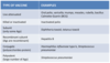
Immunological Intollerance - Central vs Peripheral

  • Central:
    • Induced in immature self‐reactive lymphocytes in the primary lymphoid organs.
    • Ensures that mature lymphocytes are NOT REACTIVE to self Ags.
    • Immature lymphocytes specific for self Ags may encounter these Ags in the generative (central) lymphoid organs and are either:
      • Deleted
      • Change BCR specificity
      • Develop into Treg cells.
  • Peripheral:
    • Induced in mature self‐reactive lymphocytes in peripheral sites.
    • Needed to prevent activation of these potentially dangerous lymphocytes in the tissues.
    • Mature self‐reactive lymphocytes in peripheral tissues may be either:
      • Inactivated (anergy)
      • Deleted (apoptosis)
      • Suppressed by the Treg cells
3
Q

Describe central T cell tollerance.

A

Central T Cell Tolerance

  • Takes place in THE THYMUS.
  • Thymocytes undergo a maturation and selection process.
  • Nonfunctional thymocytes showing NO AFFINITY at all undergo apoptosis.
  • STRONGLY SELF‐REACTIVE THYMOCYTES - as determined by interactions with MHC‐self peptide complexes - are also deleted.
  • Only thymocytes that are activated by MHC‐ self peptide complexes BELOW A CERTAIN THRESHOLD are positively selected and migrate into the periphery as mature T cells.
  • Most of these thymic emigrants develop into effector CD4+ and CD8+ T cells, and mediate both cell‐mediated and humoral (Ab‐mediated) immune responses.
  • A SMALL PERCENTAGE OF T CELLS that emigrate from the thymus express FOXP3 and develop into natural CD4+CD25+CTLA4+ Treg cells.
4
Q

Describe central B cell tolerance.

A

Central B Cell Tolerance

  • CLONAL DELETION and ANERGY were major mechanisms mediating central tolerance of developing autoreactive B cells, resulting in the elimination of autoreactive clones, and preventing immune responses against self.
  • When an immature B cell reacts with a self‐antigen with HIGH AVIDITY, such as a highly expressed membrane‐bound protein, it undergoes apoptosis within 2–3 d.
  • In contrast, LOW AVIDITY interactions of B cells with self‐antigens induce unresponsiveness to subsequent stimulation or anergy but allowed for migration into peripheral compartment. The anergic B cells fail to enter follicle and have reduced life‐span.
  • However, clonal deletion and anergy are not the only modes of selection against autoreactive immature B cells, but there operates another system, namely, RECEPTOR EDITING.
  • Autoreactive immature B cells reactivated their Ig gene rearrangement program at the Ig light chain loci resulting in the expression of a new light chain that paired with the existing H chain to form a non‐autoreactive BCR, an event that promoted the selection of these edited B cells into the periphery.
5
Q

Describe BCR editing.

A

BCR Editing

  • Precursor (pre)‐B cells, which already express rearranged IgH chains recombine the locus that encodes IgL chain, yielding a lymphocyte with an autoreactive antigen receptor.
  • BCR signaling promotes developmental arrest and continued recombination.
  • Receptor editing of the IgL chain leads to expression of a distinct IgL chain, generating cell‐surface immunoglobulin that lacks self‐reactivity
6
Q

Describe deletion of self-reactive lymphocytes.

A

Deletion of Self-Reactive Lymphocytes

7
Q

Describe the role of Treg cells in peripheral tolerance.

A

Peripheral Tolerance - Role of Treg Cells

  • Treg cells are key mediators of peripheral tolerance.
  • Treg cells may inhibit T cell activation by APCs and inhibit T‐cell differentiation into CTLs.
  • Treg cells may prevent T cells from providing help to B cells in the production of Abs.
  • FOXP3+ Treg cells can also be generated from peripheral T cells (not shown).
8
Q

Compare natural and inducible Treg cells.

A

Natural vs. Inducible Treg Cells

  • The development and survival of these regulatory T cells require IL-2 and FoxP3.
  • In peripheral tissues, Treg cells suppress the activation of self-reactive lymphocytes.
9
Q

Describe induced Treg cells.

A

Induced Treg Cells

  • Differentiate in the periphery.
  • In addition to the natural Treg cells which differentiate in the thymus, mature T cells OUTSIDE THE THYMUS can also acquire Treg phenotype and function.
  • These are called induced Treg cells (iTreg cells).
  • FoxP3 EXPRESSION can be induced in naive CD4+ cells in vitro by antigen recognition in the presence of TGF‐β.
  • There is a close developmental RELATIONSHIP between iTregs and Th17 cells.
  • Ag recognition in the presence of TGF‐β induces FoxP3 expression if IL‐6 is NOT present.
  • In contrast, Ag recognition in the presence of TGF‐β + IL‐6 prevents FoxP3 expression, induces expression of the retinoic acid receptor (RAR) related orphan nuclear receptor RORγt expression and therefore, Th17 cell DIFFERENTIATION.
10
Q

Describe peripheral B cell tolerance.

A

Peripheral B Cell Tolerance

  • Mature B cells that recognize self Ag in peripheral tissues in the absence of specific Th cells may be rendered functionally UNRESPONSIVE or die by APOPTOSIS.
  • The CD22 inhibitory receptor is phosphorylated by Lyn and then recruits SHP‐1 tyrosine phosphatase attenuating BCR signaling.
  • Therefore, DEFECTS in Lyn tyrosine kinase, SHP‐1 tyrosine phosphatase, and the CD22 inhibitory receptor lead to AUTOIMMUNITY.
11
Q

Describe the mutations breaking tolerance.

A

Mutations Breaking Tolerance

  • Incomplete induction of tolerance in the thymus (AIRE deficiency causes Autoimmune Polyendocrine Syndrome).
  • Impaired production of regulatory T cells (FoxP3 deficiency causes IPEX syndrome).
  • DECREASED CLEARANCE and impaired tolerance induction by apoptotic cells (complement deficiency of C1q and C4).
  • ALTERED IMMUNE SIGNALING thresholds (CTLA‐4 polymorphisms).
  • Loss of Self Tolerance Leads to Autoimmunity.
12
Q

Describe AIRE (AutoImmune Regulator) in central tolerance.

A

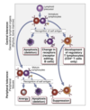
Central Tolerance - AIRE (AutoImmune Regulator)

  • The NEGATIVE SELECTION of T cells in the thymus is necessary for the maintenance of self tolerance.
  • Medullary THYMIC EPITHELIAL CELLS have a key function as APCs.
  • They EXPRESS a large number of SELF‐Ags that are presented to developing T cells.
  • MUTATIONS in AIRE (autoimmune regulator ) protein cause a breakdown of central tolerance.
  • AIRE has been proposed to function as a TRANSCRIPTION FACTOR.
  • Mutation in AIRE is associated with DECREASED EXPRESSION of self‐Ags in the thymus.
13
Q

Describe how aberrant expression of AIRE leads to autoimmunity.

A

Autoimmunity - Aberrant Expression of AIRE

  • The AIRE regulates the expression of tissue‐restricted Ags (TRAs).
  • Peptides derived from these Ags are displayed on the Medullary Thymic Epithelial Cells.
  • Ags are recognized by immature Ag‐ specific T cells, leading to the deletion of self‐reactive T cells.
  • In the absence of functional AIRE, these self‐reactive T cells are not eliminated and they can enter tissues where the Ags continue to be produced and cause injury.
14
Q

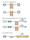
Describe the outcomes of Ag-dependent T cell activation.

A

Ag-Dependent T Cell Activation - Outcomes

15
Q

Describe the role of CTLA4 (Cytotoxic T-Lymphocyte Antigen 4) in peripheral tolerance.

A

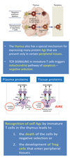
Peripheral Tolerance - Role of CTLA4 (Cytotoxic T-Lymphocyte Antigen 4)

  • Upon Ag ENCOUNTER, individual populations of T cells undergo expansion and later contraction after the elimination of Ag.
  • T cell activation is regulated by members of the B7‐CD28 family of COSTIMULATORY MOLECULES.
  • CTLA4 (Cytotoxic T‐Lymphocyte Antigen 4) is a homolog of CD28.
  • CTLA4 is an INHIBITORY RECEPTOR.
  • CTLA4 provides signals that terminate immune responses and maintain self‐tolerance.
16
Q

Describe the funcitons of CTLA-4.

A

CTLA-4 Functions

  • UNCONTROLLED LYMPHOCYTE ACTIVATION with massively enlarged LNs and spleen and fatal multi-organ lymphocytic infiltrates is seen in CTLA-4 KO mice.
  • BLOCKING of CTLA-4 with Abs also enhances autoimmune diseases in animal models.
  • POLYMORPHISMS in the CTLA-4 are associated with several autoimmune diseases in humans, including type 1 diabetes and Graves’ disease.
  • CTLA-4 has two important properties:
    • CTLA-4 expression is low on resting T cells until the cells are activated by Ag.
    • Once expressed CTLA-4, terminates continuing activation of these responding T cells.
  • CTLA-4 is expressed on REGULATORY T cells and mediates the suppressive function of these cells by inhibiting the activation of naive T cells.
17
Q

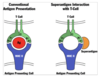
Describe the MOA of CTLA-4.

A

CTLA-4 - Mechanism of Action

  • CELL-INTRINSIC ACTION:
    • Engagement of CTLA-4 on a T cell may deliver inhibitory signals that terminate further activation of that cell.
  • CELL-EXTRINSIC ACTION:
    • CTLA-4 on Treg cells or responding T cells binds to B7 molecules on APCs or makes unavailable to CD28.
18
Q

Describe Treg cells and their role in regulating T cell responses.

A

Treg Cells

  • Treg cells develop in THE THYMUS.
  • Treg cells are POSITIVELY SELECTED in the thymus via strong TCR interactions with self‐Ags.
  • After recognition of self‐Ags they are NOT ELIMINATED by apoptosis.
  • Treg cells are able to produce ANTI‐APOPTOTIC MOLECULES which protect them from negative selection in the thymus.
  • The generation of some Treg cells requires the TGF‐β.
  • Treg cells express FOXP3 transcriptional factor and are CD4+CD25+ positive.
  • Treg cells typically express high levels of CTLA‐4.
  • CYTOKINE IL‐2 is critical for survival and functional competence of Treg cells.
  • Treg cells are endogenous LONG‐LIVED populations of self‐Ag‐specific T cells.
  • Treg cells serve to prevent potentially AUTOIMMUNE REACTIONS.
19
Q

Describe TGF-β.

A

Transforming Growth Factor - β

  • INHIBITS the proliferation and effector functions of T cells.
  • INHIBITS development of Th1 and Th2 subsets but PROMOTES Th17 in cooperation with IL‐1 and IL‐6.
  • INHIBITS activation of M1 macrophages.
  • REGULATES the differentiation of induced FoxP3+ Treg cells.
  • STIMULATES production of IgA by inducing B cells to switch to this isotype.
  • PROMOTES tissue repair after local immune and inflammatory reactions subside stimulating collagen synthesis and matrix‐modifying enzyme production by macrophages and fibroblasts.
20
Q

Describe autoimmunity.

A

Autoimmunity

  • About 5% or ~ 12‐15 million people from AUTOIMMUNE DISEASES in the US alone suffer.
  • There are 60‐70 diverse autoimmune diseases which affect various tissues of the human body.
  • There is NO known CURE or clear UNDERSTANDING the cause for any of autoimmune conditions.
  • Most autoimmune diseases are treated symptomatically.
  • The autoimmune diseases bring the PARADOX proposition that “the body both is and is not itself”.
21
Q

Describe autoimmunity in chronic disease.

A

Autoimmunity - Chronic Disease

  • There is NO FUNDAMENTAL DIFFERENCE between the structure of self auto‐Ags and non‐self Ags because Ags are all proteins composed by the same amino acids.
  • PATHOLOGIC immune RESPONSE against self Ags often clinically manifested as “immune‐mediated inflammatory diseases”.
  • CAUSED BY the activation of T cells and/or B cells in the absence of an ongoing infection or other discernible cause.
  • A result of a HYPERSENSITIVE IMMUNE SYSTEM that causes one’s own immune system to attack the self.
22
Q

Describe the prevention of autoimmunity.

A

Autoimmunity - Prevention

  • T cells that are physically separated from their specific Ag (the BBB) cannot become activated, a process termed immunologic ignorance.
  • T cells that express the Fas (CD95) can receive their signals from cells that express FasL and undergo apoptosis, a process known as deletion.
  • CTLA4 (CD152) that binds CD80 on APC and inhibits T cells activation.
  • Regulatory T cells can inhibit through the production of inhibitory cytokines such as IL-10 and TGFβ.
23
Q

Describe the factors determining Ag response vs tolerance.

A

Factors Determining Ag Response vs Tolerance

24
Q

Describe the mechanisms of autoimmunity.

A

Autoimmunity - Postulated Mechanisms

  • Various genetic loci may confer SUSCEPTIBILITY TO AUTOIMMUNITY, in part by influencing the maintenance of self‐tolerance.
  • Environmental triggers, such as infections and other inflammatory stimuli, promote the influx of lymphocytes into tissues and the activation of self‐reactive T cells, resulting in tissue injury.
25
Describe the general features of autoimmune disorders.
Autoimmune Disorders - General Features * Autoimmune diseases may be either SYSTEMIC or ORGAN SPECIFIC, depending on the distribution of the auto‐Ag that are recognized . * VARIOUS EFFECTOR MECHANISMS are responsible for tissue injury in different autoimmune diseases. * Autoimmune diseases tend to be chronic, progressive, and self‐perpetuating. * FAILURE of the mechanisms of self‐tolerance in T or B cells underlies cause of all autoimmune diseases. * INFLAMMATION or an initial innate immune response.
26
Describe the role of genetics in autoimmunity.
Autoimmunity - Genetics * Most autoimmune diseases are COMPLEX POLYGENIC TRAITS. * Affected individuals inherit multiple GENETIC POLYMORPHISMS that contribute to disease susceptibility. * Among the genes that are associated with autoimmunity, the strongest associations are with MHC genes. * Polymorphisms in NON‐HLA genes is also associated with autoimmunity. * Susceptibility genes act with ENVIRONMENTAL FACTORS to cause the diseases.
27
Describe the etiology and pathogenesis of autoimmunity.
Autoimmunity - Etiology & Pathogenesis
28
Describe environmental triggers and their role in autoimmunity.
Autoimmunity - Environmental Triggers * Microbial Ags can initiate autoimmune disorder through: * Molecular mimicry: * Rheumatic fever is triggered by streptococcal infection and mediated by cross‐reactivity between streptococcal Ags and cardiac myosin. * Multiple sclerosis - T cells react with myelin basic protein and peptides from Epstein-Barr virus, influenza virus type A, and human papillomavirus. * Polyclonal (bystander) activation: * Microbial infection can also cause polyclonal activation of autoreactive lymphocytes (cytokine field). * Release of previously sequestered Ags: * Microbes that kill cells can cause inflammation, the release of sequestered Ags, and autoimmunity.
29
Describe noninfectious triggers and their role in autoimmunity.
Autoimmunity - Noninfectious Triggers * AUTOIMMUNE DISEASES are much more common in women than in men: * Estrogens exacerbate Systemic Lupus Erythematosus (SLE) in mouse models of the disease by altering the B‐cell repertoire in the absence of inflammation. * Drugs can also alter the immune repertoire: * Penicillins and cephalosporins can bind to RBC membrane and generate a neoantigen that elicits an auto‐Ag that causes hemolytic anemia. * The blockade of TNF‐α (ENBREL or other inhibitors) can induce antinuclear Abs and even SLE and Multiple Sclerosis (MS) in certain persons. * TNF‐α has inhibitory effects on activated T cells, but it remains unknown how TNF‐α induces autoimmunity.
30
Describe immune responses to microbes.
Immune Responses to Microbes * DEFENSE AGAINST MICROBES is mediated by the effector mechanisms of innate and adaptive immunity. * The immune system responds in specialized and distinct ways to different types of microbes to most effectively combat these infectious agents. * The SURVIVAL and PATHOGENICITY of MICROBES in a host are critically influenced by the ability of the microbes to evade or resist the effector mechanisms of immunity. * In many infections, TISSUE INJURY and disease may be caused by the host response to the microbe (collateral damage) rather than by the microbe itself. * Inherited and acquired DEFECTS IN innate and adaptive IMMUNITY are important causes of susceptibility to infections. * Many microbes establish LATENT, or PERSISTENT INFECTIONS in which the immune response controls but does not eliminate the microbe and the microbe survives without propagating the infection.
31
Describe the immune response to a primary extracellular bacterial infection.
Immune Response - Primary Extracellular Bacterial Infection * A: * Break in epithelial surface allows bacterial entry and proliferation. * B: * Surface lipopolysaccharide (LPS) may activate the ALTERNATIVE complement PATHWAY or mannan‐binding protein (MBP) the LECTIN PATHWAY leading to bacterial lysis. * Other complement activators operating at this stage include acute phase proteins C‐reactive protein (CRP) and serum amyloid protein (SAP). * CRP is known to bind bacterial surfaces (phosphocholine) and to bind the globular heads of C1q and activates the CLASSICAL PATHWAY of complement. * Acute phase proteins bind bacterial coat and activate complement. * C: * C3a and C5a bind to receptors on resident MAST CELLS and activate them. * Mast cell degranulation enhances BLOOD FLOW. * The increased blood flow and LOCAL EDEMA are perceived as itchiness and irritation in the inflamed area. * D: * Locally released CYTOKINES and CHEMOKINES and bacterial‐derived molecules (e.g. ENDOTOXIN) activate both the endothelium and the neutrophils. * Rolling marginating NEUTROPHILS adhere to the vein wall. * E: * Complement fragments (C5a and C3a) and chemokines (IL‐8/CXCL8) are potent NEUTROPHIL CHEMOATTRACTANTS. * Together with bacterial products such as Formyl‐Methionyl‐Leucyl‐ Phenylalanine tripeptide (fMLP), they attract neutrophils to the site (CHEMOTAXIS). * F: * Opsonized BACTERIA are rapidly engulfed and killed by neutrophils and tissue macrophages (not shown). * IMMATURE DCs engulf and internalize bacteria (Ags) via pattern recognition receptors or PRRs (e.g. Toll‐like receptors). * Activated MATURE DCs migrate to the local LNs via the lymphatics. * G: * DCs enter local LNs, and moves to the T cell zone (shown incorrectly on the cartoon). * Local inflammation leads to up‐ regulation of adhesion molecules on high endothelial venules (HEV) of lymph node, and lymphocytes enter directly from the blood. * Many LYMPHOCYTES become trapped, activated, and proliferate in the local inflamed LN. * This leads to the consequent swelling and local hyperemia that manifested by the SYMPTOMS of swollen painful/tender LNs. * H: * NAÏVE Th CELLS are recruited and activated by DCs in the lymph node. * Naïve T cells become differentiated towards effector Th1 and Th2, according to the DC signals. * Activated Th cells migrate towards the GERMINAL CENTERS and interact with Ag‐activated B cells, promoting class switching and affinity maturation of bacteria‐ specific Abs. * Initially, IgM class Ab is produced, followed by clonal expansion and switching to other classes, e.g. IgG or IgA for mucosal pathogens. * I: * Early antibacterial Ab production is of the IgM CLASS. * This relatively low affinity interaction is enhanced by the five adhesion sites on IgM, leading to higher avidity of the binding. * IgM is a very potent complement activator. * After formation of multiple MACs, bacteria are lysed by complement. * Bacteria are also opsonized with C3b via IgM‐ACTIVATED COMPLEMENT (classical pathway) that increases phagocytosis. * J: * Upon elimination of pathogens, the IMMUNE RESPONSES is contracted and most of effector lymphocytes die via apoptosis. * Protective mechanisms for future encounters are put in place by the laying down of memory B and T cells. * K: * In the RESOLUTION of an infection, bacterial debris is removed by local phagocytes (macrophages and neutrophils) or by antibody as soluble immune complexes.
32
Describe the pathogenic mechanisms of extracellular bacteria.
Pathogenic Mechanisms of Extracellular Bacteria * EXTRACELLULAR BACTERIA are capable of replicating outside host cells in the blood, connective tissues, epithelial surfaces, the GI tract etc. * Infections caused by PATHOGENIC extracellular bacteria have two principal MECHANISMS: 1. Tissue damage is caused by Inflammation at the site of infection 2. Bacteria produce toxins which have diverse pathologic effects. * The BACTERIAL TOXINS subdivided into: * Endotoxins which are components of bacterial cell walls. * Exotoxins which are secreted by the bacteria. * The ENDOTOXIN (LPS) of Gram‐negative bacteria is as a potent activator of MΦ, DCs, and endothelial cells. * Many EXOTOXINS are cytotoxic including diphtheria toxin (shuts down protein synthesis in infected cells), cholera toxin (interferes with ion/water transport) tetanus toxin (inhibits neuromuscular transmission). * Other exotoxins interfere with normal cellular functions without killing cells, and yet other exotoxins stimulate the production of cytokines that cause disease.
33
Describe innate immunity to extracellular bacteria.
Innate Immunity - Extracellular Bacteria * The PRINCIPAL MECHANISMS of innate immunity to extracellular bacteria are: * Complement activation * Phagocytosis * Inflammation * COMPLEMENT ACTIVATION: * Bacteria that express mannose on their surface may bind mannose‐binding lectin, which activates complement by the lectin pathway. * Bacterial peptidoglycans (Gram+ bacteria) and LPS (Gram‐ bacteria) activate the alternative pathway. * Byproducts (C3a and C5a) stimulate inflammation by recruiting and activating leukocytes. * Complement activation results in opsonization and enhanced phagocytosis of the bacteria. * The MAC lyses bacteria (Neisseria) that are particularly susceptible to lysis.
34
Describe the prevention of host bystander damage.
Prevention of Host Bystander Damage * A: * The cleavage of C3b and C4b by FACTOR I prevents them from forming active convertases and requires cofactor activity. These cofactors include the membrane‐bound membrane cofactor protein (MCP) and complement receptor 1 (CR1), Factor H (FH) and C4b‐binding protein (C4BP). * B: * Proteins decay‐accelerating factor (DAF), CR1, and C4BP inhibit assembly of new C3 convertases and shorten the half‐life of the preformed convertases, limiting their ability to participate in complement activation: * Classical pathway – DAF, CR1, and C4BP * Alternative pathway – DAF, Factor H, and CR1. * C: * The MAC is the lytic complex of complement and its assembly can be inhibited by the membrane‐bound MAC‐INHIBITORY PROTEIN (CD59).
35
Describe opsonization and phagocytosis.
Opsonization & Phagocytosis
36
Describe innate immune evasion by extracellular bacteria.
Innate Immune Evasion by Extracellular Bacteria
37
Describe humoral immunity against extracellular bacteria.
Humoral Immunity Against Extracellular Bacteria * Humoral immunity is a MAJOR PROTECTIVE RESPONSE against extracellular bacteria. * It functions to block infection, to eliminate the microbes, and to neutralize their toxins. * The EFFECTOR MECHANISMS of Abs include: * Toxin neutralization * Opsonization and phagocytosis * Complement activation by the classical pathway * ANTIBODIES responses against extracellular bacteria are directed against cell wall Ags and secreted and cell‐associated toxins. * ENCAPSULATED BACTERIA rich in TI polysaccharide Ags are primarily eliminated by Ab‐mediated immunity.
38
Describe cell-mediated immunity against extracellular microbes.
Cell-Mediated Immunity Against Extracellular Microbes * The PROTEIN Ag of extracellular bacteria also activate CD4+ helper T cells. * Th17 CELLS induced by these microbes promote local inflammation and recruit neutrophils and monocytes at sites of bacterial infection. * Genetic defects in Th17 development have increased susceptibility to bacterial and fungal infections, with formation of multiple skin abscesses. * Bacteria also induce Th1 CELLS and IFN‐γ produced by the cells activates MΦ to destroy phagocytized microbes. * IFN‐γ may also stimulate production of opsonizing and COMPLEMENT‐FIXING IgG Abs.
39
Describe the injurious effects of immune response.
Immune Response - Injurious Effects * The principal INJURIOUS CONSEQUENCES of host responses to extracellular bacteria are inflammation and septic shock. * These inflammatory reactions are usually self‐limited and controlled. * SEPTIC SHOCK is a severe pathologic consequence of disseminated bacterial infection (sepsis) by some Gram‐negative and Gram‐positive bacteria. * SEPTIC SHOCK SYNDROME is characterized by circulatory collapse and disseminated intravascular coagulation. * The EARLY PHASE of sepsis and septic shock is caused by cytokines produced by MΦ that are activated by bacterial cell wall components. * Cytokines secreted cause the SYSTEMIC MANIFESTATIONS of the infection and stimulate the production of acute‐phase proteins. * The same reactions of NEUTROPHILS and MΦ that function to eradicate the infection also cause tissue damage by local production of reactive oxygen species and lysosomal enzymes.
40
Describe the mediators of septic shock.
Septic Shock - Mediators
41
Describe the mechanisms of septic shock.
Septic Shock - Mechanisms * MΦ release a diverse range of products implicated in the PATHOGENESIS OF SEPSIS. * Many MΦ PRODUCTS are involved in the regulation of each other: * TNF‐α upregulates tissue factor (TF) and nitric oxide synthase (iNOS), IL‐18 induces IFN‐γ, which in turn further activates MΦ. * IL‐10 is a global suppressor of MΦ function. * These highly COMPLEX and tightly regulated NETWORKS make it difficult to predict the outcomes of blocking or inhibiting just one pathway.
42
Describe bacterial superantigens.
Bacterial Superantigens (SAgs) * Certain bacterial toxins called superantigens (SAgs) bind to the class II MHC outside the peptide‐ binding groove. * Simultaneously, SAgs binds to the variable region of different TCR β chains, regardless of the peptide specificity of the TCR. * Because many T cells express a TCR β chain from a particular Vβ family, Sags can activate a large number of T cells causing POLYCLONAL T CELL ACTIVATION.
43
Describe the role of SAgs in human diseases.
SAgs in Human Diseases * FOOD POISONING – the staphylococcal SAgs are potent GI toxins responsible for staphylococcal food poisoning. * TOXIC SHOCK SYNDROME (TSS) is caused by S. aureus can be considered as a capillary leak syndrome. * Streptococcal TSS is caused by S. pyogenes. It is the MOST SEVERE form of invasive streptococcal infection. * ACUTE RHEUMATIC FEVER (ARF) is a post‐infection cause of preventable pediatric heart disease. * KAWASAKI DISEASE (KD) is an acute multi‐system vasculitis of unknown etiology – evidence suggests that it is a SAg‐mediated disease. * AUTOIMMUNE DISEASES – It has been proposed that SAgs might contribute to the pathogenesis of autoimmune disease by activating T cells that are specific for self Ags.
44
What is the major mechanism used by bacteria to evade humoral immunity?
The Major Mechanism Used by Bacteria to Evade Humoral Immunity Is Variation of Surface Ags.
45
Describe intracellular pathogens.
Intracellular Pathogens * INTRACELLULAR BACTERIA and viruses are able to survive and replicate within host cells where they are inaccessible to circulating Abs. * An ELIMINATION of these bacteria requires the mechanisms of cell‐mediated immunity. * In many intracellular bacterial and viral infections the host responses also cause tissue injury.
46
Describe the immune response to a primary viral infection.
Immune Response - Primary Viral Infection * A: * Virus infects epithelial cells and replicates among them. * B: * Effect of intracellular viral infection is the ACTIVATION OF CYTOKINE and cytokine‐receptor genes, especially the Type I interferons (e.g. IFN‐α). * LOCAL EFFECTS of IFN‐α are inhibition of viral gene replication, and up‐regulation of MHC class I molecules. * VIRAL PEPTIDES are expressed in the MHC class I peptide‐binding groove. * C: * NK CELLS may be recruited at two points at least during the virus infection. * They exhibit an innate (early in the course of infection) antiviral role following activation by epithelium‐ derived IFN‐α. * At a later stage of infections, NK cells are activated by cytokines IFN‐γ and IL‐2 produced by Th1 cells specific for the virus. * D: * VIRAL INFECTION results in cell death and viral replication. * COMPONENTS of viruses (e.g. single‐ stranded RNA) activate DCs and locally released cytokines and chemokines amplify the activation of MΦ and professional APCs. * These cells engulf cellular debris and present viral proteins. * Professional APCs (e.g. tissue DCs such as LANGERHANS CELLS in the skin) transport Ag to local LNs via lymphatics. * E: * CYTOKINES up‐regulate endothelial cell expression of adhesion molecules such as ICAM‐1. * CHEMOKINES (e.g. IL‐8/CXCL-8) begin to attract cells through the endothelium towards the site of infection. * IL‐1 and TNF‐α locally produced by MΦ and T cells enter bloodstream and have systemic effects of fever and arthralgia/myalgia. * F: * DCs enter local LNs, and moves to the T cell zone (shown incorrectly on the cartoon). * Local inflammation leads to up‐ regulation of adhesion molecules on high endothelial venules (HEV) of lymph node, and lymphocytes enter directly from the blood. * Many LYMPHOCYTES become trapped, activated, and proliferate in the local inflamed LN. * This leads to the consequent swelling and local hyperemia that manifested by the SYMPTOMS of swollen painful/tender LNs. * G: * NAÏVE T CELLS possessing TCRs complementary to the class II MHC molecule/viral peptide complex are activated and become Th1 cells. * NAÏVE B CELLS acquire viral Ags through attachment to surface IgM or IgD. * Antiviral IgM are produced as a result of PRIMARY Ab RESPONSE. * Ag‐ACTIVATED B CELLS process and present viral peptides to Th cells (either Th2 or Th1) from which they receive positive growth and differentiation signals. * B cells differentiate and class switch, leading later to production of high affinity antiviral IgG (SECONDARY Ab RESPONSE). * H: * A VIRAL PEPTIDE is presented by class II MHC molecules to a complementary TCR on a Th cell. * The INTERACTION IS STABILIZED by CD4/class II MHC and CD80/86 binding to CD28, which also provides co‐ stimulatory signals to the Th cell. * I: * Th 1 CELLS recruit and stimulate virus‐specific cytotoxic T lymphocytes (CTLs) by providing IL‐2 for proliferation of CD8+ T cells. * The CTLs recognize virus PEPTIDES CROSS‐PRESENTED by DCs. * The same VIRAL EPITOPES will be presented within class I MHC on the surface of infected target cells. * J: * Effector Th cells and CTLs leave the LN via the draining lymphatics and ultimately enter the blood. * At this stage their KEY ATTRIBUTES are: 1. Virus‐specific TCRs. 2. Up‐regulated adhesion molecules (LFA‐1), to allow migration into the inflamed tissues. 3. Up‐regulated production of cytokines. * K: * Virus‐specific CTLs migrate from the blood into peripheral tissue. * CTLs recognize viral Ags presented within class I MHC on virally infected cells and kill them. * In the tissue, Th1 cells, CTLs and B cells organize the local antiviral immune response: * Th1‐derived IFN‐γ activates phagocytosis by MΦ. * Abs facilitate phagocytosis via FcR and CR1. * L: * NK CELLS may be recruited at two points at least during the virus infection. * They exhibit an innate (early in the course of infection) antiviral role following activation by epithelium‐ derived IFN‐α. * At a later stage of infections, NK cells are activated by cytokines IFN‐γ and IL‐2 produced by Th1 cells specific for the virus. * M: * VIRUS‐INFECTED CELLS secrete viral proteins after their death. * These proteins may be neutralized or removed by Ab in the form of immune complexes. * Ab may guide Fc receptor‐ expressing NK CELLS that culminates in Antibody‐ Dependent Cell‐Mediated Cytotoxicity (ADCC). * N: * After resolution of the infection, virus‐specific MEMORY T and B CELLS reside long term in lymph nodes, spleen and bone marrow. * PLASMA CELLS ensure long‐ term circulation of protective virus‐neutralizing Abs.
47
Describe innate immunity to intracellular bacteria.
Innate Immunity - Intracellular Bacteria * The innate immune response to INTRACELLULAR BACTERIA is mediated mainly by phagocytes and NK cells. * Both NK cells and MΦ provide an EARLY DEFENSE against these microbes, before the development of adaptive immunity. * Products of these bacteria are recognized by TLRs and cytoplasmic proteins of the NOD‐LIKE RECEPTORS resulting in activation of DCs, MΦ and neutrophils. * PHAGOCYTES ingest and destroy intracellular microbes. * The RESISTANCE of pathogenic bacteria to degradation within phagocytes is overrun by NK cell‐produced IFN‐γ. * Activated DCs and MΦ produce IL‐12 and IL‐15 which activate NK cells. * The NK CELLS produce IFN‐γ, which in turn promotes killing of the phagocytized bacteria by MΦ.
48
Compare endogenous vs. exogenous pathways of Ag presentation.
Endogenous vs. Exogenous Pathways of Ag Presentation * ENDOGENOUS PATHWAY: * Proteins from intracellular pathogens, such as viruses, are degraded by the proteasome and the resulting peptides are shuttled into the ER by TAP proteins. * These peptides are loaded onto MHC class I molecules and the complex is delivered to the cell surface, where it stimulates CTLs that kill the infected cells. * EXOGENOUS PATHWAY: * Extracellular pathogens are engulfed by phagosomes. * Inside the phagosome, the pathogen‐derived peptides are loaded onto MHC class II molecules, which activate Th cells that stimulate the production of Abs. * Some peptides from the exogenous pathway can also be presented on MHC class I molecules.
49
Describe infection with M. tuberculosis.
Infection with M. tuberculosis * M. tuberculosis may SURVIVE in PHAGOSOMES by preventing acid‐containing lysosomes from fusing with phagosomes and creating mature phagolysosomes. * CD4+ TH1 CELLS respond to class II MHC‐associated M. tuberculosis Ags and produce IFN‐γ, which activates MΦ to destroy the microbes in phagosomes. * CD8+ T CELLS respond to class I MHC‐associated peptides derived from cytosolic Ags (cross‐presenting) and kill the infected cells.
50
Describe immune evasion by intracellular bacteria.
Immune Evasion by Intracellular Bacteria
51
Describe the role of TH1/TH2 cells in infection outcome.
Infection Outcome - Role of TH1/TH2 * NAIVE CD4+ T CELLS may differentiate into Th1 cells, which activate phagocytes to kill ingested microbes or Th2 cells, which inhibit this classical pathway of MΦ activation. * The Th1/Th2 BALANCE may influence the outcome of infections, as illustrated by Leishmania infection in mice and Mycobacterium leprae in humans.
52
Describe defenses against fungal infection.
Defenses Against Fungal Infection * FUNGI are recognized by PRRs (TLRs and C lectin‐like receptors) binding the PAMPs. * The detection of β‐GLUCAN by dectin 1 is also important. * Then occurs DIFFERENTIATION of Th1, Th2 and Th17 cells and production of cytokines. * Cytokines IL‐12 and TGFβ + IL‐6 have IMPORTANT differentiation and activation ROLES for activation of Th1 and Th17 responses. * Th1 (IFN‐γ) and Th17 (IL‐17, IL‐22) cytokines further amplify an inflammation and innate immunity. * Specific Th2 cells (Abs) are LESS IMPORTANT. * In general, Th1 RESPONSES are required for clearance of a fungal infection, while Th2 RESPONSES usually results in susceptibility to infection.
53
Describe PRR dectin 1.
Pattern Recognition Receptor Dectin 1 * Binds β-glucans. * The MΦ MANNOSE RECEPTOR (MR) has historically been considered the major receptor involved in the nonopsonic recognition of fungi. * DECTIN‐1 is a recently discovered PRR that plays an important role in antifungal innate immunity. * Recent data suggest that DECTIN‐1 and TLR2/TLR6 signaling combine to enhance the responses triggered by fungi. * Dectin‐1 is a specific receptor for β‐GLUCANS expressed on MΦ. * β‐glucans are POLYSACCHARIDE PAMPS that contain only GLUCOSE as structural components. * DECTIN‐1 binds and internalizes β‐glucans and mediates activation of NF‐κB and subsequent secretion of proinflammatory cytokines and production of reactive oxygen species (ROS).
54
Describe immune protection based on microbe class.
Immune Protection by Microbe Class
55
Summarize protective immunity.
Protective Immunity
56
Describe infections in immunocompromised patients.
Infections in Immunocompromised Patients
57
What are the types of vaccines?
Vaccines