INBR 7 - Neurobiology Flashcards
(100 cards)
1 Kondensasi dan Fragmentasi kromatin, dilatasi dan blebbing dari dinding inti, dan pengeriputan sel.
A. Apoptosis
B. Nekrosis
C. Apoptrosis dan Nekrosis
D. Bukan A, B dan C
(A)
Cellular injury, including DNA damage induced by radiation or certain chemotherapeutic drugs, can result in either necrosis or apoptosis. Apoptosis is a form of cell death that serves to eliminate unwanted host cells through preprogrammed mechanisms that result in gene expression and controlled cell death. Apoptosis can be activated by both internal and external stimuli and is characterized by a complex cascade of events that occur within a cell, involving the activation of both upstream (initiator) and downstream (effector) products known as caspases. Two major pathways of caspase-dependent apoptosis have been identified. One pathway is initiated by the formation of a death-inducing cell surface receptor signaling complex (e.g. , Fas), leading to aggregation and activation of caspase 8. A second pathway is triggered by intracellular stress, such as DNA damage, and is primarily associated with the activation of caspase 9. During this latter pathway, signals received by the mitochondria (e.g. , after DNA injury) stimulate the release of a variety of proapoptotic molecules, including cytochrome c. Release of cytochrome c induces formation of the apoptosome, a multiprotein complex composed of APAF- 1 , caspase 9, cytochrome c, and ATP. This, in turn, leads to activation of caspase 9 via allosteric regulation by APAF- 1 . Once activated, the initiator caspases, caspases 8 and 9, activate downstream caspases, such as 3 and 7, by cleavage. These downstream effector caspases, in turn, cleave multiple cellular proteins, triggering a range of apoptotic events such as nuclear membrane blebbing, DNA condensation and fragmentation, and phagocytosis (avoiding an inflammatory response) . Necrosis, o n the other hand, results in rapid cell lysis and a widespread inflammatory reaction without the activation of internal cell death pathways. Sometimes it is referred to as “extrinsic cell death ,” as opposed to apoptosis, which is the result of endogenous cell death pathways. A characteristic biochemical feature of apoptosis is DNA fragmentation into multiple smaller fragments, which are readily detected by agarose gel electrophoresis as a characteristic “DNA ladder” formation. In contrast, necrosis causes random cleavage of DNA, resulting in a diffuse smear on DNA electrophoresis. The annexin V (A V)/propidium iodide (PI) assay appears to be the most sensitive, specific, and user-friendly method for measuring apoptosis but also concurrently provides quantitative data about the number of vital and necrotic cells. In the early stages of apoptosis, phosphatidyl serine (PS) is externalized to the outer plasma membrane. Fluorescein isothiocvanate (FITC)-labeled AV, in the presence of calcium ions, immediately adheres to PS, which results in green fluorescence of the cells. This binding serves as a specific indicator of early-stage apoptosis in cells whose cell membrane is still intact, as demonstrated by the exclusion of the nuclear stain propidium iodide. (PI ) . In cells that have lost their membrane integrity (necrotic cells), P I readily traverses the leaky membrane and binds to the DNA, inducing red fluorescence of the nucleus. The AV/PI assay can, therefore, not only measure the extent of early apoptosis (Av+;pr-) but also concurrently provides information about the number of vital cells (Av-;pi-) and necrotic cells (AV+/PI+) . Of note, differentiating between necrotic (AV+fPI+) and late apopfotic (AV+fPI+) cells may be difficult with this assay. The terminal deoxynucleotidyl transferase nick-end labeling (TUNEL) method also measures cellular apoptosis (the method traditionally used), but it has proven to be less specific and sensitive and more time-consuming and expensive than the AV/PI assay, as described in the literature ( Kandel, pp. 1058-1061; Overbeeke, pp. 115-121; Ross, pp. 41-44; Schwartz, p p . 1268-12 79) .
2 Memobilisasikan sistem kekebalan A. Apoptosis B. Nekrosis C. Apoptrosis dan Nekrosis D. Bukan A, B dan C
(B) ;Cellular injury, including DNA damage induced by radiation or certain chemotherapeutic drugs, can result in either necrosis or apoptosis. Apoptosis is a form of cell death that serves to eliminate unwanted host cells through preprogrammed mechanisms that result in gene expression and controlled cell death. Apoptosis can be activated by both internal and external stimuli and is characterized by a complex cascade of events that occur within a cell, involving the activation of both upstream (initiator) and downstream (effector) products known as caspases. Two major pathways of caspase-dependent apoptosis have been identified. One pathway is initiated by the formation of a death-inducing cell surface receptor signaling complex (e.g. , Fas), leading to aggregation and activation of caspase 8. A second pathway is triggered by intracellular stress, such as DNA damage, and is primarily associated with the activation of caspase 9. During this latter pathway, signals received by the mitochondria (e.g. , after DNA injury) stimulate the release of a variety of proapoptotic molecules, including cytochrome c. Release of cytochrome c induces formation of the apoptosome, a multiprotein complex composed of APAF- 1 , caspase 9, cytochrome c, and ATP. This, in turn, leads to activation of caspase 9 via allosteric regulation by APAF- 1 . Once activated, the initiator caspases, caspases 8 and 9, activate downstream caspases, such as 3 and 7, by cleavage. These downstream effector caspases, in turn, cleave multiple cellular proteins, triggering a range of apoptotic events such as nuclear membrane blebbing, DNA condensation and fragmentation, and phagocytosis (avoiding an inflammatory response) . Necrosis, o n the other hand, results in rapid cell lysis and a widespread inflammatory reaction without the activation of internal cell death pathways. Sometimes it is referred to as “extrinsic cell death ,” as opposed to apoptosis, which is the result of endogenous cell death pathways. A characteristic biochemical feature of apoptosis is DNA fragmentation into multiple smaller fragments, which are readily detected by agarose gel electrophoresis as a characteristic “DNA ladder” formation. In contrast, necrosis causes random cleavage of DNA, resulting in a diffuse smear on DNA electrophoresis. The annexin V (A V)/propidium iodide (PI) assay appears to be the most sensitive, specific, and user-friendly method for measuring apoptosis but also concurrently provides quantitative data about the number of vital and necrotic cells. In the early stages of apoptosis, phosphatidyl serine (PS) is externalized to the outer plasma membrane. Fluorescein isothiocvanate (FITC)-labeled AV, in the presence of calcium ions, immediately adheres to PS, which results in green fluorescence of the cells. This binding serves as a specific indicator of early-stage apoptosis in cells whose cell membrane is still intact, as demonstrated by the exclusion of the nuclear stain propidium iodide. (PI ) . In cells that have lost their membrane integrity (necrotic cells), P I readily traverses the leaky membrane and binds to the DNA, inducing red fluorescence of the nucleus. The AV/PI assay can, therefore, not only measure the extent of early apoptosis (Av+;pr-) but also concurrently provides information about the number of vital cells (Av-;pi-) and necrotic cells (AV+/PI+) . Of note, differentiating between necrotic (AV+fPI+) and late apopfotic (AV+fPI+) cells may be difficult with this assay. The terminal deoxynucleotidyl transferase nick-end labeling (TUNEL) method also measures cellular apoptosis (the method traditionally used), but it has proven to be less specific and sensitive and more time-consuming and expensive than the AV/PI assay, as described in the literature ( Kandel, pp. 1058-1061; Overbeeke, pp. 115-121; Ross, pp. 41-44; Schwartz, p p . 1268-12 79) .
3 Mekanisme kematian sel setelah terapi radiasi
A. Apoptosis
B. Nekrosis
C. Apoptrosis dan Nekrosis
D. Bukan A, B dan C
(C)
Cellular injury, including DNA damage induced by radiation or certain chemotherapeutic drugs, can result in either necrosis or apoptosis. Apoptosis is a form of cell death that serves to eliminate unwanted host cells through preprogrammed mechanisms that result in gene expression and controlled cell death. Apoptosis can be activated by both internal and external stimuli and is characterized by a complex cascade of events that occur within a cell, involving the activation of both upstream (initiator) and downstream (effector) products known as caspases. Two major pathways of caspase-dependent apoptosis have been identified. One pathway is initiated by the formation of a death-inducing cell surface receptor signaling complex (e.g. , Fas), leading to aggregation and activation of caspase 8. A second pathway is triggered by intracellular stress, such as DNA damage, and is primarily associated with the activation of caspase 9. During this latter pathway, signals received by the mitochondria (e.g. , after DNA injury) stimulate the release of a variety of proapoptotic molecules, including cytochrome c. Release of cytochrome c induces formation of the apoptosome, a multiprotein complex composed of APAF- 1 , caspase 9, cytochrome c, and ATP. This, in turn, leads to activation of caspase 9 via allosteric regulation by APAF- 1 . Once activated, the initiator caspases, caspases 8 and 9, activate downstream caspases, such as 3 and 7, by cleavage. These downstream effector caspases, in turn, cleave multiple cellular proteins, triggering a range of apoptotic events such as nuclear membrane blebbing, DNA condensation and fragmentation, and phagocytosis (avoiding an inflammatory response) . Necrosis, o n the other hand, results in rapid cell lysis and a widespread inflammatory reaction without the activation of internal cell death pathways. Sometimes it is referred to as “extrinsic cell death ,” as opposed to apoptosis, which is the result of endogenous cell death pathways. A characteristic biochemical feature of apoptosis is DNA fragmentation into multiple smaller fragments, which are readily detected by agarose gel electrophoresis as a characteristic “DNA ladder” formation. In contrast, necrosis causes random cleavage of DNA, resulting in a diffuse smear on DNA electrophoresis. The annexin V (A V)/propidium iodide (PI) assay appears to be the most sensitive, specific, and user-friendly method for measuring apoptosis but also concurrently provides quantitative data about the number of vital and necrotic cells. In the early stages of apoptosis, phosphatidyl serine (PS) is externalized to the outer plasma membrane. Fluorescein isothiocvanate (FITC)-labeled AV, in the presence of calcium ions, immediately adheres to PS, which results in green fluorescence of the cells. This binding serves as a specific indicator of early-stage apoptosis in cells whose cell membrane is still intact, as demonstrated by the exclusion of the nuclear stain propidium iodide. (PI ) . In cells that have lost their membrane integrity (necrotic cells), P I readily traverses the leaky membrane and binds to the DNA, inducing red fluorescence of the nucleus. The AV/PI assay can, therefore, not only measure the extent of early apoptosis (Av+;pr-) but also concurrently provides information about the number of vital cells (Av-;pi-) and necrotic cells (AV+/PI+) . Of note, differentiating between necrotic (AV+fPI+) and late apopfotic (AV+fPI+) cells may be difficult with this assay. The terminal deoxynucleotidyl transferase nick-end labeling (TUNEL) method also measures cellular apoptosis (the method traditionally used), but it has proven to be less specific and sensitive and more time-consuming and expensive than the AV/PI assay, as described in the literature ( Kandel, pp. 1058-1061; Overbeeke, pp. 115-121; Ross, pp. 41-44; Schwartz, p p . 1268-12 79) .
4 Jenis kematian sel dideteksi dengan pemeriksaan anneksin V/propidium iodida .
A. Apoptosis
B. Nekrosis
C. Apoptrosis dan Nekrosis
D. Bukan A, B dan C
(C)
Cellular injury, including DNA damage induced by radiation or certain chemotherapeutic drugs, can result in either necrosis or apoptosis. Apoptosis is a form of cell death that serves to eliminate unwanted host cells through preprogrammed mechanisms that result in gene expression and controlled cell death. Apoptosis can be activated by both internal and external stimuli and is characterized by a complex cascade of events that occur within a cell, involving the activation of both upstream (initiator) and downstream (effector) products known as caspases. Two major pathways of caspase-dependent apoptosis have been identified. One pathway is initiated by the formation of a death-inducing cell surface receptor signaling complex (e.g. , Fas), leading to aggregation and activation of caspase 8. A second pathway is triggered by intracellular stress, such as DNA damage, and is primarily associated with the activation of caspase 9. During this latter pathway, signals received by the mitochondria (e.g. , after DNA injury) stimulate the release of a variety of proapoptotic molecules, including cytochrome c. Release of cytochrome c induces formation of the apoptosome, a multiprotein complex composed of APAF- 1 , caspase 9, cytochrome c, and ATP. This, in turn, leads to activation of caspase 9 via allosteric regulation by APAF- 1 . Once activated, the initiator caspases, caspases 8 and 9, activate downstream caspases, such as 3 and 7, by cleavage. These downstream effector caspases, in turn, cleave multiple cellular proteins, triggering a range of apoptotic events such as nuclear membrane blebbing, DNA condensation and fragmentation, and phagocytosis (avoiding an inflammatory response) . Necrosis, o n the other hand, results in rapid cell lysis and a widespread inflammatory reaction without the activation of internal cell death pathways. Sometimes it is referred to as “extrinsic cell death ,” as opposed to apoptosis, which is the result of endogenous cell death pathways. A characteristic biochemical feature of apoptosis is DNA fragmentation into multiple smaller fragments, which are readily detected by agarose gel electrophoresis as a characteristic “DNA ladder” formation. In contrast, necrosis causes random cleavage of DNA, resulting in a diffuse smear on DNA electrophoresis. The annexin V (A V)/propidium iodide (PI) assay appears to be the most sensitive, specific, and user-friendly method for measuring apoptosis but also concurrently provides quantitative data about the number of vital and necrotic cells. In the early stages of apoptosis, phosphatidyl serine (PS) is externalized to the outer plasma membrane. Fluorescein isothiocvanate (FITC)-labeled AV, in the presence of calcium ions, immediately adheres to PS, which results in green fluorescence of the cells. This binding serves as a specific indicator of early-stage apoptosis in cells whose cell membrane is still intact, as demonstrated by the exclusion of the nuclear stain propidium iodide. (PI ) . In cells that have lost their membrane integrity (necrotic cells), P I readily traverses the leaky membrane and binds to the DNA, inducing red fluorescence of the nucleus. The AV/PI assay can, therefore, not only measure the extent of early apoptosis (Av+;pr-) but also concurrently provides information about the number of vital cells (Av-;pi-) and necrotic cells (AV+/PI+) . Of note, differentiating between necrotic (AV+fPI+) and late apopfotic (AV+fPI+) cells may be difficult with this assay. The terminal deoxynucleotidyl transferase nick-end labeling (TUNEL) method also measures cellular apoptosis (the method traditionally used), but it has proven to be less specific and sensitive and more time-consuming and expensive than the AV/PI assay, as described in the literature ( Kandel, pp. 1058-1061; Overbeeke, pp. 115-121; Ross, pp. 41-44; Schwartz, p p . 1268-12 79) .
5 Strategi-strategi farmakologi yang menghambat kaspase 8 bisa menurunkan bentuk kematian sel semacam ini.
A. Apoptosis
B. Nekrosis
C. Apoptrosis dan Nekrosis
D. Bukan A, B dan C
(A)
Cellular injury, including DNA damage induced by radiation or certain chemotherapeutic drugs, can result in either necrosis or apoptosis. Apoptosis is a form of cell death that serves to eliminate unwanted host cells through preprogrammed mechanisms that result in gene expression and controlled cell death. Apoptosis can be activated by both internal and external stimuli and is characterized by a complex cascade of events that occur within a cell, involving the activation of both upstream (initiator) and downstream (effector) products known as caspases. Two major pathways of caspase-dependent apoptosis have been identified. One pathway is initiated by the formation of a death-inducing cell surface receptor signaling complex (e.g. , Fas), leading to aggregation and activation of caspase 8. A second pathway is triggered by intracellular stress, such as DNA damage, and is primarily associated with the activation of caspase 9. During this latter pathway, signals received by the mitochondria (e.g. , after DNA injury) stimulate the release of a variety of proapoptotic molecules, including cytochrome c. Release of cytochrome c induces formation of the apoptosome, a multiprotein complex composed of APAF- 1 , caspase 9, cytochrome c, and ATP. This, in turn, leads to activation of caspase 9 via allosteric regulation by APAF- 1 . Once activated, the initiator caspases, caspases 8 and 9, activate downstream caspases, such as 3 and 7, by cleavage. These downstream effector caspases, in turn, cleave multiple cellular proteins, triggering a range of apoptotic events such as nuclear membrane blebbing, DNA condensation and fragmentation, and phagocytosis (avoiding an inflammatory response) . Necrosis, o n the other hand, results in rapid cell lysis and a widespread inflammatory reaction without the activation of internal cell death pathways. Sometimes it is referred to as “extrinsic cell death ,” as opposed to apoptosis, which is the result of endogenous cell death pathways. A characteristic biochemical feature of apoptosis is DNA fragmentation into multiple smaller fragments, which are readily detected by agarose gel electrophoresis as a characteristic “DNA ladder” formation. In contrast, necrosis causes random cleavage of DNA, resulting in a diffuse smear on DNA electrophoresis. The annexin V (A V)/propidium iodide (PI) assay appears to be the most sensitive, specific, and user-friendly method for measuring apoptosis but also concurrently provides quantitative data about the number of vital and necrotic cells. In the early stages of apoptosis, phosphatidyl serine (PS) is externalized to the outer plasma membrane. Fluorescein isothiocvanate (FITC)-labeled AV, in the presence of calcium ions, immediately adheres to PS, which results in green fluorescence of the cells. This binding serves as a specific indicator of early-stage apoptosis in cells whose cell membrane is still intact, as demonstrated by the exclusion of the nuclear stain propidium iodide. (PI ) . In cells that have lost their membrane integrity (necrotic cells), P I readily traverses the leaky membrane and binds to the DNA, inducing red fluorescence of the nucleus. The AV/PI assay can, therefore, not only measure the extent of early apoptosis (Av+;pr-) but also concurrently provides information about the number of vital cells (Av-;pi-) and necrotic cells (AV+/PI+) . Of note, differentiating between necrotic (AV+fPI+) and late apopfotic (AV+fPI+) cells may be difficult with this assay. The terminal deoxynucleotidyl transferase nick-end labeling (TUNEL) method also measures cellular apoptosis (the method traditionally used), but it has proven to be less specific and sensitive and more time-consuming and expensive than the AV/PI assay, as described in the literature ( Kandel, pp. 1058-1061; Overbeeke, pp. 115-121; Ross, pp. 41-44; Schwartz, p p . 1268-12 79) .
6 Lisis sel terjadi cepat
A. Apoptosis
B. Nekrosis
C. Apoptrosis dan Nekrosis
D. Bukan A, B dan C
(B)
Cellular injury, including DNA damage induced by radiation or certain chemotherapeutic drugs, can result in either necrosis or apoptosis. Apoptosis is a form of cell death that serves to eliminate unwanted host cells through preprogrammed mechanisms that result in gene expression and controlled cell death. Apoptosis can be activated by both internal and external stimuli and is characterized by a complex cascade of events that occur within a cell, involving the activation of both upstream (initiator) and downstream (effector) products known as caspases. Two major pathways of caspase-dependent apoptosis have been identified. One pathway is initiated by the formation of a death-inducing cell surface receptor signaling complex (e.g. , Fas), leading to aggregation and activation of caspase 8. A second pathway is triggered by intracellular stress, such as DNA damage, and is primarily associated with the activation of caspase 9. During this latter pathway, signals received by the mitochondria (e.g. , after DNA injury) stimulate the release of a variety of proapoptotic molecules, including cytochrome c. Release of cytochrome c induces formation of the apoptosome, a multiprotein complex composed of APAF- 1 , caspase 9, cytochrome c, and ATP. This, in turn, leads to activation of caspase 9 via allosteric regulation by APAF- 1 . Once activated, the initiator caspases, caspases 8 and 9, activate downstream caspases, such as 3 and 7, by cleavage. These downstream effector caspases, in turn, cleave multiple cellular proteins, triggering a range of apoptotic events such as nuclear membrane blebbing, DNA condensation and fragmentation, and phagocytosis (avoiding an inflammatory response) . Necrosis, o n the other hand, results in rapid cell lysis and a widespread inflammatory reaction without the activation of internal cell death pathways. Sometimes it is referred to as “extrinsic cell death ,” as opposed to apoptosis, which is the result of endogenous cell death pathways. A characteristic biochemical feature of apoptosis is DNA fragmentation into multiple smaller fragments, which are readily detected by agarose gel electrophoresis as a characteristic “DNA ladder” formation. In contrast, necrosis causes random cleavage of DNA, resulting in a diffuse smear on DNA electrophoresis. The annexin V (A V)/propidium iodide (PI) assay appears to be the most sensitive, specific, and user-friendly method for measuring apoptosis but also concurrently provides quantitative data about the number of vital and necrotic cells. In the early stages of apoptosis, phosphatidyl serine (PS) is externalized to the outer plasma membrane. Fluorescein isothiocvanate (FITC)-labeled AV, in the presence of calcium ions, immediately adheres to PS, which results in green fluorescence of the cells. This binding serves as a specific indicator of early-stage apoptosis in cells whose cell membrane is still intact, as demonstrated by the exclusion of the nuclear stain propidium iodide. (PI ) . In cells that have lost their membrane integrity (necrotic cells), P I readily traverses the leaky membrane and binds to the DNA, inducing red fluorescence of the nucleus. The AV/PI assay can, therefore, not only measure the extent of early apoptosis (Av+;pr-) but also concurrently provides information about the number of vital cells (Av-;pi-) and necrotic cells (AV+/PI+) . Of note, differentiating between necrotic (AV+fPI+) and late apopfotic (AV+fPI+) cells may be difficult with this assay. The terminal deoxynucleotidyl transferase nick-end labeling (TUNEL) method also measures cellular apoptosis (the method traditionally used), but it has proven to be less specific and sensitive and more time-consuming and expensive than the AV/PI assay, as described in the literature ( Kandel, pp. 1058-1061; Overbeeke, pp. 115-121; Ross, pp. 41-44; Schwartz, p p . 1268-12 79) .
7 Translokasi posfatidil-serin ke membran eksternal plasma luar merupakan karakteristik awal model kematian sel ini
A. Apoptosis
B. Nekrosis
C. Apoptrosis dan Nekrosis
D. Bukan A, B dan C
(A)
Cellular injury, including DNA damage induced by radiation or certain chemotherapeutic drugs, can result in either necrosis or apoptosis. Apoptosis is a form of cell death that serves to eliminate unwanted host cells through preprogrammed mechanisms that result in gene expression and controlled cell death. Apoptosis can be activated by both internal and external stimuli and is characterized by a complex cascade of events that occur within a cell, involving the activation of both upstream (initiator) and downstream (effector) products known as caspases. Two major pathways of caspase-dependent apoptosis have been identified. One pathway is initiated by the formation of a death-inducing cell surface receptor signaling complex (e.g. , Fas), leading to aggregation and activation of caspase 8. A second pathway is triggered by intracellular stress, such as DNA damage, and is primarily associated with the activation of caspase 9. During this latter pathway, signals received by the mitochondria (e.g. , after DNA injury) stimulate the release of a variety of proapoptotic molecules, including cytochrome c. Release of cytochrome c induces formation of the apoptosome, a multiprotein complex composed of APAF- 1 , caspase 9, cytochrome c, and ATP. This, in turn, leads to activation of caspase 9 via allosteric regulation by APAF- 1 . Once activated, the initiator caspases, caspases 8 and 9, activate downstream caspases, such as 3 and 7, by cleavage. These downstream effector caspases, in turn, cleave multiple cellular proteins, triggering a range of apoptotic events such as nuclear membrane blebbing, DNA condensation and fragmentation, and phagocytosis (avoiding an inflammatory response) . Necrosis, o n the other hand, results in rapid cell lysis and a widespread inflammatory reaction without the activation of internal cell death pathways. Sometimes it is referred to as “extrinsic cell death ,” as opposed to apoptosis, which is the result of endogenous cell death pathways. A characteristic biochemical feature of apoptosis is DNA fragmentation into multiple smaller fragments, which are readily detected by agarose gel electrophoresis as a characteristic “DNA ladder” formation. In contrast, necrosis causes random cleavage of DNA, resulting in a diffuse smear on DNA electrophoresis. The annexin V (A V)/propidium iodide (PI) assay appears to be the most sensitive, specific, and user-friendly method for measuring apoptosis but also concurrently provides quantitative data about the number of vital and necrotic cells. In the early stages of apoptosis, phosphatidyl serine (PS) is externalized to the outer plasma membrane. Fluorescein isothiocvanate (FITC)-labeled AV, in the presence of calcium ions, immediately adheres to PS, which results in green fluorescence of the cells. This binding serves as a specific indicator of early-stage apoptosis in cells whose cell membrane is still intact, as demonstrated by the exclusion of the nuclear stain propidium iodide. (PI ) . In cells that have lost their membrane integrity (necrotic cells), P I readily traverses the leaky membrane and binds to the DNA, inducing red fluorescence of the nucleus. The AV/PI assay can, therefore, not only measure the extent of early apoptosis (Av+;pr-) but also concurrently provides information about the number of vital cells (Av-;pi-) and necrotic cells (AV+/PI+) . Of note, differentiating between necrotic (AV+fPI+) and late apopfotic (AV+fPI+) cells may be difficult with this assay. The terminal deoxynucleotidyl transferase nick-end labeling (TUNEL) method also measures cellular apoptosis (the method traditionally used), but it has proven to be less specific and sensitive and more time-consuming and expensive than the AV/PI assay, as described in the literature ( Kandel, pp. 1058-1061; Overbeeke, pp. 115-121; Ross, pp. 41-44; Schwartz, p p . 1268-12 79) .
8 Pembentukan tangga DNA pada ‘gel electrophoresis’.
A. Apoptosis
B. Nekrosis
C. Apoptrosis dan Nekrosis
D. Bukan A, B dan C
(A)
Cellular injury, including DNA damage induced by radiation or certain chemotherapeutic drugs, can result in either necrosis or apoptosis. Apoptosis is a form of cell death that serves to eliminate unwanted host cells through preprogrammed mechanisms that result in gene expression and controlled cell death. Apoptosis can be activated by both internal and external stimuli and is characterized by a complex cascade of events that occur within a cell, involving the activation of both upstream (initiator) and downstream (effector) products known as caspases. Two major pathways of caspase-dependent apoptosis have been identified. One pathway is initiated by the formation of a death-inducing cell surface receptor signaling complex (e.g. , Fas), leading to aggregation and activation of caspase 8. A second pathway is triggered by intracellular stress, such as DNA damage, and is primarily associated with the activation of caspase 9. During this latter pathway, signals received by the mitochondria (e.g. , after DNA injury) stimulate the release of a variety of proapoptotic molecules, including cytochrome c. Release of cytochrome c induces formation of the apoptosome, a multiprotein complex composed of APAF- 1 , caspase 9, cytochrome c, and ATP. This, in turn, leads to activation of caspase 9 via allosteric regulation by APAF- 1 . Once activated, the initiator caspases, caspases 8 and 9, activate downstream caspases, such as 3 and 7, by cleavage. These downstream effector caspases, in turn, cleave multiple cellular proteins, triggering a range of apoptotic events such as nuclear membrane blebbing, DNA condensation and fragmentation, and phagocytosis (avoiding an inflammatory response) . Necrosis, o n the other hand, results in rapid cell lysis and a widespread inflammatory reaction without the activation of internal cell death pathways. Sometimes it is referred to as “extrinsic cell death ,” as opposed to apoptosis, which is the result of endogenous cell death pathways. A characteristic biochemical feature of apoptosis is DNA fragmentation into multiple smaller fragments, which are readily detected by agarose gel electrophoresis as a characteristic “DNA ladder” formation. In contrast, necrosis causes random cleavage of DNA, resulting in a diffuse smear on DNA electrophoresis. The annexin V (A V)/propidium iodide (PI) assay appears to be the most sensitive, specific, and user-friendly method for measuring apoptosis but also concurrently provides quantitative data about the number of vital and necrotic cells. In the early stages of apoptosis, phosphatidyl serine (PS) is externalized to the outer plasma membrane. Fluorescein isothiocvanate (FITC)-labeled AV, in the presence of calcium ions, immediately adheres to PS, which results in green fluorescence of the cells. This binding serves as a specific indicator of early-stage apoptosis in cells whose cell membrane is still intact, as demonstrated by the exclusion of the nuclear stain propidium iodide. (PI ) . In cells that have lost their membrane integrity (necrotic cells), P I readily traverses the leaky membrane and binds to the DNA, inducing red fluorescence of the nucleus. The AV/PI assay can, therefore, not only measure the extent of early apoptosis (Av+;pr-) but also concurrently provides information about the number of vital cells (Av-;pi-) and necrotic cells (AV+/PI+) . Of note, differentiating between necrotic (AV+fPI+) and late apopfotic (AV+fPI+) cells may be difficult with this assay. The terminal deoxynucleotidyl transferase nick-end labeling (TUNEL) method also measures cellular apoptosis (the method traditionally used), but it has proven to be less specific and sensitive and more time-consuming and expensive than the AV/PI assay, as described in the literature ( Kandel, pp. 1058-1061; Overbeeke, pp. 115-121; Ross, pp. 41-44; Schwartz, p p . 1268-12 79) .
9 Ion channel manakah yang bertanggung jawab untuk membawa aliran listrik dalam fase repolarisasi pada sel-sel rambut koklear ?
A. Saluran NA+
B. Saluran Ca2+
C. Saluran Ca2+ -saluran K+ sensitif
D. Saluran Cll-
E. Saluran Mg2+
(C).
The origin of electrical resonance during hearing has been determined by recording isolated hair cells using voltage-clamp techniques. A positive deflection of the hair bundle or injection of current into the cell with a microelectrode allows W influx into the cell and depolarization. Depolarization opens voltage-sensitive C a2+ channels , which augments depolarization by allowing Ca2+ entry into the cell. As C a2+ accumulates in the cytoplasm, it activates Ca2+sensitive rz+ channels, which along with voltage-sensitive K+ channels allow for K+ efflux and repolarization of hair cells ( Kandel, pp. 620-622 ) .
- Manakah diantara penyebab-penyebab di bawah ini yang menambah rigiditas deserebrasi ?
A. Pemotongan radiks dorsalis
B. Inaktivasi kimiawi nukleus vestibularis lateralis
C. Pemotongan γ n motor neuron
D. Aktivasi formasio retikular modula
E. Merusak lobe flokulonodularis dari serebelum
(E)
Decerebrate rigidity occurs following isolation o f the brainstem from more rostral regions of the brain. This was demonstrated in animals that underwent surgical transection between the superior and inferior colliculi, which resulted in hyperreflexia and increased extensor tone due to loss of descending inhibitory tracts. Transection results in disruption of at least three key descending pathways. First, the lateral vestibular nucleus and pontine reticular formation are released from the inhibitory control of the cerebral cortex, which facilitates extensor motor neurons of the anns and legs. Second, projections from the red nucleus to the spinal cord are disrupted; these normally inhibit extensor motor neurons of the arms and legs. And last, the medullary reticular formation, which also inhibits extensor tone, is 9 / 10 Intensive Neurosurgery Board Review inoperative because of the loss of excitatory input from the cerebral cortex. The net effect is profound facilitation of extensor motor neurons of the arms and legs by the lateral vestibular nuclei and pontine reticular formation. Destruction of the vestibulocerebellum (flocculonodular lobe) also increases contraction of tonic extensors by releasing the lateral vestibular nucleus from tonic inhibition, which facilitates extensor motor neurons of the anns and legs. Sectioning the dorsal roots, chemically inactivating the lateral vestibular nucleus, acute injury in the thoracic spine, and sectioning of the y motor neurons all decrease decerebrate rigidity. Patients with significant brain injury above the level of the red nucleus (or at its rostral margin) exhibit a postural state called decorticate rigidity, characterized by contraction of extensors in the l egs and flexors of the arms. One reason for this is that the rubrospinal tract in humans projects only as far as the cervical spine, which may counteract vestibulospinal facilitation of arm extensors but not leg extensors ( Kandel, p p . 654-656, 717, 841; Green berg, pp. 118-119; Pritchard, pp. 254-259; Merritt, p. 18) .
- Pelepasan neurotransmitter pada terminal sinaptik terutama dipicu oleh ion?
A. Na+
B. K+
C. Cl-
D. Ca2+
E. Mg2+
(D) The quanta] release of neurotransmitter by synaptic vesicles occurs by a specialized method of exocytosis at the active zones of the presynaptic terminal requiring calcium. Synaptic vesicles are bound to cytoskeletal elements near the active zone by synapsins. With depolarization, calcium/ calmodulin-dependent protein kinase phosphorylates these synapsin proteins, resulting in the release of the synaptic vesicle (Kandel, pp. 262-2 7 4 ) .
- Manakah yang akan menyebabkan hiperpolarisasi atas neuron yg istirahat (resting neuron)
A. Kenaikan konduktan Cl-
B. Kenaikan konduktan Na+
C. Kenaikan konduktan Ca2+
D. Penurunan konduktan K+
E. Kenaikan konduktan K+
- E.
A typical neuron has a resting membrane potential of -65 m V. The equilibrium potential for K+ is -86 m V, and an increase in conductance of this ion would result in movement of the neurons membrane potential toward -86 mV and hyperpolarization. The E(;1 ( -66 m V) is very similar to the resting membrane potential of a neuron (-65 mV), and an increase in conductance of this anion would not result in any drastic change in the resting membrane potential of a cell. Increasing Na+ and ea2+ conductance would lead to depolarization of the neuron instead of hyperpolarization• ( Ka ndel, p p . 150-170)
- Manakah diantara hal-hal yang berikut ini yang akan meningkatkan kecepatan konduksi axon?
- Menaikkan diameter axon
- Menaikkan resistensi trans-membran (Rm)
- Menurunkan kapasitansi membran (Cm)
- Menurunkan konstanta panjang membran (α)
- A.
How rapidly an action potential travels through an axon depends on a number of factors, including the internal resistance of an axon (RJ; the transmembrane resistance of the plasma membrane (R,n), (inversely related to the number of ion channels), and membrane capacitance (em). To better understand the relationship between these properties, we can use the analogy of a leaky straw. There are two paths that the water can take: one, down the inside of the straw, and the other, through the leaky holes along the straw. How much water flows along each of these paths depends on the relative resistance of each of these pathways, as most of the water will tend to go down the path of least resistance. The same principles apply to current flowing down an axon. The current can either continue to flow down the axon or exit the axon through a leaky plasma membrane (ion channels). Increasing the diameter of the axon will decrease the R, and allow the action potential to be conducted down the axon with increased conduction velocity. Increasing the Rm by myelination facilitates flow down the axon as well, just as wrapping tape around a leaky straw would also facilitate water flow down the inside of the straw. The ratio of Rm to R, is called the membrane length constant (A) and represents the distance between the point of peak depolarization produced by Na+ influx and the point where the depolarization has declined to approximately 3 7% of peak value. A indicates that Na+ current is more likely to spread further along the axon if the membrane resistance is higher than the cytoplasmic resistance (increasing A) . I n terms of em, this property indicates how well the plasma membrane can hold positive and negative charges. Thinner membranes generally hold charges better than thicker ones because the electrostatic attraction between ions on opposite sides of the plasma membrane increases with decreased membrane thiclmess. Therefore thinner axons with increased membrane capacitance have decreased conduction velocity because it takes more time for current traveling clown an axon to change the electrical potential of the adjacent membrane (and continue current propagation clown the axon). The addition of myelin around an axon increases conduction velocity because it decreases em (increases membrane thickness). Decreasing the relative refractory period does not affect conduction velocity, but decreasing the diameter of the
- Manakah pernyataan di bawah ini yang benar untuk utrikulus dan sakulus ?
A. Dengan kepala pada posisi tegak, orientasi utrikulus yaitu vertikal pada dinding medial dari vestibulum
B. Utrikulus dan sakulus merespon akselerasi angular.
C. Di dalam makula utrikulus, sel-sel rambut tersusun dengan orientasi kinosilium menjauh dari striola
D. Permukaan makula memanjang ke dalam membran labirin dan terendam dalam perilim
E. Ujung sel-sel rambut ditutup oleh membran otolitik, yang mana dilekati kristal kalsium karbonat (otokonia).
- E.
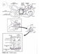
Refer to Figure 1 . 14A. The utricle and saccule are located in the vestibule, a large chamber that separates the semicircular canals and the cochlea. The sensory epithelia of the saccule and utricle are called the maculae. Each macula consists of numerous hair cells surrounded by supporting cells resting on a connective tissue base. The orderly arrangement of hair cells within the macula gives the appearance of a curved equatorial line called the striola. In the utricle, the hair cells are arranged with the kinocilium oriented toward the striola, whereas in the saccule, the hair cells are polarized away from the striola. This anatomic polarity ensures that the two otolith organs can respond to linear acceleration or head tilt in any direction. The surface of the macula extends into the membranous labyrinth, which is bathed in endolymph, not perilymph. The macular surface is covered with a gelatinous structure, the otolithic membrane, which has calcium carbonate crystals (otoliths or otoconia) embeclclecl on its surface. Relative movement between the otolithic membrane - and the surface of hair cells is the essential macular stimulus, since this produces movement (bending) of hair cells, which results in ionic current flow at the base of hair cells and neurotransmitter release. With the head in a neutral position, the macula of the utricle lies in the horizontal plane (on the floor of the vestibule) and the macula of the saccule lies in the vertical plane (on the medial wall of the vestibule) . Linear acceleration is detected by the maculae, whereas angular acceleration is detected by the specialized hair cells of the semicircular canals, called the cristae ampullaris ( Ka ndel , pp. 802-814; Pritchard, pp. 250-253)
Tn. X, 52 tahun menjalani reseksi subtotal glioblastoma multiforme yang berasal dari lobus fronalis kanan dan meluas ke nukleus didalam dari hemisfir tersebut. Pasca-operasi, dia menjalani terapi radiasi pada otak secara keseluruhan, dan menerima 1, 3-bis-2¬kloroetil-1-nitrourea (BCNU), Delapan bulan kemudian pasien yang bersangkutan terkena proses penyakit ini kembali. 15. Resistensi tumor ini terhadap BCNU kemungkinan disebabkan oleh:
A. Konsentrasi tinggi O6-alkilguanin-DNA alkil-transferase (O6-AGAT) pada sel-sel tumor.
B. Tumor ini berada dalam S-fase dari siklus sel (fase resistensi) pada waktu pemberian BCNU
C. Sel-sel tumor ini tidak memiliki topoisomerase II, yang menyebabkan strand DNA transien pecah selama induksi kemoterapi.
D. Sel-sel tumor ini tidak memiliki protein permukaan sel yang mengenal BCNU
E. Obat yang mengganggu sawar darah¬otak yang tidak diberikan bersamaan dengan pemberian BCNU
A
The nitrosoureas (BCNU, CCNU) are alkylating agents and are the most widely used drugs for patients with malignant brain tumors. They alkylate DNA in multiple locations, primarily on guanine but also on adenine and cytosine. The resultant DNA cross links often produce single- or doublestranded DNA breaks and eventual tumor cell death. 06- AGAT is a repair enzyme that mediates repair of alkylation products of nitrosoureas. It has been noted that approximately 70% of tumors have high levels of 06-AGAT and are often resistant to nitrosourea chemotherapy ( Bernstein, p p . 231-232)
n. X, 52 tahun menjalani reseksi subtotal glioblastoma multiforme yang berasal dari lobus fronalis kanan dan meluas ke nukleus didalam dari hemisfir tersebut. Pasca-operasi, dia menjalani terapi radiasi pada otak secara keseluruhan, dan menerima 1, 3-bis-2¬kloroetil-1-nitrourea (BCNU), Delapan bulan kemudian pasien yang bersangkutan terkena proses penyakit ini kembali. 16. Bahan manakah yang memiliki potensi menaikkan tingkat respons terhadap kemoterapi BCNU? A. Irinotecan (CPT-11) B. Tamoxifen C. Suramin D. O6-benzylguanine E. 1-(2-kloroetil)=3=sikloetil-1-nitrosourea (OCNU)
D Attempts t o modify resistance t o nitrosoureas are ongoing. As stated in the previous discussion (question 1 5), 06-AGAT mediates the repair of alkylating products of nitrosoureas. I nhibition of this repair protein has been the subject of a number of clinical trials using 06-benzylguanine, a methylating agent. Tamoxifen inhibits protein kinase C, CPT- 1 1 is a topoisomerase I inhibitor, and suramip works by inhibiting growth factors (FGF, I GF-1 , PDGF) . These agents do not modify resistance to alkylating agents. The addition of CCNU can potentially increase the risk of nitrosoureainduced side effects ( Bernstein, pp. 229-332 )
- Penelitian eksperimental dengan menggunakan pendekatan transfer gen bunuh diri HSV-tk/GCV pada berbagai model binatang membuktikan adanya regresi tumor dan survival memanjang meskipuh efisiensi transduksi kurang dari 10 persen. Keberhasilan mengaplikasikan dengan terapi gen bunuh diri kanker (suicide gene cancer theraphy) pada penelitian ini, terlepas dari tidak tuntasnya pengiriman vektor genetika ke semua sel tumor, kemungkinan disebabkan oleh:
A. Transfer GCV posforilase (pGCV) ke dalam sel-sel tumor yang tidak tertransdusi melalui gap junctions.
B. Reaksi inflamasi yang disebabkan vektor virus, mengakibatkan aktifnya proses pengiriman sinyal kematian pada sel (Fas/APO-1)
C. Regulasi-ke atas dari p53 yang serta merta menyebabkan pelepasan mediator-mediator apoptotis (misalnya kaspase 8) dari mitokondria
D. Regulasi-ke atas dari cAMP, pembawa pesan kedua yang telah dipastikan dapat menghentikan proliferasi pada fase G1 siklus sel.
E. Transfer vektor-vektor biral ke dalam sel sel tumor non-transduksi melalui lubang-lubang berselaput klatrin.
A
The mechanism \•hereby untransd1,1ced tumor cells die during gene therapy is called the “bystander effect.” Until recently, this mechanism was poorly understood; it requires the presence of gap junctions that allow the transfer of toxic metabolites into untransduced tumor cells. In the I-ISVtk/ GCV approach, the nucleoside analogue GCV becomes cytotoxic after being converted to its triphosphorylated form by HSV-tk and host cellular kinases. It acts as a chain terminator and interrupts DNA synthesis in replicating cells. Phosphorylated GCV can then be transported into surrounding untransduced cells via gap junctions and induce cell death. The degree of bystander effect in individual tumors depends on the cell type and its capability to express gap junctions, the vector used, and the enzymatic activity of the therapeutic gene. The other choices have not been sho\‘11 to propagate toxicity from transduced to untransduced cells (Bernste i n , pp. 280-281).
- Apa yang menjadi satu-satunya neurotransmitter yang disintesis dalam vesikel sinaptik?
A. Dopamin
B. Norepineprin
C. Asetilkolin
D. Serotonin
E. Senyawa P
B
Acetylcholine (Ach) is synthesized from cholin and acetyl-CoA by the enzyme choline acetyltransferas::c ACh is utilized by spinal cord motor neurons at the neurc.muscular junction, all preganglionic autonomic neu postganglionic parasympathetic neurons, postganglioni s:- pathetic neurons to sweat glands, and within the basalis of Meynert. ACh is metabolized in the synaptic “by acetylcholinesterase into acetate and choline. then recycled b y reuptake into the terminal bourreceptor-mediated endocytosis. Dopamine epinephrine (NE), and epinephrine are all synthesized he same precursor molecule, the amino acid L-cyr Tyrosine hydroxylase synthesizes L-DOPA from r:-r “ - = and is the rate-limiting enzyme for both DA and :sis. Aromatic amino acid decarboxylase then symhesizrom L-DOPA. Dopamine is synthesized by neuronssubstantia nigra and arcuate nucleus of the hyporh ‘ and is also active in some mesolimbic and me tracts. Reserpine prevents the uptake of DA into vesicles. Dopamine ex-hydroxylase is located on rh brane of synaptic vesicles, where it converts DA w synaptic vesicle itself. NE is the only neurotransmme synthesized within the synaptic v’esicle. NE exerrs feedback on tyrosine hydroxylase. NE is the neurmra:ter of most postganglionic sympathetic neurons and - 12 I ntensive Neurosurgery Board Review found in the locus ceruleus. After NE is released into the synaptic cleft, the termination of its bioactivity is primarily accomplished by reuptake into the presynaptic neuron. NE reuptake is blocked by cocaine. NE is also metabolized by catechol 0-methyltransferase (COMT) and monoamine oxidase (MAO) in the cytoplasm of numerous cells. The medications tropolone and selegiline inhibit the enzymes COMT and :tvfA08, respectively. Serotonin (an indole) is synthesized from the amino acid tryptophan. Tryptophan is initially converted into 5-hydroxytryptophan by the enzyme tryptophan hydroxylase, which represents the rate-limiting step. Then 5-hydroxytryptophan is converted into serotonin by the enzyme 5-hydroxytryptophan decarboxylase. Serotonergic neurons are primarily found in the raphe nuclei of the brainstem reticular formation. Serotonin reuptake is inhibited by several antidepressants, including the selective serotonin reuptake inhibitors (SSRis; e.g. , fluoxetine) and the tricyclic antidepressants ( Kandel, p p . 280-295; Pritchard, pp. 32-45)
- Paling peka terhadap peregangan kulit
A. Ujung-ujung saraf bebas
B.Korpuskel MEISSNER
C. Korpuskel PACINIAN
D. Korpuskel RUFFINI
E. Diskus MERKEL
F. Bukan salah satu diatas
D
Sensory endings of the skin can be classified on a structural basis i n to encapsulated and nonencapsulated receptors. Nonencapsulated receptors include free nerve endings, Merkel’s discs, and hair follicle receptors. Encapsulated endings include Meissner’s corpuscles, pacinian corpuscles, and Ruffini’s corpuscles. Free nerve endings are widely distributed throughout the body. They line the alimentary tract and are found between epitheli al cells of the skin, in the cornea, and in a variety of connective tissues including the dermis, fascia, ligaments, joint capsules, periosteum, and muscle. They are either myelinated or unmyelinated, and most detect pain; however, some detect crude touch, pressure, and tickling sensations. Merkel’s discs are found i n hairless regions of the body including the fingertips. They terminate in the deeper aspects of the epidermis, are slowly adapting, and transmit information about pressure and texture. l’vlerkel’s disc receptors also provide the sharpest resolution of spatial patterns of all the sensory endings of the skin. 1vleissner’s corpuscles also provide sharp resolution of spatial patterns, but the image is generally not as sharp as the one produced by l’l’lerkel’s endings because they have slightly larger receptive fields. lvierkel’s discs are normally found in clusters at the center of the papillary ridge. Hair-follicle receptors ll’ind around hair follicles adjacent to a sebaceous gland. Some surround the hair follicle and others run parallel to it. These receptors are rapidly adapting and respond to the bending of hair follicles. Encapsulated receptors include lv leissner’s corpuscles, pacinian corpuscles, and Ruffini corpuscles. Jvleissner’s corpuscles are located in the dermal papillae of the skin, especially in the palms and soles of the feet. They are oval in shape and consist of a stack of flattened Schwann cells arranged transversely along their long axis. They are very sensitive to touch (especially stroking, fluttering), are rapidly adapting, and allo\1’ people to distinguish between two pointed structures placed together on the skin. Pacinian corpuscles are very similar physiologically to Meissner’s corpuscles, are widely distributed, and are numerous in the dermis, subcutaneous tissues, joint capsules, pleura, pericardium, and nipples. Each pacinian corpuscle is ovoid shape, measuring 2 mm long and about 1 00-500 J.. l!n across (largest sensory receptor). The capsule consists of concentric lamellae of flattened cells. A large myelinated nerve enters the corpuscle, loses the myelin sheath, and then passes through the central core before terminating i n an expanded fashion. Pacinian corpuscles are rapidly adapting and sensitive mainly to vibration. Ruffini’s corpuscle is located in the dermis of hairy areas, is a slowly adapting mechanoreceptor, and responds mainly when the skin is stretched. Muscle spindles and group Ia fibers i nnervate the afferent limb of the stretch reflex ( Kandel, p p . 430-450, 565).
- Terutama peka terhadap getaran (600stimuli/detik)
A. Ujung-ujung saraf bebas
B.Korpuskel MEISSNER
C. Korpuskel PACINIAN
D. Korpuskel RUFFINI
E. Diskus MERKEL
F. Bukan salah satu diatas
C
Sensory endings of the skin can be classified on a structural basis i n to encapsulated and nonencapsulated receptors. Nonencapsulated receptors include free nerve endings, Merkel’s discs, and hair follicle receptors. Encapsulated endings include Meissner’s corpuscles, pacinian corpuscles, and Ruffini’s corpuscles. Free nerve endings are widely distributed throughout the body. They line the alimentary tract and are found between epitheli al cells of the skin, in the cornea, and in a variety of connective tissues including the dermis, fascia, ligaments, joint capsules, periosteum, and muscle. They are either myelinated or unmyelinated, and most detect pain; however, some detect crude touch, pressure, and tickling sensations. Merkel’s discs are found i n hairless regions of the body including the fingertips. They terminate in the deeper aspects of the epidermis, are slowly adapting, and transmit information about pressure and texture. l’vlerkel’s disc receptors also provide the sharpest resolution of spatial patterns of all the sensory endings of the skin. 1vleissner’s corpuscles also provide sharp resolution of spatial patterns, but the image is generally not as sharp as the one produced by l’l’lerkel’s endings because they have slightly larger receptive fields. lvierkel’s discs are normally found in clusters at the center of the papillary ridge. Hair-follicle receptors ll’ind around hair follicles adjacent to a sebaceous gland. Some surround the hair follicle and others run parallel to it. These receptors are rapidly adapting and respond to the bending of hair follicles. Encapsulated receptors include lv leissner’s corpuscles, pacinian corpuscles, and Ruffini corpuscles. Jvleissner’s corpuscles are located in the dermal papillae of the skin, especially in the palms and soles of the feet. They are oval in shape and consist of a stack of flattened Schwann cells arranged transversely along their long axis. They are very sensitive to touch (especially stroking, fluttering), are rapidly adapting, and allo\1’ people to distinguish between two pointed structures placed together on the skin. Pacinian corpuscles are very similar physiologically to Meissner’s corpuscles, are widely distributed, and are numerous in the dermis, subcutaneous tissues, joint capsules, pleura, pericardium, and nipples. Each pacinian corpuscle is ovoid shape, measuring 2 mm long and about 1 00-500 J.. l!n across (largest sensory receptor). The capsule consists of concentric lamellae of flattened cells. A large myelinated nerve enters the corpuscle, loses the myelin sheath, and then passes through the central core before terminating i n an expanded fashion. Pacinian corpuscles are rapidly adapting and sensitive mainly to vibration. Ruffini’s corpuscle is located in the dermis of hairy areas, is a slowly adapting mechanoreceptor, and responds mainly when the skin is stretched. Muscle spindles and group Ia fibers i nnervate the afferent limb of the stretch reflex ( Kandel, p p . 430-450, 565).
- Hampir semuanya ditemukan sebagi klusterdi pusat sulkus papilaris.
A. Ujung-ujung saraf bebas
B.Korpuskel MEISSNER
C. Korpuskel PACINIAN
D. Korpuskel RUFFINI
E. Diskus MERKEL
F. Bukan salah satu diatas
E
Sensory endings of the skin can be classified on a structural basis i n to encapsulated and nonencapsulated receptors. Nonencapsulated receptors include free nerve endings, Merkel’s discs, and hair follicle receptors. Encapsulated endings include Meissner’s corpuscles, pacinian corpuscles, and Ruffini’s corpuscles. Free nerve endings are widely distributed throughout the body. They line the alimentary tract and are found between epitheli al cells of the skin, in the cornea, and in a variety of connective tissues including the dermis, fascia, ligaments, joint capsules, periosteum, and muscle. They are either myelinated or unmyelinated, and most detect pain; however, some detect crude touch, pressure, and tickling sensations. Merkel’s discs are found i n hairless regions of the body including the fingertips. They terminate in the deeper aspects of the epidermis, are slowly adapting, and transmit information about pressure and texture. l’vlerkel’s disc receptors also provide the sharpest resolution of spatial patterns of all the sensory endings of the skin. 1vleissner’s corpuscles also provide sharp resolution of spatial patterns, but the image is generally not as sharp as the one produced by l’l’lerkel’s endings because they have slightly larger receptive fields. lvierkel’s discs are normally found in clusters at the center of the papillary ridge. Hair-follicle receptors ll’ind around hair follicles adjacent to a sebaceous gland. Some surround the hair follicle and others run parallel to it. These receptors are rapidly adapting and respond to the bending of hair follicles. Encapsulated receptors include lv leissner’s corpuscles, pacinian corpuscles, and Ruffini corpuscles. Jvleissner’s corpuscles are located in the dermal papillae of the skin, especially in the palms and soles of the feet. They are oval in shape and consist of a stack of flattened Schwann cells arranged transversely along their long axis. They are very sensitive to touch (especially stroking, fluttering), are rapidly adapting, and allo\1’ people to distinguish between two pointed structures placed together on the skin. Pacinian corpuscles are very similar physiologically to Meissner’s corpuscles, are widely distributed, and are numerous in the dermis, subcutaneous tissues, joint capsules, pleura, pericardium, and nipples. Each pacinian corpuscle is ovoid shape, measuring 2 mm long and about 1 00-500 J.. l!n across (largest sensory receptor). The capsule consists of concentric lamellae of flattened cells. A large myelinated nerve enters the corpuscle, loses the myelin sheath, and then passes through the central core before terminating i n an expanded fashion. Pacinian corpuscles are rapidly adapting and sensitive mainly to vibration. Ruffini’s corpuscle is located in the dermis of hairy areas, is a slowly adapting mechanoreceptor, and responds mainly when the skin is stretched. Muscle spindles and group Ia fibers i nnervate the afferent limb of the stretch reflex ( Kandel, p p . 430-450, 565).
- Memberikan sensasi paling tajam untuk kesan ruang.
A. Ujung-ujung saraf bebas
B.Korpuskel MEISSNER
C. Korpuskel PACINIAN
D. Korpuskel RUFFINI
E. Diskus MERKEL
F. Bukan salah satu diatas
E
Sensory endings of the skin can be classified on a structural basis i n to encapsulated and nonencapsulated receptors. Nonencapsulated receptors include free nerve endings, Merkel’s discs, and hair follicle receptors. Encapsulated endings include Meissner’s corpuscles, pacinian corpuscles, and Ruffini’s corpuscles. Free nerve endings are widely distributed throughout the body. They line the alimentary tract and are found between epitheli al cells of the skin, in the cornea, and in a variety of connective tissues including the dermis, fascia, ligaments, joint capsules, periosteum, and muscle. They are either myelinated or unmyelinated, and most detect pain; however, some detect crude touch, pressure, and tickling sensations. Merkel’s discs are found i n hairless regions of the body including the fingertips. They terminate in the deeper aspects of the epidermis, are slowly adapting, and transmit information about pressure and texture. l’vlerkel’s disc receptors also provide the sharpest resolution of spatial patterns of all the sensory endings of the skin. 1vleissner’s corpuscles also provide sharp resolution of spatial patterns, but the image is generally not as sharp as the one produced by l’l’lerkel’s endings because they have slightly larger receptive fields. lvierkel’s discs are normally found in clusters at the center of the papillary ridge. Hair-follicle receptors ll’ind around hair follicles adjacent to a sebaceous gland. Some surround the hair follicle and others run parallel to it. These receptors are rapidly adapting and respond to the bending of hair follicles. Encapsulated receptors include lv leissner’s corpuscles, pacinian corpuscles, and Ruffini corpuscles. Jvleissner’s corpuscles are located in the dermal papillae of the skin, especially in the palms and soles of the feet. They are oval in shape and consist of a stack of flattened Schwann cells arranged transversely along their long axis. They are very sensitive to touch (especially stroking, fluttering), are rapidly adapting, and allo\1’ people to distinguish between two pointed structures placed together on the skin. Pacinian corpuscles are very similar physiologically to Meissner’s corpuscles, are widely distributed, and are numerous in the dermis, subcutaneous tissues, joint capsules, pleura, pericardium, and nipples. Each pacinian corpuscle is ovoid shape, measuring 2 mm long and about 1 00-500 J.. l!n across (largest sensory receptor). The capsule consists of concentric lamellae of flattened cells. A large myelinated nerve enters the corpuscle, loses the myelin sheath, and then passes through the central core before terminating i n an expanded fashion. Pacinian corpuscles are rapidly adapting and sensitive mainly to vibration. Ruffini’s corpuscle is located in the dermis of hairy areas, is a slowly adapting mechanoreceptor, and responds mainly when the skin is stretched. Muscle spindles and group Ia fibers i nnervate the afferent limb of the stretch reflex ( Kandel, p p . 430-450, 565).
- Membatasi saluran pencernaan. A. Ujung-ujung saraf bebas
B.Korpuskel MEISSNER
C. Korpuskel PACINIAN
D. Korpuskel RUFFINI
E. Diskus MERKEL
F. Bukan salah satu diatas
A
Sensory endings of the skin can be classified on a structural basis i n to encapsulated and nonencapsulated receptors. Nonencapsulated receptors include free nerve endings, Merkel’s discs, and hair follicle receptors. Encapsulated endings include Meissner’s corpuscles, pacinian corpuscles, and Ruffini’s corpuscles. Free nerve endings are widely distributed throughout the body. They line the alimentary tract and are found between epitheli al cells of the skin, in the cornea, and in a variety of connective tissues including the dermis, fascia, ligaments, joint capsules, periosteum, and muscle. They are either myelinated or unmyelinated, and most detect pain; however, some detect crude touch, pressure, and tickling sensations. Merkel’s discs are found i n hairless regions of the body including the fingertips. They terminate in the deeper aspects of the epidermis, are slowly adapting, and transmit information about pressure and texture. l’vlerkel’s disc receptors also provide the sharpest resolution of spatial patterns of all the sensory endings of the skin. 1vleissner’s corpuscles also provide sharp resolution of spatial patterns, but the image is generally not as sharp as the one produced by l’l’lerkel’s endings because they have slightly larger receptive fields. lvierkel’s discs are normally found in clusters at the center of the papillary ridge. Hair-follicle receptors ll’ind around hair follicles adjacent to a sebaceous gland. Some surround the hair follicle and others run parallel to it. These receptors are rapidly adapting and respond to the bending of hair follicles. Encapsulated receptors include lv leissner’s corpuscles, pacinian corpuscles, and Ruffini corpuscles. Jvleissner’s corpuscles are located in the dermal papillae of the skin, especially in the palms and soles of the feet. They are oval in shape and consist of a stack of flattened Schwann cells arranged transversely along their long axis. They are very sensitive to touch (especially stroking, fluttering), are rapidly adapting, and allo\1’ people to distinguish between two pointed structures placed together on the skin. Pacinian corpuscles are very similar physiologically to Meissner’s corpuscles, are widely distributed, and are numerous in the dermis, subcutaneous tissues, joint capsules, pleura, pericardium, and nipples. Each pacinian corpuscle is ovoid shape, measuring 2 mm long and about 1 00-500 J.. l!n across (largest sensory receptor). The capsule consists of concentric lamellae of flattened cells. A large myelinated nerve enters the corpuscle, loses the myelin sheath, and then passes through the central core before terminating i n an expanded fashion. Pacinian corpuscles are rapidly adapting and sensitive mainly to vibration. Ruffini’s corpuscle is located in the dermis of hairy areas, is a slowly adapting mechanoreceptor, and responds mainly when the skin is stretched. Muscle spindles and group Ia fibers i nnervate the afferent limb of the stretch reflex ( Kandel, p p . 430-450, 565).
- Serat-serat aferen pada refleks peregangan.
A. Ujung-ujung saraf bebas
B.Korpuskel MEISSNER
C. Korpuskel PACINIAN
D. Korpuskel RUFFINI
E. Diskus MERKEL
F. Bukan salah satu diatas
E
Sensory endings of the skin can be classified on a structural basis i n to encapsulated and nonencapsulated receptors. Nonencapsulated receptors include free nerve endings, Merkel’s discs, and hair follicle receptors. Encapsulated endings include Meissner’s corpuscles, pacinian corpuscles, and Ruffini’s corpuscles. Free nerve endings are widely distributed throughout the body. They line the alimentary tract and are found between epitheli al cells of the skin, in the cornea, and in a variety of connective tissues including the dermis, fascia, ligaments, joint capsules, periosteum, and muscle. They are either myelinated or unmyelinated, and most detect pain; however, some detect crude touch, pressure, and tickling sensations. Merkel’s discs are found i n hairless regions of the body including the fingertips. They terminate in the deeper aspects of the epidermis, are slowly adapting, and transmit information about pressure and texture. l’vlerkel’s disc receptors also provide the sharpest resolution of spatial patterns of all the sensory endings of the skin. 1vleissner’s corpuscles also provide sharp resolution of spatial patterns, but the image is generally not as sharp as the one produced by l’l’lerkel’s endings because they have slightly larger receptive fields. lvierkel’s discs are normally found in clusters at the center of the papillary ridge. Hair-follicle receptors ll’ind around hair follicles adjacent to a sebaceous gland. Some surround the hair follicle and others run parallel to it. These receptors are rapidly adapting and respond to the bending of hair follicles. Encapsulated receptors include lv leissner’s corpuscles, pacinian corpuscles, and Ruffini corpuscles. Jvleissner’s corpuscles are located in the dermal papillae of the skin, especially in the palms and soles of the feet. They are oval in shape and consist of a stack of flattened Schwann cells arranged transversely along their long axis. They are very sensitive to touch (especially stroking, fluttering), are rapidly adapting, and allo\1’ people to distinguish between two pointed structures placed together on the skin. Pacinian corpuscles are very similar physiologically to Meissner’s corpuscles, are widely distributed, and are numerous in the dermis, subcutaneous tissues, joint capsules, pleura, pericardium, and nipples. Each pacinian corpuscle is ovoid shape, measuring 2 mm long and about 1 00-500 J.. l!n across (largest sensory receptor). The capsule consists of concentric lamellae of flattened cells. A large myelinated nerve enters the corpuscle, loses the myelin sheath, and then passes through the central core before terminating i n an expanded fashion. Pacinian corpuscles are rapidly adapting and sensitive mainly to vibration. Ruffini’s corpuscle is located in the dermis of hairy areas, is a slowly adapting mechanoreceptor, and responds mainly when the skin is stretched. Muscle spindles and group Ia fibers i nnervate the afferent limb of the stretch reflex ( Kandel, p p . 430-450, 565).