Inflammation Flashcards
(5 cards)
Explain the pathology and histological features of acute inflammation
Inflammation: reactions of living vascularized tissue to sub-lethal cellular injury
- A protective response geared towards removing the cause and consequences of the injury
- Sets stage for potential healing
- Tightly regulated process consisting mainly of leukocyte and vascular responses
- Triggered by various cell types and soluble mediators
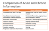
Acute inflammation is a rapid non-specific response to cellular injury
Key Features
- Histamine release
- Hours/days
- May be prominent necrosis
Histology
- Lots of neutrophils
- May also be mast cells and eosinophils
Recognised on examination by the cardinal signs:
- Rubor (Redness)
- Calor (Heat)
- Tumor (Swelling)
- Dolor (Pain)
•Rapid delivery of leukocytes and plasma proteins to the site of injury
•Three main components:
- Alteration in the calibre of blood vessels to increase flow
- Structural changes to the microvasculature to allow proteins and leukocytes to leave the circulation
- Emigration, accumulation and activation of leukocytes at the focus of injury
Vasodilation
- Vasodilation is one of the earliest manifestations
- May be preceded by brief arteriolar constriction
- Causes the heat and redness of acute inflammation
- Induced by several mediators including histamine and nitric oxide
- Affect vascular smooth muscle
- Quickly followed by increased permeability of microvasculature
- Increased diameter and loss of fluid slow down flow and lead to stasis
- Histamine is a major vasoactive amine
- Richest source is mast cells
- Preformed and released as the cell degranulates
- Triggered by binding of surface IgE, trauma, heat, cold, complement C3a/C5a, cytokines IL-1 / IL-8
- Leads to vasodilation and also increased vascular permeability
- Dysregulation can be seen in allergic reactions
- Type 1 Hypersensitivity
Increased Vascular Permeability
- Endothelial cells contract; increased interendothelial spacing
- Immediate Transient Response
- Histamine and Nitric Oxide
Exudate:
- Result of increased vascular permeability
- High protein content (fibrin, antibodies)
- High specific gravity
- Contains cells and cell debris
- May be purulent (leukocyte-rich)
Exudate serves to:
- Dilute pathogens
- “Wall off” pathogens
- Permit spread of soluble inflammatory mediators
- Provide substrate for inflammatory cell migration
- Intravascular fluid losses can be very high
- Life threatening in severe burns
Transudate:
- Ultrafiltrate of blood plasma caused by increased hydrostatic pressure or decreased osmotic pressure
- Low protein content (albumin)
- Low specific gravity
- Low cell content
Leukocyte Response:
- The most important leukocytes in the initial phase of typical acute inflammation are those capable of phagocytosis
- Neutrophils
- Macrophages
- Kill bacteria and eliminate foreign and necrotic material
- Produce multiple factors and mediators that interact with other cells
- Overactivation may prove harmful in the long term
Neutrophils
- Neutrophils are produced in bone marrow
- Circulate in blood and migrate towards damaged tissues
- Often the first cell into a damaged area
- Rapid response
- Main roles are to kill bacteria and recruit additional cells
- Phagocytosis
- Degranulation – enzymes, free radicals, soluble mediators
Leukocyte Response
- Leukocytes first need to be recruited to the site of injury
- The process of exiting the vessel lumen (extravasation) has the following steps:
- Margination
- Rolling
- Adhesion to activated endothelium
- Transmigration (diapedesis) across endothelium through vessel wall
- Migration through tissues towards chemotactic stimulus
- Once recognised, microbes and necrotic tissues need to be destroyed
- Phagocytosis requires:
- Attachment
- Engulfment and formation of phagocytic vacuole
- Degradation by various substances
- Reactive oxygen species; myeloperoxidase (neutrophils)
- Lysozyme (antibacterial)
- Lactoferrin (iron binding; prevents bacterial reproduction)
- Major Basic Protein (produced by eosinophils; antiparasitic)
Termination of the Acute Inflammatory Response
- Inflammatory mediators and neutrophils have a short half life
- Macrophages release a number of anti-inflammatory products
- Mast cells and lymphocytes produce anti-inflammatory products
- Lipoxins
- The cause of the injury (e.g. bacteria) is removed
- Under normal conditions, process comes to a stop

Explain the pathology and histological features of chronic inflammation
Chronic inflammation is a persistent inflammatory response
- Ongoing inflammation and repair over weeks to years
- May arise form acute inflammation
Key Features
- Cytokines
- Caused by persistent damage (e.g. persistent infection, autoimmunity)
- Form granulation tissue
Histology
•Lots of macrophages, lymphocyes and plasma cells
Granulomatous inflammation is a specific subtype of chronic inflammation
Characterised by:
- Mononuclear cell infiltrate (macrophages, lymphocytes, plasma cells)
- Tissue destruction, induced by persistent inflammatory agent or by the inflammatory cells themselves
- Attempts at healing by replacement of damaged tissue with connective tissue
- Accomplished by fibrosis and accompanied by angiogenesis
- “Granulation tissue”
Macrophages
- In the acute phase of inflammation macrophages destroy the offending agent either directly or by stimulating other pathways that do so
- When the offending agent is cleared, the macrophages fade away
- In chronic inflammation macrophages persist and cause significant tissue destruction
- Ongoing tissue destruction can trigger the inflammatory cascade in of itself
- Acute and Chronic inflammation may co-exist
A number of other cell types are also involved:
- T-Lymphocytes (can be stimulated by macrophages; regulated immune reaction and can be cytotoxic)
- Plasma cells (develop from activated B-lymphocytes and produce antibodies)
- Eosinophils (in response to parasites or IgE mediated inflammation)
- Mast cells
- Neutrophils if co-existing acute inflammation
- There is prominent angiogenesis
- VEGF from macrophages and endothelial cells
Explain the pathology and histological features of granulomatous inflammation
Granulomatous inflammation:
- Distinctive pattern of chronic inflammation showing granuloma formation
- A granuloma is an aggregate of activated macrophages; an attempt to eliminate a resistant offending agent
- Triggered by strong and specific T-lymphocyte reaction
- Several causes
- Infections (TB, leprosy, syphilis, fungi)
- Foreign material (foreign body granuloma)
- Tumour reaction
- Granulomatous diseases (Sarcoidosis, Crohn’s disease)
Histology
- Granuloma: ball of activated lymphocytes and macrophages
- Giant cells: fused macrophages with horseshoe-shaped nuclei
List the sequelae of inflammation
Positive outcomes:
- Removal of offending agent
- Cessation of the inflammatory response
- Healing of tissue damage with preservation of integrity and function (resolution)
Undesirable Outcomes
- Excessive tissue damage and scarring
- Possibly with detrimental effect on adjacent tissue
- Systemic involvement with multiorgan failure
- Septic shock, amyloid
Inflammation cuts both ways
Wound healing: explain the sequential changes in wound healing
Wounds may heal either by resolution or scarring
Resolution
- involves regeneration of parenchymal cells with restoration of function
- Only occurs if tissue can regenerate and there is little structural damage
- Example: lobar pneumonia
Repair by scarring
- involves angiogenesis, migration and repair of fibroblasts, scar formation and connective tissue remodelling
- Occurs when there is significant tissue loss and tissue is unable to regenerate; results in loss of function
Process
- Fibroblasts lay down collagen
- Collagen is remodeled for maximal tensile strength
- Normal tissues is replaced by non-functional scar tissue
Wound healing may be impaired by:
- Poor nutrition (protein, energy)
- Vitamin deficiency (Vitamin C, Vitamin A)
- Mineral deficiency (Zinc)
- Suppressed inflammation (Steroids, Old Age)
- Poor local blood supply (Peripheral vascular disease)
- Persistent foreign body
- Movement
Complications of Wound Healing
Keloid
•Due to excess collagen deposition
Contracture
- Fibrous tissue contracts
- Can cause reduced joint mobility
Impaired Organ Function
•Due to replacement of functional parenchymal tissue by scar tissue
