Inflammatory Bowel Disease Flashcards
(75 cards)
What is Crohn’s Disease?
Chronic GRANULOMATOUS inflammatory disease than can affect any part of the GI tract. Can affect any segment of the GI tract from the mouth to anus. Characterised by transmural inflammation.
What is the aetiology of Crohn’s disease?
Unknown, though believed to be a mixture of environmental and genetic. Theories suggest that it is an inappropriate immune response against (?abnormal) gut flora in a genetically susceptible individual. Associated with altered neutrophil function and abnormality in epithelial cell integrity.
Where is the location of Crohn’s disease in the GI tract as a proportion of cases?
20% colic, 30% ileal, 50% ileocolic.
What are the risk factors of Crohn’s disease?
Mutation of NOD2 (aka CARD15) gene increases risk, smoking (x4 increased risk), altered cell mediated immunity, NSAIDs may exacerbate the disease, increased animal protein intake, contraception.
What is the epidemiology of Crohn’s disease: Age? Gender? Location? Prevalence?
Any age, but peak incidence 10-30 years, F>M, lower prevalence in Asian countries and populations. 50-80/100 000 prevalence in the UK.
What are the symptoms of Crohn’s disease? (x5)
- Crampy abdominal pain due to inflammation, fibrosis or obstruction
- Diarrhoea with urgency
- Blood/steatorrhea
- Fever and malaise
- Decreased weight and anorexia
What are the signs of Crohn’s disease? (x9)
- Abdominal tenderness/mass
- Abdominal distension
- Aphthous ulceration of mouth
- Perianal manifestations such as skin tags, anal strictures and signs of complications
- Signs of extra-intestinal complications
- Clubbing
- Signs of anaemia
- Weight loss
- Fever and tachycardia
What is anal stricture?
Narrowing of anal canal. Also called anal stenosis.
What are the investigations for Crohn’s disease? (x10) How is diagnosis made?
- BLOODS: decreased Hb, increased WCC, decreased Albumin (LFT), increased ESR/CRP, hypocholesterolaemia, hypocalcaemia.
- HAEMATINICS: look for deficiencies such as B12, folate (malabsorption) and ferritin (bleeding)
- STOOL: microscopy, culture and sensitivity and CDT (Clostridium difficile toxin) – exclude infective colitis. Can also look at FAECAL CALPROTECTIN as a simple test for GI inflammation with high sensitivity.
- AXR: look for complications, in particular, toxic megacolon
- ERECT CXR: if perforation risk
- CT/MRI: assess pelvic disease and fistulae, small bowel disease activity and strictures
- COLONSCOPY and BIOSPY: 70% effective in diagnosis. Not 100% as would miss disease when exclusively small bowel.
- Consider SMALL BOWEL BARIUM FOLLOW-THROUGH
- Consider CAPSULE ENDOSCOPY to visualise the small intestines
- Consider US: can provide small bowel imaging
- Diagnosis made by combination of investigations.
What does faecal calprotectin measure? Sensitivity?
Elevated levels is indicative of migration of neutrophils to the intestinal mucosa and is highly sensitive in IBD.
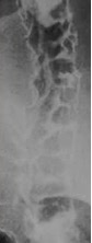
What is the purpose of small bowel barium follow-through in Crohn’s disease? (x3)
Look for fibrosis/strictures (you would see string sign of Kantor), deep ulceration (you would see rose thorn), or cobblestone mucosa in the SMALL INTESTINE.
What is the pathology of string sign?
Oedema or fibrosis associated with ulcerated mucosa
What does string sign look like in barium follow-through?
.

What does cobblestone mucosa look like in barium follow-through?
.
What is the clinical use of small bowel barium follow-through in Crohn’s disease?
Rarely used; useful when disease involves small intestine.
What is the purpose of colonoscopy in Crohn’s disease? (x4)
DEFINITIVE DIAGNOSIS, biopsy tissue, visualise mucosa to differentiate between UC and CD, monitor for disease progression or malignancy. Note that a biopsy is CONFIRMATORY rather than DIAGNOSTIC
!!! What do you look for in colonoscopy (or capsule endoscopy) for Crohn’s disease?
- Mucosal oedema
- Mucosal inflammation and discrete deep ulcers located transversely (penetrating deep; rose thorn fissures) and longitudinally (across sections of mucosal wall), creating a COBBLESTONE appearance
- Skip lesions: because of the transmural chronic inflammation
- Hyperaemia
- Fistulae and abscesses
- Involvement of the colon and ileum but NOT the rectum is suggestive of Crohn’s.
What are the histological findings in Crohn’s disease?
- Widening of submucosa
- Lymphoid aggregates in the submucosa associated with inflammation
- Cryptitis accompanied by abundant lymphatic and plasma cells
- Non-caseating granulomas (aggregation of macrophages from chronic inflammation) with epithelioid giant cells (aka epithelioid histiocyes; these describe macrophages and dendritic cells)
- Granulomas with epithelioid giant cells may be seen in blood vessels or lymphatics.
What does rose thorn fissures refer to?
Deep penetrating linear ulcers or fissuring typically seen within stenosed terminal ileum with a thickened wall.
What are the gastrointestinal complications of Crohn’s disease? (x10)
- Small bowel obstruction
- Toxic megacolon (rare complication relative to UC)
- Haemorrhage
- Bowel strictures
- Perforation
- Fistulae (between bowel, bladder and vagina)
- Perianal fistulae
- Abscesses (abdominal, pelvic or perianal)
- GI carcinoma
- Malabsorption
What is the risk of GI carcinoma in Crohn’s disease?
5% risk in 10 years.
What is toxic megacolon?
Bowel dilatation associated with inflammation and increased risk of stasis, perforation and haemorrhage. It is an acute presentation of Crohn’s with significant risk of death if left untreated.
What are the extraintestinal complications of Crohn’s disease? (x12)
- Uveitis: inflammation of uvea (middle pigmented layer of the eye comprising of iris, ciliary bodies and choroid)
- Episcleritis
- Gallstones
- Kidney stones
- Arthropathy (disease of joints) including arthritis
- Sacroiliitis (sacroiliac join inflammation)
- Ankylosing spondylitis
- Osteoporosis (from steroid treatment)
- Erythema nodosum (redness and swelling of skin arising from inflammation of subcutaneous adipose tissue)
- Pyoderma gangrenosum: immune system dysfunction leading to ulcer formation, typically on legs
- Amyloidosis
- DVT/PE from hypercoagulability associated with inflammation
What is amyloidosis?
Abnormal amyloid protein aggregates (fibrils) build-up in tissues leading to diarrhoea, weight loss, macroglossia, bleeding presenting as bruising, and more.