Innate Immunity I and III Flashcards
(41 cards)
Monocytes/Macrophages
- Phagocytosis and activation of bacteriocidal mechanisms
- Monocytes circulate through blood, then enter tissues and differentiate into macrophages 3 important functions:
- engulf and kill invading organisms
- help induce inflammation to trigger the immune system
- secrete signaling proteins to recruit/activate other immune cells
- Classically Activated M1
- Alternatively Activated M2
- Macrophages express numerous PRRs to aid their role in pathogen detection – triggering of these receptors leads primarily into the production of classical activated / M1 macrophages.
- TLRs, NLRs, RLRs, CLRs.
- Complement receptors to detect Complement-bound pathogens.
- Scavenger receptors to detect oxidized/acetylated low density lipoproteins (LDL) (sign of tissue damage).
- Cytokine receptors (especially the IFNg receptor) to allow activation even in the absence of direct pathogen detection. In other words, if surrounding cells are producing IFNg (in response to infection), nearby macrophages will become activated as well. Upon their activation in tissues, macrophages are responsible for:
- Cytokine and chemokine production to initiate and regulate inflammation, and recruit other cells to the site of infection. CLASSICAL/M1
- Ingesting pathogens through phagocytosis, followed by pathogen killing via the oxidative burst within the phagosome. CLASSICAL/M1
- Clearing dead cells and tissues and initiating wound repair. ALTERNATIVE/M2

PMNs
- neutrophils
- basophils
- eosinophils
aka granulocytes
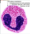
Neutrophils
- Phagocytosis and activation of bactericidal mechanisms
- Most abundant leukocyte in circulation and rapidly recruited to infection
- Excellent phagocytosis, express high levels of CR1 and CR3
- VERY SHORT lifespan (hours upon recruitment to tissues)
- Major component of pus upon their death
Basophils and Eosinophils
•Defense against parasites: release cytotoxic granules

Mast Cells
- Defense against parasites: release histamine granules
- basophils in blood, mast cells in tissue and mucous membranes
- Key roles in wound repair, immunity to parasitic worms and allergy

Natural Killer Cells
- Anti-viral & anti-tumor responses by release of cytotoxic granules
- Typically held in check by normal ratio of activating and inhibitory proteins on host cell surface.
- Infection/transformation changes this ratio, makes host cell a target.
- Many virus target NK receptor proteins to avoid immune detection.

Dendritic Cells
- Antigen uptake and presentation – LINKS INNATE & ADAPTIVE
- • Phagocytic cells
- Reside in tissues in an “immature” state – maturation is triggered upon pathogen recognition
- Migrate to nearest secondary lymphoid organ to present antigens to the adaptive system

PAMPs
•pathogen-associated molecular patterns, or PAMPs
PRRs
- pattern recognition receptors (PRRs)
- recognize PAMPs
- TLRs
- NLRs
- RLRs
- CLRs
TLRs
- Toll-Like Receptors (TLRs)
- There are at least 13 different TLRs identified to date.
- These receptors function to detect different components shared by microbes that are highly distinct from host molecules, including bacterial cell wall components and pathogen-derived nucleic acids.
- They are membrane bound and are distributed both on cell surfaces as well as intracellularly in the membranes of endosomal compartments.
- Although different intracellular adaptor proteins and signaling pathways are used, the end goals are the same, control infection and buy time while the adaptive response develops:
- Trigger the inflammatory response via NF-kB activation
- Produce antiviral Type-I IFNs via IRFs

NLRs
- NOD-Like Receptors (NLRs)
- These cytosolic receptors recognize PAMPs as well as host molecules released in the “wrong location” following cell stress or damage, such as ATP, glucose, urate, and cholesterol (Damageassociated molecular patterns; DAMPs).
- Their activation leads to formation of the inflammasome and promotes the inflammatory response.

Inflammasome
- NLR stimulation by PAMPs or DAMPs activates the inflammasome, a multi-component complex to produce pro-inflammatory cytokines.
- Upon final assembly of the inflammasome, (pro) caspase-1 is cleaved into active caspase-1, which then cleaves and activates precursors of the pro-inflammatory IL-1b family (IL-1b and IL-18).
RLRs
- RIG-I-Like Receptors (RLRs)
- These cytosolic receptors recognize the presence of viral RNA in the cytosol as a consequence of viral replication, leading to the production of key antiviral Type-I interferons (IFNa and IFNb).

CLRs
- C-type Lectin Receptors (CLRs)
- These cell surface receptors recognize carbohydrate PAMPs (bacterial mannose and fungal dectins). Pathogen binding to lectins improves phagocytosis and inflammatory responses to those pathogens.
The downstream consequences of PAMP recognition by PRRs…
- Activate transcription of pro-inflammatory cytokines, adhesion molecules, and costimulatory molecules.
- Activate transcription to stimulate production of Type-I IFNs (IFN a/b; primarily in response to virus detection).
*NLR stimulation by PAMPs or DAMPs activates the inflammasome, a multi-component complex to produce pro-inflammatory cytokines.
Phagocytosis
•Phagocytosis is the process of ingesting extracellular items by engulfing them within the host membrane. For pathogen clearance, the phagocytosed vesicle containing the extracellular pathogen is fused with lysosomes within the cytosolic compartment that contain numerous proteases. This fusion event results in the dramatic lowering of the pH within the vesicle. Ultimately these events cause breakdown of the ingested matter and destruction of the pathogen.
3 Professional Phagocytic Cell Types
- Neutrophils
- Macrophages
- Dendritic Cells
M1 Macrophages
•• Classically activated macrophages (M1) are activated through pattern recognition receptors (PRRs) and/or IFNg in response to microbial products. These are proinflammatory, and secrete cytokines that stimulate the production of reactive oxygen species within phagosomes to destroy ingested microbes.
M2 Macrophages
•Alternatively, activated macrophages (M2) are induced by non-inflammatory cytokines produced in the absence of a strong PRR signal. They promote fibrosis and wound repair, helping to control and resolve inflammation. These cells are key players in the resolution of the infection and in restoring tissue homeostasis.
Cytokines
- In response to PAMP detection, macrophages and dendritic cells are key secretors of cytokines that signal and coordinate the immune response to the infection.
- Cytokines are soluble proteins that enable communication between leukocytes, thus many are called interleukins.
- Cytokines are secreted in small amounts and bind to very high affinity receptors on their target cells. They can function in 3 manners:
- Autocrine, acting on the cell that originally secreted the cytokine.
- Paracrine, acting on adjacent cells.
- Endocrine, acting on distant cells.
Cytokines are critical coordinators of the innate and adaptive immune responses:
- These soluble mediators recruit circulating leukocytes to the site of infection.
- They activate innate cells (NK and macrophages) to become more potent killers.
- Their systemic effects include signaling the hypothalamus to induce fever, the liver to produce acute phase proteins, and the bone marrow to increase neutrophil production.
- The nature of the innate cytokine response (which cytokines are made? how much?) influences the subsequent adaptive response that develops. This is dictated by which PRRs are triggered during the initial detection of the pathogen. In other words, the type of PRRs triggered during innate recognition (dependent on the pathogen type) dictates which early inflammatory cytokines are produced, which then dictates the type of adaptive response that develops – ensuring the right adaptive response (TH1, TH2, CD8, B cell isotype) for the type of pathogen encountered (extracellular bacteria, virus, parasite).
- TNFa and IL-1 family members are the primary pro-inflammatory cytokines that activate endothelial cells to promote leukocyte recruitment to the site of infection in tissues. Systemically, these cytokines induce the fever response in the hypothalamus. TNF-a also stimulates production of acute phase proteins by the liver.
Chemokines
•Chemokines are soluble mediators that control adhesion, chemotaxis and activation of many leukocyte populations. They are the major regulators of cell trafficking.
Tumor necrosis factor a (TNFa)
- Pro-Inflammatory
- Macrophages, T cells
- Endothelial cells: activation (upregulate adhesion molecules to recruit other cells)
- Neutrophils: activation leading to their secretion of various effector molecules
- Hypothalamus: fever
- Liver: synthesis of acute-phase proteins
Interleukin-1 (IL-1)
- Pro-Inflammatory
- Macrophages, endothelial cells
- Endothelial cells: activation (upregulate adhesion molecules to recruit other cells)
- Hypothalamus: fever
- Liver: synthesis of acute-phase proteins

