Instruments & Imaging Flashcards

1
Q
A

These are two blood culture bottles, one for aerobic bacteria and one for anaerobic bacteria. The blood is injected in a sterile manner into the bottles using a different needle from the one the blood was drawn with.
Blood cultures are a useful investigation in a case of pyrexia or suspected systemic sepsis

2
Q
A

This is the bottle to which the chest drain is attached.
If you look carefully on these there will be a line called prime level which is filled with sterile water. The chest drain tubing is connected to a tube which is under the sterile water and therefore acts as a water seal.
After a chest drain has been inserted you can see bubbling in the water as the air leaves the pleural space. The chest drain bottle can also be used to collected blood, fluid and pus from the pleural space. The system can be driven by attaching suction to the top of the bottle making it an example of a active closed drainage system.

3
Q
A

Denver’s Retractors

This is a type of retractor which is used in open abdominal surgery to allow the surgeon to operate.

There are different sizes and types of retractors available, you may frequently be asked to use one during you clinical training when you are assisting in theatre

4
Q
A

This is a disposable rigid sigmoidoscope, which is used for the inspection of the rectum and lower sigmoid colon. WILL NOT REACH SIGMOID, SHOULD BE CALLED A RECTOSCOPE.

After explaining to the patient what you are about to do, you must attach a light source and a air pumping device. The patient is placed in the left lateral position and a digital rectal examination is performed. The sigmoidoscope is then lubricated with jelly and inserted pointing towards the umbilicus. Air is pumped into the rectum to allow you see the direction of the rectal lumen.

Biopsies can also be taken of rectal mucosa through the sigmoidoscope e.g. in a case of ulcerative colitis

5
Q
A

This is a drainage bag which can be connected top either a nasogastric tube or a drain coming out of the abdomen. Drainage relies on gravity so this is an example of a closed passive drainage system

6
Q
A

This is an adult endotracheal tube which is used to provide a definitive airway for patients for example during long operations e.g. laparotomies and during cardiac arrests or trauma.

The endotracheal tube is inserted using a laryngoscope, through the laryngeal folds. The end of the tube should lie just above the carina to allow ventilation of both lungs. After inserting the tube a balloon at the end of the tube is inflated with air through the blue side port. Position of the tube is checked by looking for symmetrical rising of the chest on ventilation breath sounds bilaterally and no gurgling over the epigastrium indicating oesophageal intubation. The tube is then tied into place.

7
Q
A

This is a feeding nasogastric tube (clinifeed tube) which is used to long term enteral nutrition in patients. It is thin bore and soft making it more comfortable for patients, it is also made of silastic which blocks less often.

After explaining to the patient what you are about to do the tube is inserted into the nostril after it has been lubricated. These tubes come with a wire inside them to aid their introduction, you advance the tube as the patient swallows. Correct position of the tube is checked by x-raying for the wire. When you are happy with the position of the tube the wire is removed and the feed attached in a sterile manner.

8
Q
A

This is a bag of 5% dextrose, which can be used in conjunction with normal saline to provide the normal daily fluid requirement for a patient.
One litre of 5% dextrose contains 50g of dextrose in 1 litre of water.
It should be remembered that because the sugar in this fluid is metabolised to carbon dioxide and water you are essentially giving them water

9
Q
A

This is a plate that is used in conjunction with screws to internally fix a bone fracture.

Need at least two screws either side of the fracture

10
Q
A

This is 500ml of gelofusin which is an example of an artificial colloid solution.

Colloid solutions raise the plasma oncotic pressure and hence expand the intravascular compartment. There are other colloids and some available are natural e.g. albumin and blood. Colloids are useful in cases of shock e.g. due to sepsis or hypovolaemia.

Bascially synthetic albumin

11
Q
A

This is an example of a crystalloid solution, which contains sodium, chloride, bicarbonate and lactate.

It has a similar composition to the extracellular fluid (acutally the closest you can get!)

It can be used to provide the normal daily fluid requirement of a patient or to supplement the patient for additional loses.

Hartmann’s solution is a favorite solution of anaesthetists and is the fluid advocated to be given initially in trauma in the Advanced Trauma and Life Support (ATLS) guidelines.

12
Q
A

This is a hemiarthoplasty hip prosthesis.

It is used in cases of intracapsular fractures of the neck of femur, as these fractures are prone to avascular necrosis of the femoral head.

13
Q
A

This is a an example of a long term central venous line which is inserted in a similar way to a central line (usually subclavian). Large bore veins.

The remnant of the line is tunnelled subcutaneously which decreases the incidence of line infection.

These are indicated for longterm parenteral nutrition, longterm intravenous antibiotic therapy and chemotherapy.

14
Q
A

This is a total hip replacement which articulates with an plastic acetabular cup.

The main indication for a hip replacement is pain from osteoarthtis of the hip.

15
Q
A

This is an intramedullary femoral nail which is used to internally fix femoral shaft fractures. Long bone fractures.

Interlocking screws are used to fix the nail. They are usually removed after 12 / 18 months.

16
Q
A

This is an intravenous cannula which can be used to give intravenous fluids and drugs.
If you wish to give fluid quickly the cannula must be short and large bore. I.e. brown or grey. Note with central lines - these tend to be fine bore!

Orange/Brown 14G

Grey 16G

Green 18G

Pink 20G

Blue 22G

Yellow 24G

17
Q
A

This is a large bore irrigation type foley urinary catheter which is used to irrigate the bladder of patients at risk of clot retention e.g. after a TURP. hey bleed a lot so tend to need irrigation. If it blocks, can back up and cause hydronephrosis.

18
Q
A

This is a Laparoscopic port which is used during Laparoscopic procedures e.g. Laparoscopic cholecystectomy.

These ports allow the surgeon to insert a telescope and instruments in the patient. Has a trochar in the middle.

19
Q
A

This is a laryngeal mask airway which can be used to provide an airway during short operations e.g. day cases. It does not protect the airway.

20
Q
A

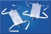
This is a leg bag which is attached to urinary catheter.
The Bag is strapped to the leg of the patient and is indicated for patients who are mobile and have either a short or long term indwelling urinary catheter.
A man who suffers with incontinence following sphincter damage after multiple TURPs.

21
Q
A

Mannitol is an osmotic diuretic which can be used to lower raised intracranial pressure or drive the urine output in a patient with obstructive jaundice to prevent hepato renal syndrome.

22
Q
A

This is a nasopharyngeal airway which is inserted into the nose using a rotational action.

It is used to provide an airway in people with a decreased level of consciousness or decreased gag reflex. The diameter tube should be sized against the patients own little finger distal phalanx. A safety pin is placed in the end of the tube to prevent it being inhaled.

23
Q
A

These are special forceps designed to hold the needle to allow the surgeon to suture accurately.

24
Q
A

Normal (0.9%) Saline. Normal saline is an example of a crystalloid solution which contains 153mmol of NaCl.

It can be used to provide the normal daily fluid requirement for a patient or to replace additional losses e.g. vomit or diarrhoea. Has more sodium than Hartmann’s.

25
Q
A

This an example of a synthetic non absorbable monofilament suture. This suture can be used to close skin wounds.

26
Q
A

This is an oral pharengeal airway used to provide an airway for a patient where there is an impaired level of consciousness.

It is sized my measuring the distance the from the angle of the mouth to the angle of the jaw. It is inserted into the mouth upside down and rotated within the oral cavity. It is inserted the correct way up in children.

27
Q
A

This is a paediatric oxygen mask.

28
Q
A

This is a proctoscope, it is used to visualise the anal canal and lower rectum, it is also used when injecting or banding haemorrhoids.

After explaining the procedure to the patient, the patient is placed in the left lateral position and a digital rectal examination is performed. The proctoscope is then attached to a light source and lubricated prior to its insertion into the rectum.

29
Q
A

This is a Ryles nasogastric tube which is primarily used for draining the stomach but can also be used to insert drug, feed or contrast into the GI.

After explaing what you are about to do to the patient , you will require a NG tube which has been in the fridge as it is stiffer, some lubricant, a bladder syringe , a drainage bag, litmus paper and tape.

The tip of the NG tube is lubricated and inserted into the nostril of the patient pointing it towards the occiput. The tube is then advanced as the patient swallows. The correct position of the tube is checked by aspirating gastric contents and checking for acidity on litmus paper, if this is unavailable then air can be inserted to the tube and the epigastrium auscultated for bubbling. Finally an x ray can be taken to identify the tube, however this is time consuming and often does not show the tube.
Once the tube is in the correct position a bag is attached and it is taped to the patients face

30
Q
A

This is a type of retractor used to hold wounds open e.g. during a hernia repair or an appendicectomy.

31
Q
A

This is a drainage type silastic Foley urinary catheter.

Silastic catheters are made of silicone and are more appropriate than latex one for long term catheterization.

32
Q
A

Central venous cannulas are inserted in the superior vena can usually via either the internal jugular or subclavian veins.

They can be single or triple lumen lines. They are primarily used to measure the central venous pressure. They can also be used for the insertion of drugs e.g. amiodarone, dopamine or chemotherapy.

33
Q
A

This is a stiff neck collar which is used to stabilise the cervical spine in a trauma patient when used in conjunction with 2 sand bags and tape.

They are sized by measuring the number of fingers from the clavicle to the angle of the mandible, and this is then compared to the measuring peg on the stiff neck collar.

34
Q
A

This is a Swan Ganz catheter which is used to measure the pressures in the heart, the pulmonary artery wedge pressure and can also be used to measure the cardiac output.

These are used in the intensive care setting and are typically inserted into a central vein.

35
Q
A

This is an example of a synthetic absorbable suture which can be used for bowel anastomosis or tying off vessels.

36
Q
A

This is a thromboembolic deterrent stocking which should be used in all patients undergoing surgery and those who are immobile to help prevent deep vein thromboses.

They are available in different sizes and are used in conjunction with low dose subcutaneous heparin. They are contraindicated in patients with arterial disease of the lower limb.

37
Q
A

This is a temporary tracheostomy; it is an example of a definitive airway as it protects the patients lungs from aspiration.

One of the most common occasions you will see a tracheostomy is on a patient being ventilated on the intensive care unit.

A tracheostomy allows more efficient ventilation of the patient with a decreased dead space and also allows more effective suctioning of the airways. Tracheostomy can also be used in patients with upper airway obstruction or after laryngeal surgery

38
Q
A

Central venous cannulas are inserted in the superior vena can usually via either the internal jugular or subclavian veins.

They can be single or triple lumen lines. They are primarily used to measure the central venous pressure. They can also be used for the insertion of drugs e.g. amiodarone, dopamine or chemotherapy.

39
Q
A

This is a trucut biopsy needle which is used to take histological specimens from lesions e.g. breast lumps or liver.

The procedure can be performed under local anaesthetic.

40
Q
A

Acute subdural haematoma

The bleed in subdural haemorrhages usually comes from tearing of “bridging veins” found in the subdural space 3. These veins can be subject to shearing or tearing when the head is subject to a change in velocity (as in head injury). Tearing of these veins may occur with less force in the elderly where the effect of cerebral atrophy can leave these stretched veins more vulnerable to damage 2.

Acute subdurals are usually associated with head injury, particularly in younger patients who may have evidence of head injury, e.g. extradural haemorrhage, cerebral contusions. There may be underlying abnormalities such as arteriovenous malformations in spontaneous bleeds.

Subacute or chronic subdural haemorrhages can present in the elderly as confusion or with vague neurological change. SDH is a classic cause of pseudo-dementia and there may only be a history of minor head injury (sometimes there is no history of head trauma) 2. Special attention should be given to patients on oral anticoagulants such as warfarin.

Differential diagnosis

subdural empyema: similar appearance on CT but usually febrile and generally unwell, can enhance with contrast with evidence of abscess or underlying infarction

extradural haemorrhage: difficult if small to differentiate, EDH are bi-convex rather than crescentic, EDH limited by sutures, SDH by dural reflections, usually clear history of head trauma/skull fracture

subdural hygroma: very similar appearance of CT, same density as CSF, no evidence of previous haemorrhage

enlarged subarachnoid space (due to cerebral atrophy or changes in infancy)

41
Q
A

Chronic Subdural Haematoma

The bleed in subdural haemorrhages usually comes from tearing of “bridging veins” found in the subdural space 3. These veins can be subject to shearing or tearing when the head is subject to a change in velocity (as in head injury). Tearing of these veins may occur with less force in the elderly where the effect of cerebral atrophy can leave these stretched veins more vulnerable to damage 2.

Acute subdurals are usually associated with head injury, particularly in younger patients who may have evidence of head injury, e.g. extradural haemorrhage, cerebral contusions. There may be underlying abnormalities such as arteriovenous malformations in spontaneous bleeds.

Subacute or chronic subdural haemorrhages can present in the elderly as confusion or with vague neurological change. SDH is a classic cause of pseudo-dementia and there may only be a history of minor head injury (sometimes there is no history of head trauma) 2. Special attention should be given to patients on oral anticoagulants such as warfarin.

Differential diagnosis

subdural empyema: similar appearance on CT but usually febrile and generally unwell, can enhance with contrast with evidence of abscess or underlying infarction

extradural haemorrhage: difficult if small to differentiate, EDH are bi-convex rather than crescentic, EDH limited by sutures, SDH by dural reflections, usually clear history of head trauma/skull fracture

subdural hygroma: very similar appearance of CT, same density as CSF, no evidence of previous haemorrhage

enlarged subarachnoid space (due to cerebral atrophy or changes in infancy)

42
Q
A

Epidural haemorrhage

Extradural haemorrhages (EDHs) represent collections of blood in the extradural (epidural) space. The haemorrhage sits between the skull superficially and the dura which overlies the brain parenchyma.

Patients with EDH often describe headache, although this may be the result of head injury and underlying fracture.

EDH may result in mass effect which can cause localising signs (sixth cranial nerve palsy) or autonomic changes, followed by focal neurology and loss of consciousness if the mass effect is significant.

Some patients have an initial loss of consciousness followed by a return to normal conscious level which is accompanied by a persistent severe headache. This is termed the “lucid interval” 1 and is followed by subsequent drop in conscious level.

Extradural haemorrhages necessitate urgent consultation with neurosurgical services 2. Surgical treatment involves evacuation of the clot through a cranial burr hole. Smaller bleeds may be managed conservatively.

EDHs generally have a good prognosis if appropriately treated 3. In high impact trauma, morbidity and mortality may be related to other associated injuries including brain injury.

The source of haemorrhage tends to be arterial and the result of an associated skull fracture. The middle meningeal artery is particularly at risk because it sits under the thinnest part of the skull between the ears and the eyes in the squamous temporal bone.

The headache that patients experience is the result of the dura mater is stripped off the skull vault as the collection grows.

43
Q
A

Epidural haemorrhage

Extradural haemorrhages (EDHs) represent collections of blood in the extradural (epidural) space. The haemorrhage sits between the skull superficially and the dura which overlies the brain parenchyma.

Patients with EDH often describe headache, although this may be the result of head injury and underlying fracture.

EDH may result in mass effect which can cause localising signs (sixth cranial nerve palsy) or autonomic changes, followed by focal neurology and loss of consciousness if the mass effect is significant.

Some patients have an initial loss of consciousness followed by a return to normal conscious level which is accompanied by a persistent severe headache. This is termed the “lucid interval” 1 and is followed by subsequent drop in conscious level.

Extradural haemorrhages necessitate urgent consultation with neurosurgical services 2. Surgical treatment involves evacuation of the clot through a cranial burr hole. Smaller bleeds may be managed conservatively.

EDHs generally have a good prognosis if appropriately treated 3. In high impact trauma, morbidity and mortality may be related to other associated injuries including brain injury.

The source of haemorrhage tends to be arterial and the result of an associated skull fracture. The middle meningeal artery is particularly at risk because it sits under the thinnest part of the skull between the ears and the eyes in the squamous temporal bone.

The headache that patients experience is the result of the dura mater is stripped off the skull vault as the collection grows.

44
Q
A

Subarachnoid haemorrhage (SAH)

One of the types of extra-axial intracranial haemorrhage and denotes the presence of blood within the subarachnoid space.

Patients typically present with a thunderclap headache, usually the worst headache of their lives. It is often associated with photophobia and meningism. In a substantial number of patients (almost half 2), it is associated with collapse and loss of consciousness, even in those patients who subsequently regain consciousness and have a good grade.

Focal neurological deficits often present either at the same time as the headache or soon thereafter 2.

Causes include:

trauma (with associated cerebral contusion): traumatic subarachnoid haemorrhage

spontaneous

  • ruptured berry aneurysm: 85% 1
  • perimesencephalic haemorrhage (PMSAH): 10% 4
  • arteriovenous malformation (AVM)
  • dural arteriovenous fistula (DAVF)
  • spinal arteriovenous malformation
  • venous infarction 1
  • intradural arterial dissection
  • pituitary apoplexy
  • cocaine use

Risk factors include:

  • family history
  • hypertension
  • heavy alcohol consumption
  • abnormal connective tissue

—autosomal dominant polycystic kidney disease (ADPKD)

—Ehlers-Danlos disease type IV

—neurofibromatosis type 1 (NF1)

  • female gender: 1.6 x baseline risk
  • African race: 2.1 baseline risk
  • Japanese or Finnish descent

Treatment will vary according to the underlying cause, however regardless of the source of subarachnoid blood, a number of treatment principles and potential complications are encountered:

-elevated intracranial pressure

—often require ICP monitoring

—hydrocephalus may require extra-ventricular drain placement

-cerebral vasospasm

—triple H therapy (Haemodilution, Hypertension, Hypervolaemia)

—calcium channel blockers (e.g. nimodipine)

—endovascular intervention (e.g. intra-arterial delivery of vasodilating agents (such as NO) and/or balloon angioplasty)

  • hyponatremia
  • coronary spasm
  • neurogenic pulmonary oedema

Prognosis varies greatly depending on:

  • cause of subarachnoid
  • grade of subarachnoid
  • presence of other injuries/pathologies/co-morbidities
45
Q
A

Subarachnoid haemorrhage (SAH)

One of the types of extra-axial intracranial haemorrhage and denotes the presence of blood within the subarachnoid space.

Patients typically present with a thunderclap headache, usually the worst headache of their lives. It is often associated with photophobia and meningism. In a substantial number of patients (almost half 2), it is associated with collapse and loss of consciousness, even in those patients who subsequently regain consciousness and have a good grade.

Focal neurological deficits often present either at the same time as the headache or soon thereafter 2.

Causes include:

trauma (with associated cerebral contusion): traumatic subarachnoid haemorrhage

spontaneous

  • ruptured berry aneurysm: 85% 1
  • perimesencephalic haemorrhage (PMSAH): 10% 4
  • arteriovenous malformation (AVM)
  • dural arteriovenous fistula (DAVF)
  • spinal arteriovenous malformation
  • venous infarction 1
  • intradural arterial dissection
  • pituitary apoplexy
  • cocaine use

Risk factors include:

  • family history
  • hypertension
  • heavy alcohol consumption
  • abnormal connective tissue

—autosomal dominant polycystic kidney disease (ADPKD)

—Ehlers-Danlos disease type IV

—neurofibromatosis type 1 (NF1)

  • female gender: 1.6 x baseline risk
  • African race: 2.1 baseline risk
  • Japanese or Finnish descent

Treatment will vary according to the underlying cause, however regardless of the source of subarachnoid blood, a number of treatment principles and potential complications are encountered:

-elevated intracranial pressure

—often require ICP monitoring

—hydrocephalus may require extra-ventricular drain placement

-cerebral vasospasm

—triple H therapy (Haemodilution, Hypertension, Hypervolaemia)

—calcium channel blockers (e.g. nimodipine)

—endovascular intervention (e.g. intra-arterial delivery of vasodilating agents (such as NO) and/or balloon angioplasty)

  • hyponatremia
  • coronary spasm
  • neurogenic pulmonary oedema

Prognosis varies greatly depending on:

  • cause of subarachnoid
  • grade of subarachnoid
  • presence of other injuries/pathologies/co-morbidities
46
Q
A

Subarachnoid haemorrhage (SAH)

One of the types of extra-axial intracranial haemorrhage and denotes the presence of blood within the subarachnoid space.

Patients typically present with a thunderclap headache, usually the worst headache of their lives. It is often associated with photophobia and meningism. In a substantial number of patients (almost half 2), it is associated with collapse and loss of consciousness, even in those patients who subsequently regain consciousness and have a good grade.

Focal neurological deficits often present either at the same time as the headache or soon thereafter 2.

Causes include:

trauma (with associated cerebral contusion): traumatic subarachnoid haemorrhage

spontaneous

  • ruptured berry aneurysm: 85% 1
  • perimesencephalic haemorrhage (PMSAH): 10% 4
  • arteriovenous malformation (AVM)
  • dural arteriovenous fistula (DAVF)
  • spinal arteriovenous malformation
  • venous infarction 1
  • intradural arterial dissection
  • pituitary apoplexy
  • cocaine use

Risk factors include:

  • family history
  • hypertension
  • heavy alcohol consumption
  • abnormal connective tissue

—autosomal dominant polycystic kidney disease (ADPKD)

—Ehlers-Danlos disease type IV

—neurofibromatosis type 1 (NF1)

  • female gender: 1.6 x baseline risk
  • African race: 2.1 baseline risk
  • Japanese or Finnish descent

Treatment will vary according to the underlying cause, however regardless of the source of subarachnoid blood, a number of treatment principles and potential complications are encountered:

-elevated intracranial pressure

—often require ICP monitoring

—hydrocephalus may require extra-ventricular drain placement

-cerebral vasospasm

—triple H therapy (Haemodilution, Hypertension, Hypervolaemia)

—calcium channel blockers (e.g. nimodipine)

—endovascular intervention (e.g. intra-arterial delivery of vasodilating agents (such as NO) and/or balloon angioplasty)

  • hyponatremia
  • coronary spasm
  • neurogenic pulmonary oedema

Prognosis varies greatly depending on:

  • cause of subarachnoid
  • grade of subarachnoid
  • presence of other injuries/pathologies/co-morbidities
47
Q

What is the image?

What does it show?

A

Ba swallow

Dilated oesophagus with food particles and a narrowing of the GOJ junction with a ‘bird’s beak’ appearance.

This is consistent with achalasia.

48
Q

What is the image?

What does it show?

A

Pelvic Xray.

Very large calcified AAA.

49
Q

What is the image?

What position is the patient lying in and how can you tell?

What does it show?

A

Double contrast Ba enema.

Patient is lying on Right side, you can tell from the air-fluid levels.

Shows an apple-core stricture on the right side in the ascending colon. Consistent with a colon carcinoma.

50
Q

What is it?

What does it show?

A

Ba follow-through (not done very often any more)

Image shows rose thorn ulcers and cobblestoning. This is consistent with Crohn’s colitis.

51
Q

What is the image?

What does it show?

A

Double contrast Ba enema.

Diverticular disease with an inflammatory stricture.

52
Q

What is the image?

What does it show?

A

Double contrast Ba enema

Image shows loss of haustration of large bowel with lead piping.

This is consistent with ulcerative colitis.

53
Q

What is the image?

What does it show?

A

ERCP

Dilated Common Bile Duct with multiple gallstones.

54
Q

What is the image?

What does it show? What substance has made them radiopaque?

A

This is an abdominal Xray.

Shows 4 gallstones in the gallbladder. They are radiopaque and therefore visible due to being calcified.

55
Q

What is the image?

What does it show?

A

This is a Ba meal.

Showing linitis plastica. Consistent with gastric Ca.

(Can’t see it? Me neither)

56
Q

What is the image?

What 2 things does it show?

A

This is a Ba meal.

Shows a paraoesophageal hiatus hernia as well as a gastric ulcer in the lesser curve of the stomach.

57
Q

What is the image?

What does it show?

A

2 views of a Ba swallow.

Showing a stricture of the middle 1/3 of the oesophagus.

This is consistent with oesophageal Ca.

58
Q

What is the image?

What does it show?

A

2 views of a Ba swallow.

Shows multiple filling defects along the length of the oesphagus.

Consistent with oesophageal varices.

59
Q

What is the image?

What does it show?

A

Erect AP Chest Xray

Air under both hemi-diaphragms. This is suggestive of a perforated intra-abdominal viscus.

60
Q

What is the image?

What does it show?

A

Percutaneous transhepatic cholangiogram (PTC): note catheter in upper third on left side

This shows a dilated Common Bile Duct (CBD) due to a stone at the Ampulla of Vater.

61
Q

What is the image?

What does it show?

A

Supine abdominal X ray

Dilated small bowel loops showing valvulae conniventes.

62
Q

What is the image?

What does it show?

A

60 minute IV Urogram

This shows a horseshoe kidney

63
Q

What is the image?

On what side is the abnormality?

What does it show?

A

This is a 20 minute IVU

Abnormality on the patient’s Left side. It shows a dilated left ureter and pelvis with clubbing of the calyces.

This is suggestive of ureteric obstruction, probably due to a ureteric stone.

64
Q

What is the organ being shown?

What is the pathology and likely mechanism?

A

Pancreas.

Calcified pancreas due to chronic pancreatitis.

65
Q

What is the image?

What does it show?

A

This is a chest xray.

This shows a diaphragmatic hernia, probably post-traumatic.

66
Q

What is the image?

What does it show?

A

Left sided lower zone opacity with an air-fluid line. This is suggestive of a left sided pleural effusion.

The trachea is deviated to the left and there are absent left lung markings. This is suggestive of a left pneumonectomy.

67
Q

What is the image?

What does it show?

A

Supine AXR

Both small and large bowel dilatation.

68
Q

What is the image?

What does it show?

A

CXR

Right side pneumothorax with a left upper lobe consolidaiton.

69
Q

What does it show?

A

Lung mets